A mole, birthmark, pigmented nevus (lat. nevus) is a congenital or acquired defect of the skin, a benign tumor consisting of cells of the epidermis (the surface layer of the skin), the dermis (the deep layer of the skin) and melanocytes - cells saturated with the pigment melanin.
Moles appear in children starting from 2-3 months of age. The largest number of them is formed during puberty under the influence of the pituitary hormone – melanotropic hormone. Also, new formations may appear in women during pregnancy, and old ones may change in color and size.
The medical classification of moles (nevi) is based on their histological origin, i.e. tissues that take part in the formation of a birthmark. So, moles are divided into:
— Moles of epidermal-melanocytic origin
• Nevus border – a congenital or acquired at an early age type of birthmark. Mainly localized on the feet, palms and genitals. Clinically, it has the form of a spot, less often - papules with a smooth surface of dark brown, dark gray or black. The diameter of such a neoplasm usually does not exceed 1-2 mm. Has a tendency to malignancy (transformation into a malignant tumor).
• Intradermal nevus (birthmark) – the most common type of nevus. It has no localization features. Clinically, it has the shape of a hemisphere rising above the surface of the skin. The color of the formation can vary from brownish to almost black. The diameter of such a spot is about 1 cm.
• Complex nevus – a transitional form of congenital melanocytic nevus. Does not have a specific localization. Clinically presented as a papule or a group of papules (papulomatosis).
• Epithelioid nevus (Spitz nevus, juvenile melanoma) – refers to benign myelocytic tumors. Most often, this type of mole is represented by a single formation with a smooth or bumpy surface of a red-brown color. Cases of multiple formations are also observed. The most common localization is the face, legs. Such moles are characterized by rapid growth up to 1-2 cm in diameter, are easily injured and bleed. They exist for quite a long time and can spontaneously decrease in size.
• Balloon cell nevus - an extremely rare type of mole that does not have a characteristic clinical picture. Such moles look like brown spots or papules with a yellow rim. The main feature of such moles is enlarged cells and the presence of balloon cells, the number of which varies.
• Nevus of Setton – a mole surrounded by an area of depigmentation (discolored skin). There are both single and multiple formations. Most often they appear at an early age and during pregnancy in women, in people with veteligo and in people with autoimmune processes. This type of mole is prone to spontaneous regression (reduction to the point of disappearance).
— Moles of dermal-melanocytic origin
• "Mongolian Spot" - a type of birthmark in newborns in the sacrum area, less often - buttocks and thighs. It is formed due to the presence of melanin in the deep layers of the skin. The spot is smooth, does not rise above the skin level, and has green, blue and black shades of color. About 6-10 centimeters in diameter. It is presented either as one spot or as a scattering of spots in the buttock area.
• Nevus Ota - a type of skin defect represented by a single spot or a group of merging smooth spots of dark blue color. Pigmentation is located around the eye, in the cheekbone, cheek and upper jaw. It is one-sided. Spots may also appear on the mucous membrane of the nose, pharynx and sclera of the eye. Prone to malignancy, although cases of malignant degeneration are extremely rare.
• Nevus Ito - a type of skin pigmentation that has a similar clinical picture to nevus of Ota. It differs in its localization. Most often located in the supraclavicular, cervical region, in the area of the scapula and deltoid muscle.
• Blue nevus . There are two types of this skin defect:
A simple blue nevus is a single dense nodule with a smooth, hairless surface. Its color varies from light gray to almost black. The diameter of the neoplasm rarely exceeds 1 cm.
Cellular blue nevus is clinically different from a simple nevus in its size. The formation reaches 3 cm in diameter.
The main localization of blue nevus is the back of the hands and feet, the lumbosacral region and buttocks. It is characterized by slow growth and is prone to malignant degeneration.
— Moles of mixed origin
• Combined nevus - a skin defect that combines the features of blue and limited or complex nevi.
• Congenital nevus – a benign pigment formation that occurs in the process of disruption of the specialization of melanoblasts (melanin-producing cells) in the prenatal period of development. Congenital moles can be either regular or irregular in shape, with clear or blurred edges, light or dark brown. Their surface can be smooth, but warty, papular, folded forms of congenital nevus are also found. Often the skin defect is covered with hair. There is no specific localization of congenital nevus.
Externally, congenital birthmarks are practically indistinguishable from acquired ones. The main difference is the size of the spot. Most often, the size of a congenital nevus exceeds 1.5 cm. There are also large nevi (about 20 cm) and giant ones - occupying an entire anatomical area (torso, neck, limb). Another difference between a congenital nevus and an acquired one is that this type of birthmark is located in all layers of the skin at once and does not have a tendency to spontaneously decrease and disappear.
— Moles of melanocytic origin (precursors of melanoma).
• Dysplastic (atypical nevus, Clark's nevus) – acquired pigment formation. Clinically presented as a spot or group of spots of a round or oval shape, with uneven edges and a central papular element - a part protruding above the surface of the skin. The coloring of such formations is uneven. Color ranges from various shades of brown to reddish and light red. The size of an atypical nevus exceeds 6 mm in diameter, which is often larger than the size of other acquired moles.
Typical localization of Clark's nevus is the torso, arms, legs, dorsum of the feet, and buttocks. Less commonly, such moles appear on the face.
Dysplastic - from the word "dysplasia". Dysplasia is the pathological development of tissues: changes in the shape, structure of cells, entire tissues or organs. In the context of pigmented nevi, dysplasia implies a violation of the cellular structure of the epidermal tissue (the surface layer of the skin). Depending on the severity of dysplasia, three forms are defined: mild, moderate and severe. Such formations are most prone to malignant transformation.
Moles in normal and pathological conditions
How to distinguish normal moles from pathological ones? What is the difference between “safe” and “dangerous” pigment formations?
Most types of birthmarks do not have a tendency to become malignant. However, there are a number of signs indicating malignant degeneration of a mole. Among them:
- painful sensations in the area of the mole;
- itching;
- increase in size of the mole;
- the appearance of additional elements of the mole, the so-called “setelites”.
However, it is worth distinguishing between pathological and normal changes in moles. Changes in color, slow growth, and raised spots above the surface of the skin are common phenomena during adolescence due to the influence of hormones. The process of malignant degeneration is characterized by fairly rapid changes in the size and surface relief of the birthmark.
In what cases can a mole become malignant? Often, malignancy is observed with active exposure to ultraviolet radiation and injury to the mole.
To remove or not to remove a mole?
Surgical intervention is indicated not only for malignant degeneration of birthmarks. Often pigmented skin defects are purely cosmetic in nature. However, it is worth remembering that removing moles yourself is contraindicated due to the high risk of subsequent malignancy.
So, what can be considered indications for removal? First of all, this is an injury to the pigmented nevus, a change in its size and shape. The second is the localization of the nevus in a vulnerable, open area. Often, a nevus can be injured by friction with clothing and other everyday conditions, which can also lead to its malignancy. In any case, the issue of removing a birthmark should not be decided on your own. Different types of moles have their own methods for solving the situation, and therefore it is necessary to seek advice from a specialist.
There are no absolute contraindications to mole removal. However, the procedure may be postponed due to relative (temporary) contraindications. These include: exacerbations of chronic diseases, inflamed or unhealthy appearance of the skin area on which the intervention is supposed to be carried out, as well as diseases of the cardiovascular system.
How will moles be removed?
First of all, it should be noted that the removal of moles is carried out by a specialized doctor, since each type of mole requires a specific approach. The method of removal primarily depends on the clinical case and indications, since patients address this issue mainly in two cases: in the presence of a cosmetic defect and in the case of malignant degeneration of a congenital or acquired formation.
At the moment, medicine knows several methods for removing moles, including:
Removing a mole using nitrogen (cryodestruction). This operation may require several sessions, since the depth of exposure of nitrogen to the tissue is impossible to control accurately. This method is dangerous due to complications in the form of a burn, which slows down healing after the intervention and can lead to scar formation.
Electrocoagulation. This type of operation involves thermal exposure of tissue with high-frequency current generated around the area to be removed. After removing the mole, a wound is formed that heals under the crust. This type of healing, when properly cared for, does not prevent the formation of a noticeable scar.
Laser mole removal. This option for removing age spots is most suitable for patients with complaints of aesthetic defects, since it allows removal without leaving a trace. The procedure does not take much time and most laser removal operations do not take more than 2 minutes.
Radiosurgery. This is a non-contact tissue excision method in which the mole is removed using radio waves. The method, like laser removal, does not leave scars on the skin, and therefore is widely used in cases of complaints of cosmetic defects. Also indicated in cases of malignant moles.
Surgical removal of moles. This method is considered most suitable for removing large or deep moles, those involving several layers of tissue, as well as moles with a malignant course. In this case, the operation is performed using a scalpel. The formation is excised within healthy tissue, which results in the appearance of a scar. However, with proper surgery and proper post-operative care, the scar becomes almost invisible over time.
Precancerous and malignant moles should not undergo any cosmetic procedures. In these cases, only surgical intervention with complete excision of the formation is required, after which the removed material must be sent for histological examination.
It should be noted that cosmetologists and surgeons have not reached a consensus on whether it is possible to use cosmetic methods for removing moles, including the widespread and patient-friendly laser removal, as well as cryodestruction and electrocoagulation. The fact is that even a mole that is harmless in your opinion and in the opinion of a cosmetologist can turn out to be malignant or precancerous. By removing a cosmetic defect in this case, you will provoke the growth of a cancerous tumor and the prognosis for you may be very unfavorable. Therefore, any mole should first be shown to an oncologist surgeon, not a cosmetologist, and if necessary, resort to surgical removal.
Complications after operations to remove age spots can include scars, scars and burns if the cryogenic method is used, as well as malignancy after incomplete removal of a mole.
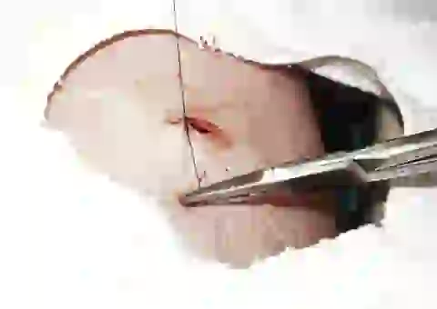
Surgical removal of moles
Depending on the size and depth, moles are removed either with a scalpel or with a special instrument. The mole in the photo below needs deep removal, let’s look at the process in more detail.
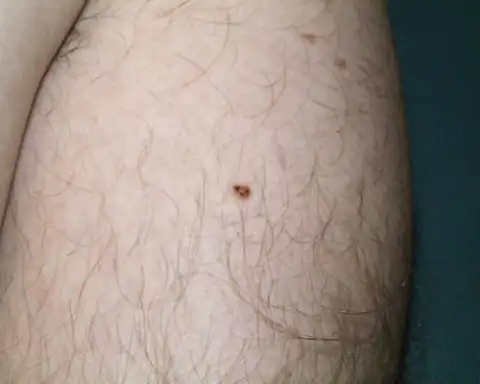
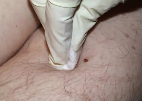
The surface of the skin is treated with an antiseptic:
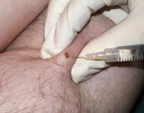
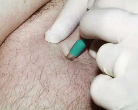
An anesthetic injection is given:
Mole removal tool:
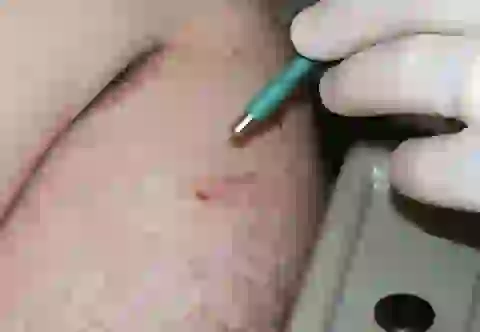
Excision of a mole to take its cells for biopsy:
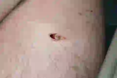
To remove a mole, tweezers are used:

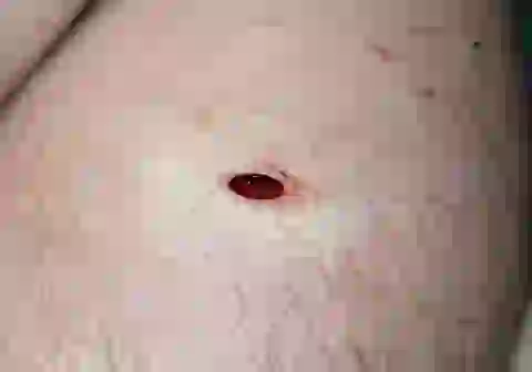
Wound after mole removal:
The edges of the wound are sutured:
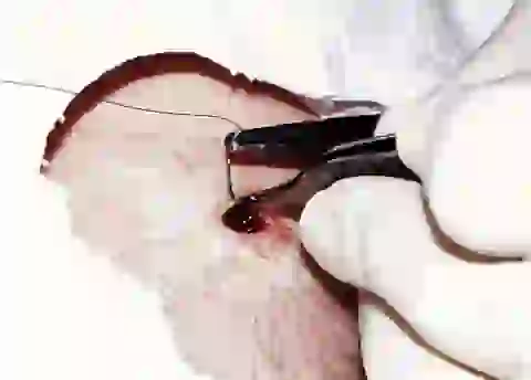
The wound is stitched up and ready for dressing:

The area is treated with hydrogen peroxide to remove dried blood and disinfect:


Dress the bandage daily for a week, then the stitches are removed.
Moles that are not deep or large in size are excised with a scalpel after anesthesia:

Care after mole removal
After removing a mole, the skin does not need any special care. However, in order to avoid the appearance of pigment spots at the site of the operation, you should follow some rules.
Firstly, after removing a mole, a crust remains on the skin. It is strictly forbidden to wet or tear it off, as this may affect the tissue healing process.
Secondly, you should avoid getting cosmetics on the wound.
Thirdly, you should protect the wound area from aggressive ultraviolet rays, especially in the summer. However, after healing, you should also protect the area where the removal surgery was performed as much as possible until the removal area no longer differs from the surrounding skin. To do this, it is recommended to apply a cream with high UV protection to this area.
It's rare to see a person without small dark marks on their body. Is it worth paying attention to these points? Only a doctor will distinguish between dangerous and normal moles - malignant melanoma or harmless nevus - and give recommendations on what to do with them. Is it worth worrying about the appearance of new formations, when immediate contact with specialists is required, what are the signs of cancer development - the answers to these questions remain to be found out. No one is immune from disaster, and early diagnosis will protect you from severe consequences.
What is a mole
The first tiny spots may appear in children in infancy. A mole is a small formation on the skin - a nevus - that is considered benign and harmless. The basis for their appearance is melanocyte cells that accumulate the natural pigment melanin. Depending on its quantity, a difference in color is observed. Available colors:
The shape of the tumors depends on the location and concentration of melanin. They may have a stalk or be located under the skin, be flat and convex. The most common type is round, but there are exceptions. The development of neoplasms is provoked by ultraviolet radiation - natural from the sun, in a solarium. Hereditary factors cannot be excluded. A common cause of growth is hormonal imbalance, characteristic of periods:
- puberty;
- pregnancy;
- menopause.
What types of moles are there?
One person may discover very different tumors. Types of moles are classified according to several criteria. This helps in correct diagnosis in case of changes. They differ in:
- origin– congenital, newly acquired;
- structure– pigment, vascular;
- place of education – in depth, on the surface, in the boundary layer;
- raised above the skin – flat – even, protruding as a hemisphere, pedunculated, larger birthmarks;
- potential threats – dangerous, degenerating into melanoma, non-dangerous.

Safe moles
Those who have dark spots on their skin should be wary of their changes. In time, detected signs of degeneration into melanoma contribute to the timely removal of the formation and preservation of health. Safe moles are different:
- the presence of a stalk – it cannot be formed by malignant cells that grow randomly;
- long-term condition without changes.
Spots that appear shortly after birth are not considered dangerous. It is important that they are small in size. Good – non-dangerous – signs of neoplasms include:
- flesh tone;
- unchanged pattern of the skin of the nevus and adjacent tissues;
- soft consistency;
- hair on the surface of the neoplasm - growing from the skin, indicates the absence of pathologies;
- diameter no more than 5 mm;
- symmetry;
- nevus in the form of a spot.
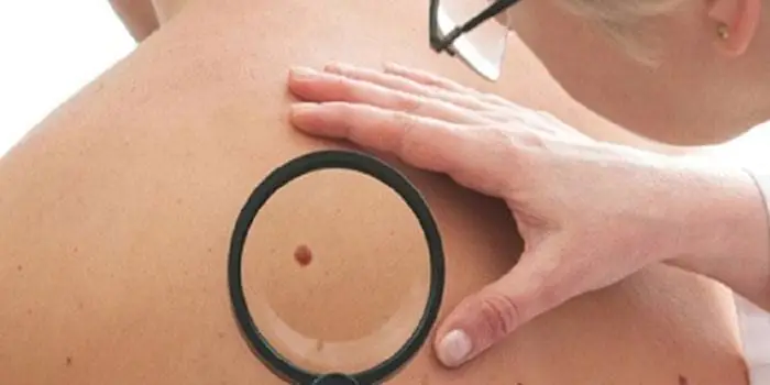
Which moles are dangerous?
Why do people with nevi on their bodies need to monitor their changes? There is always a threat of degeneration of non-dangerous tumors into a cancerous tumor. What moles are dangerous to health? Key signs you need to know:
- change in shades towards the dark side, the appearance of multi-color;
- rapid increase in size - exceeds two millimeters per year;
- occurrence of cracks;
- the formation of asymmetry due to uneven growth;
- lack of elasticity;
- the appearance of itching, burning;
- presence of discomfort.
The appearance of dangerous moles requires an immediate visit to a specialist to clarify the nature of the changes and the likelihood of developing skin cancer. Pathological transformations provoke:
- injury to the nevus due to negligence;
- self-removal;
- abuse of exposure to the sun, use of a solarium;
- location of the formation in places of frequent contact with clothing - on the neck, head, genitals, legs;
- placement in the hair, on the face, palms - where there is a high probability of injury;
- previously removed melanoma.
Why are moles dangerous?
Not a single person is protected from the sudden proliferation of cells of a harmless mole. Melanoma is an extremely serious disease. Changes not detected at the initial stage can result in death. The provoking factor is unsuccessful independent removal of tumors. Moles are dangerous because of their ability to:
- transform into an atypical – precancerous form;
- grow to large sizes;
- turn into cancerous;
- with minor external changes, metastases actively spread throughout the body through the circulatory and lymphatic channels.
How quickly does melanoma develop from a mole?
The transformation of a nevus into a cancerous formation can occur in different ways. The process depends on the stage of the disease and the type of tumor. Instant metastases are dangerous. Begins:
- growth of cancer (oncological) cells in the deep layers of the epidermis;
- their entry into the blood and lymph;
- penetration into the lungs, liver, kidneys;
- growth in these organs;
- complete damage to the body;
- death.
The growth phases of pigment cells are observed, along which melanoma develops from a mole. There are varieties:
- horizontal– damage to the upper layers of the skin occurs, lasting up to 10 years, but metastases do not appear;
- vertical– accompanied by the spread of cancer cells throughout the organs, can last two years, has an unfavorable prognosis;
- nodal – especially dangerous – characterized by deep spread within two months.
The first signs of melanoma
The patient can be assisted only when suspicious changes begin to be identified. The diagnosis, research, and referral for surgical treatment save a person’s life. The first signs of melanoma:
- increase in the height of the tumor;
- bleeding;
- the appearance of discharge;
- redness;
- burning, itching;
- swelling of tissues;
- softening of the nevus;
- the appearance of a crust;
- thickening;
- hair loss;
- expansion of pigmentation around the lesion.
With the further development of dangerous melanoma, the following are observed:
- significant change in size;
- the appearance of pain;
- enlarged lymph nodes;
- surface ulceration;
- formation of new foci;
- bleeding from places of pigmentation;
- liquid separation;
- skin thickening;
- the appearance of an earthy tint;
- signs of metastases are chronic cough, weight loss, cramps, headaches.
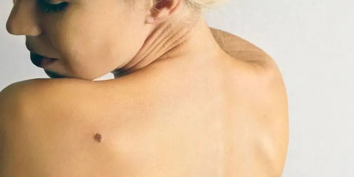
How to distinguish a mole from melanoma
To recognize which moles are dangerous and which are not dangerous, you need to know what they look like. A person with nevi, in order to avoid dire consequences, must constantly monitor the appearance of new formations and changes that occur. You can distinguish a mole from melanoma by its signs. Non-dangerous neoplasm:
- symmetrical;
- with smooth edges;
- uniform in color;
- with dimensions not exceeding 6 millimeters.
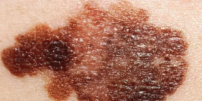
Features of dangerous melanoma that require seeking help from dermatologists:
- growth in a short time;
- pronounced asymmetry of shape;
- heterogeneity in color - the presence of inclusions of several shades;
- lack of clear boundaries - the contour line is blurred, jagged, and looks like a coastline on a geographical map;
- increased diameter over six millimeters;
- variability of any parameters - color, size, shape.
What dangerous moles look like
What do nevi that are subject to pathological changes look like? Only a doctor can correctly distinguish between non-dangerous tumors. Dangerous formations look like this:
- blue– compactions under the skin with clear boundaries, with dimensions no more than 10 mm;
- nodal– round, flat in shape, color – brown, black;
- cutaneous– often pale, convex;
- halo nevus – pigment surrounded by a light or white rim;
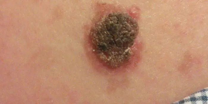
- spitz- looks like a dome-shaped tumor of pink shades, with the possible presence of a hole through which blood and liquid leak;
- connecting- connect individual entities into a whole.
Mole with jagged edges
One of the signs of a non-hazardous formation turning into a dangerous one is a change in contours. It often has blurred edges and scalloped borders. There are non-dangerous types of nevi - dysplastic. Only a specialist can make a correct diagnosis. A mole with uneven edges can be dangerous if there are additional signs of melanoma:
- accelerated changes in size;
- the presence of clearly defined asymmetry;
- the appearance of highly indented boundaries.
Rough mole
Such a neoplasm is harmless if its diameter is no more than 5 mm and remains constant in size. Often its appearance signals a lack of vitamins and nutritional disorders. Doctors advise coming for a consultation if it is discovered that:
- the smooth nevus turned into a rough one;
- bothered by burning, itching, tingling;
- irregularities and compactions appeared in the middle;
- areas with different shades formed;
- diameter has increased significantly.
A dangerous rough mole requires immediate examination if:
- the appearance of bleeding;
- development of the inflammatory process;
- rapid change in size;
- formation of asymmetry;
- formation of purulent discharge;
- the occurrence of painful sensations when touched;
- the emergence of an irregular shape, blurred boundaries, along the edges of the neoplasm.
Large moles
Large formations on the skin are pigment spots. When they remain unchanged and do not cause inconvenience, this is not a dangerous phenomenon. It is important to constantly monitor their appearance, color, and size. To eliminate worries, you need to consult a dermatologist. During the visit, the specialist will conduct a diagnosis and give a forecast of the risk of developing a malignant neoplasm. Large moles become dangerous if they:
- injured;
- thickened;
- started to itch;
- were unsuccessfully removed independently;
- changed in size, shape;
- are bleeding.
What moles can be removed
Often nevi cause trouble for women when they are in a visible place - the face, neck. Even if they do not bother you, using removal will be the right decision - the appearance will improve significantly. After the procedure, the doctor must necessarily send the tissue for histological analysis to decide whether the mole is malignant or not. If the neoplasm is not dangerous, does not bother you, and does not change in size, surgery is not required. What moles cannot be removed? Experts believe:
- there are no contraindications;
- It is important to choose the right excision technique.
You should be careful about skin growths; it is unacceptable to remove them yourself. Only the doctor will determine whether a nevus is dangerous or not and decide what to do with it. You can delete it if:
- injured from clothing - on the neck, in the groin area, under the armpits;
- cause pain when touched;
- are located under the hair on the head and can be damaged when combing or cutting;
- change color, shape, outline;
- significantly increase in size;
- characterized by the presence of burning, itching;
- accompanied by inflammation and bleeding.

A birthmark is a pigmented spot on or under the skin that appears mainly during a person's birth or immediately after birth. A birthmark is not dangerous. A mole is a nodular formation, in most cases protruding above the skin, formed not only after birth, but throughout a person’s life. Moles are dangerous, they can develop into a malignant tumor, it is dangerous to sunbathe a lot with a mole and injure it.

A mole, birthmark is a congenital or acquired defect of the skin, a benign tumor that consists of epidermal cells (the deep layer of the skin) and melanocytes - these are cells saturated with the pigment melanin. Moles appear in the first years of life, but a larger number of them are formed during puberty under the influence of the pituitary hormone, and new formations can appear in women during pregnancy, and old ones can change in color and size. How is a mole different from a birthmark? The fact that a birthmark on the skin is present from the moment of birth, and moles form or disappear throughout life. This is the main difference between a mole and a birthmark. They can also be differentiated, for example by size, color or shape.
