The accumulation of melanocytes leads to visible pigmented formations - moles. Their location on the body has no patterns. The presence of such formations does not cause discomfort to a person, but can serve as an aesthetic defect. There are also situations when the nevus is located on the body in traumatic places. Patients, having discovered such a tumor, begin to worry and think about getting rid of it. Whether moles can be removed or not is decided by the doctor individually in each case. He discusses the method of removal with the patient. There are dangerous and safe formations. Not every one of them is recommended to get rid of.
Signs of dangerous moles
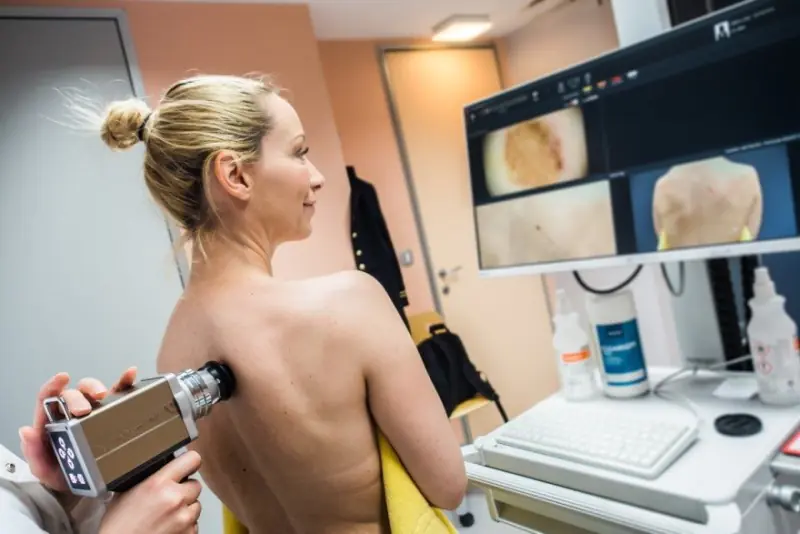
Pigmented formations that can develop into a malignant form are considered dangerous. A thorough examination is required before their removal. But already at the first examination, the doctor can assume the presence of cancer based on external signs.
The distinctive features of a dangerous nevus are:
- its sudden occurrence in adults;
- active growth;
- change in shape and thickening of the spot;
- glossy shine or flaking;
- emerging asymmetry;
- the occurrence of itching, burning sensation;
- loss of hair in the pigmentation zone;
- multi-colored area;
- bleeding;
- swelling and redness in the affected area;
- the appearance of nodules on the spot;
- exudative discharge from the formation.
If one or more signs of pathology are detected, it is strictly prohibited to remove pigment formations in beauty salons. This type of mole requires only professional examination in specialized clinics. Oncologists or surgeons prescribe tests. This is necessary to prevent infection, as well as for accurate diagnosis of moles.
Pros and cons of mole removal
All doctors are inclined to believe that any pigmented tumors that cause concern should be removed. Previously, it was assumed that a complication occurred at the site of the removed mole. But later it was found that compliance with all postoperative measures completely eliminates the risk of adverse consequences.
Hygiene rules after removal of a nevus:
- disinfect the wound at least once a day;
- abstain from water procedures for at least five days;
- do not tear off the formed crust from the wound;
- avoid ultraviolet radiation.
In rare cases, removal is considered preventive. It is indicated when there is a high probability of injury to the nevus.
If the cause of the nevus is a hereditary factor and similar formations are located in the same places in other relatives, then removal is contraindicated. Statistics show that most often such moles appear again in the same place.
Is it possible to remove moles for pregnant and lactating women?
Pregnancy and breastfeeding are a special time for a woman. Her body is being rebuilt, hormonal levels are changing, and the work of all systems and organs is intensifying. Therefore, nevus removal is not recommended.
Doctors can only authorize the procedure if there is a threat to the mother’s life. In this case, the doctor will insist on terminating the pregnancy. If treatment can be postponed to a later date, then the operation is postponed until postpartum.
Women carrying a child should not resort to home methods of getting rid of pigmented formations. This in most cases ends in complications, which is very dangerous for the mother and child.
Moles that appear on the mammary glands can interfere with the baby’s feeding process. In such a situation, you should trust a dermatologist, who will determine the degree of risk of injury.
What moles can be removed
Doctors recommend removing nevi if they:
- are located on the scalp, since there is a high probability of injury when combing;
- appeared on the face, lips and spoil the patient’s appearance;
- located in the armpits and intimate area, as they are susceptible to injury during shaving;
- are located in the neck, décolleté and are injured by jewelry;
- sharply begin to change, grow and become asymmetrical.

Removal of tumors should be carried out in specialized medical institutions. After removal, the mole is submitted for histological examination. Getting rid of a nevus on your own can trigger the development of melanoma.
Which nevi cannot be removed
Not all birthmarks can be removed. It is forbidden to get rid of large formations, as this is fraught with the development of inflammation. Before performing such an operation, the doctor conducts an extensive examination to ensure that the risks of complications are minimal.
If a dermatologist has even the slightest suspicion of melanoma or papilloma, then you need to show the nevus to an oncologist. Removal is carried out in an oncology clinic, where the patient remains after the operation for subsequent therapeutic treatment.
Any intervention without examination often leads to the degeneration of pigmented formations into a malignant form.
What are the dangers of mole removal?
The big risk during surgery is the transformation of the mole into cancer. With any incorrect action regarding a tumor, the risks of degeneration into oncology increase sharply. In this case, atypical cells spread throughout the body through the bloodstream and enter any organ and tissue.

Regular preventive examinations allow you to determine the malignancy of tumors at an early stage. This helps to avoid sudden complications.
Mole removal methods
If the examination shows that removal of the nevus is possible, then the doctor and the patient choose the method of excision. Each method has its own advantages and disadvantages.
The main methods for removing pigment formations are:
- Laser. It is performed under local anesthesia. A laser beam is used for burning. The operation is practically bloodless, since the coagulation of blood vessels occurs during the process. Very short recovery period. The formation is removed in one go. The disadvantage of the method is that the tumor is excised layer by layer, and this excludes the possibility of postoperative histological examination. It is impossible to determine the nature of the mole.
- Cryodestructive. Exposure to liquid nitrogen at low temperatures is used. Anesthesia is carried out locally. This method can remove hemangioma. It is difficult to control the depth of cauterization and it is impossible to select material for histology. Removal of large tumors results in noticeable scars.
- Electrocoagulative. Low frequency currents are used. It is used less and less due to the high risk of injury. There is a plus - you can take a nevus for examination.
- Radio wave. Small superficial nevi are removed. During the operation, a high-frequency radio knife is used. Does not injure the skin and does not leave noticeable marks.
- Surgical. Best suited for excision of large pigmented formations, as well as nevi with deep penetration into the skin. During the operation, a scalpel is used. The nevus and part of the skin are excised. The material for further research is taken in full. Long rehabilitation period. After removal, scars and scars often remain.

At any age, it is necessary to be attentive to skin tumors. Parents are required to carefully monitor moles on their children's skin. Timely contacting a dermatologist eliminates the adverse effects of nevi on the body.
Probably, each of us has heard a story about someone who removed or injured a “mole” and died a month later. Let's figure out what is true in this story and what is from the realm of “horror stories”.
There really is such a tumor as melanoma. It develops from pigment cells of the skin; in rare cases, melanoma may develop in the cells of the membranes of the brain or eye. Old school oncologists call melanoma the “queen” of tumors. In stages 3 and 4, the patient's death occurs in 80-90% of cases. But we should also not forget that with stage 1 of the disease, with adequate treatment, the patient’s recovery can be guaranteed in 95 cases out of 100.
As already mentioned, in most cases, melanoma develops from pigmented nevi or what are popularly called “moles.” A pigmented nevus is a cluster of pigment cells - melanocytes, which on the skin look like formations from light brown to black-blue. It is also necessary to pay attention to the fact that there are a lot of other formations of different colors on the skin that have nothing to do with pigmented nevi and melanomas. In many cases, only an oncologist can determine their nature.
What are the predisposing factors that cause a harmless “mole” to turn into melanoma?
1. Constant trauma to the nevus, especially when it is located in areas of friction with clothing.
2. The presence of a large number of pigmented nevi on the human body (the risk of developing melanoma increases several times if there are more than 15 pigmented nevi on the human body).
3. Prolonged exposure to ultraviolet radiation (for example, in Australia, skin melanoma is an occupational disease among people working outdoors). Solar insolation is especially dangerous for children and adolescents.
Let's look at the first signs of malignancy of a pigmented nevus, or what should make you urgently contact an oncologist:
1. Itching and peeling in the area of the nevus.
2. Change in the size, shape, color of the nevus, and the color can change in both dark and light directions.
3. Bleeding and destruction of the nevus.
4. The appearance of a blue or purple rim around the “mole”.
5. The appearance of small brown or black formations around the main tumor.
Remember that timely consultation with a doctor will save your life.
If the patient does not consult a specialist when the first symptoms appear, the malignant transformation of the nevus will take its course, in accordance with the biological laws of tumor growth. Manifestations of the disease will sooner or later lead the patient to the doctor, but as already mentioned, at 3-4 tbsp. recovery occurs in only 5-20% of cases. There will be another reason to tell your friends a terrible story that after the removal of a “mole” a person died.
Measures to prevent malignant transformation of pigmented nevus:
1. Wearing loose clothing that does not compress the nevi.
2. Avoid exposure to ultraviolet radiation on nevi (when in the sun, cover the “moles” with an adhesive plaster or use sunscreen with a protection factor of at least 50 IU).
3. Preventive removal of nevi (it is especially recommended to remove nevi in areas of friction with clothing and in open areas of the body).
A radically removed nevus will never become a source of melanoma development.
Methods for removing nevi:
1. Surgical method: includes excision of the formation with a scalpel followed by suturing. The most reliable method for removing any skin formations. Allows you to radically remove the formation and obtain material for histological examination. The method is well suited for removing large formations, especially on the body. When used on the face, it can cause significant cosmetic defects.
2. Cryodestruction (removal with liquid nitrogen): a good method for small formations, as well as for the removal of vascular tumors in children. Not a bad cosmetic effect. Disadvantages - it is impossible to obtain material for histological examination.
3. Electrocogulation (removal using an electric loop). The method is quite easy to use, bloodless, there are no stitches, a short rehabilitation period, it is possible to take a biopsy. Disadvantages - poor cosmetic effect: white spots remain, high risk of keloid scars.
4. Laser destruction (removal using a high-energy laser). An excellent method for removing small skin formations. When removing large nevi, there is a high risk of scar formation. It is difficult to take material for a biopsy.
5. Radio wave excision (removal using devices generating high frequency radio waves). Allows you to remove both small and large formations. Very good cosmetic effect. The risk of scarring is minimal. Short rehabilitation period (10-15 days). Allows you to take a biopsy.
In any case, no matter what method you choose to remove a nevus, it is important that it be done by a specialist, preferably an oncologist or a dermatologist with oncological training. Remote lesions must be sent for histological examination. You also need to know that pigment formations are always present. are completely removed. and then sent for histological examination. It is unacceptable to remove the pigmented nevus in parts, because if there are areas of malignancy in the nevus, this will be enough for metastasis.
If a histological examination reveals areas of a malignant tumor in a remote formation, then within 1 month from the date of intervention it is necessary to perform surgical excision of the formation with sufficient deviation from the edges of the tumor. This will prevent recurrence of melanoma.
Do not forget to contact specialists in a timely manner, even if your worries are completely groundless.
A mole is an acquired or congenital formation on the body, accompanied by a change in skin color (pigmentation). Birthmarks can have different diameters and shapes. For many patients, the urgent question is: what is the danger of removing moles? After all, many have heard frightening stories about the development of skin cancer after removing or damaging a spot. Is this really true?
Causes of moles and indications for removal
A birthmark (mole, nevus) is a benign formation on the human dermis. The mechanism of development of moles is the degeneration of dermal cells into melanocytes (skin cells that synthesize the pigment melanin).
Causes of nevi:
- The influence of ultraviolet radiation. Prolonged exposure to the open sun not only contributes to the formation of new birthmarks, but also causes the risk of transformation of benign formations into malignant ones. According to statistics, people whose skin is regularly exposed to ultraviolet radiation are more likely to suffer from cancer.
- Hormonal imbalance in the body. It has been proven that hormonal surges often provoke the appearance of new nevi or their disappearance.
- Skin injuries lead to the formation of certain types of formations.
- Infectious diseases. In children and adolescents, spots often occur due to damage to the body by viral or bacterial infections.
- Hereditary predisposition. If parents have many birthmarks on their skin, the baby is also more likely to develop nevi.
- Other diseases. Pathologies of the thyroid, pancreas, liver, vitamin deficiency, radiation exposure, and hormonal disorders can provoke the development of moles.
The danger of moles lies in their ability to degenerate into malignant tumors. Experts advise paying close attention to birthmarks and regularly visiting an oncologist and dermatologist.
