Hemangiomas, better known as red moles, are benign growths that originate from blood vessels.
Red moles appear mainly in children of both sexes, less often in adults. It is impossible to prevent or predict their occurrence.
There is still debate as to which pathology a hemangioma is classified as - a vascular tumor or a congenital malformation. Recent data confirming the occurrence of tumors due to proliferation of vascular endothelium allow the neoplasms to be classified as vascular tumors.
What is a red mole?
Why are moles and dots red? Because it is actually vascular tissue filled with blood. If ordinary moles are skin growths, then red moles are several small (or single) overgrown blood vessels. In a pronounced process, the accumulation of blood vessels merges into a blue or burgundy spot.
Prevalence and localization
In most cases, vascular tumors are detected immediately after birth (87%), and 70% of the total mass are girls, who, accordingly, fall into the highest risk group. This pathology accounts for about 48% of all soft tissue and skin tumors in childhood. 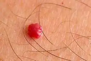
On the body, a red mole can be localized in any part; about 80% of tumors occur in the upper part of the body. Very rarely found in internal organs - liver, brain, lungs, bones.
- about 95% of all diagnosed vascular tumors are simple formations
- about 3% are cavernous
- and another 2% are mixed and combined variants of the disease.
Causes
No doctor can still give an exact answer as to why these formations appear. Why there are many red moles in the facial area is also difficult to explain. This is probably due to the abundant vascular network of facial tissues.
In children
It is generally accepted that a congenital tumor develops as a result of an intrauterine disorder in the formation of vascular tissue, which occurs against the background of incompetence and the processes of development and growth.
How does this happen? During the formation of organs and systems, vascular tissue penetrates all parts of the body without exception along a certain chain of pericytic cells. These cells, being a kind of conductors of information, react to the slightest lack of oxygen: if the fetal tissues experience hypoxia, the synthesis of special proteins that attract pericyte cells is immediately launched. These cells begin to pave new blood supply routes, thus eliminating hypoxia. In some cases, even after the cessation of hypoxia, the synthesis of specific proteins does not stop; the vascular system continues to develop, turning into voluminous tumor-like formations.
The second name for red moles is vascular hyperplasia. This means that the tumor arises as a result of disruption of the growth processes of vascular tissue, which lead to an increase in its quantity. How and in what way this process occurs is difficult to answer with 100% accuracy, since this requires monitoring the characteristics of intrauterine tissue development. The data presented are based on autopsy results of aborted and stillborn fetuses.
In adults
- Acquired pathology is associated with hormonal disorders, explaining the appearance of hemangiomas in adults (pregnancy, menopause, diseases of the endocrine system, as well as hormonal therapy or oral contraception).
- There are suggestions about the negative impact of ultraviolet and radiation exposure, viruses and chemicals that provoke tumor growth in adults.
- Microtraumas and skin cracks with permanent damage to the capillary network lead to such neoplasms.
- Long-term and uncompensated hypovitaminosis C, leading to thinning and fragility of capillaries, is also relevant among the causes.
- Red moles accompany the course of other diseases (for example, diseases of the liver, pancreas, cancer of internal organs). It is not uncommon for a cluster of red moles in a certain area of the body to indicate a predisposition to cancer in this area, a nearby organ.
Red moles in newborns
This is a common occurrence in babies, and if such a mole is noticeable in a newborn, then most often by the age of 3-5 the red mole may disappear. Since this is a benign tumor, it is not dangerous if:
- Does not bother the baby (skin itching, irritation, pain)
- Does not increase in size (in a month, for example, it doubled)
- Located in a non-hazardous place (if it is located under the eye, on the nose, genitals, on the face, then its removal is indicated)
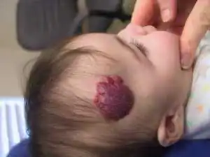
Red moles are characterized by rapid peripheral growth, especially intense in the first months of a child’s life. Therefore, 10-12% of hemangiomas in children are removed for medical reasons. During the growth process, the tumor destroys tissue and leads to a cosmetic and sometimes functional defect, especially when located near or on vital organs (eyes, ears, brain). Impaired function of organs and tissues occurs due to compression of them by the tumor.
Features in adults
Primary hemangiomas do not occur in adults, i.e. They arise from existing, undiagnosed tumors. As a rule, visible formations are treated even before school age, so in adulthood either untreated superficial moles or tumors on internal organs are discovered.
Of particular danger is a vascular tumor on the spine, which is localized in the vertebral body and weakens its structure, sometimes leading to fractures.
Classification
According to morphology
Capillary. The histological structure of the neoplasm is compact layers or concentric groups of capillary vessels, closely adjacent one to one. The wall of each vessel consists of a basement membrane and 1 or several layers of epithelial-like cells. The lumens of fused capillaries are filled with formed elements of blood. In some cases, groups of vessels form lobules separated by stroma.
Cavernous. It consists of multiple cavities of various shapes and sizes, which are lined with 1 layer of endothelial cells, similar in structure to the endothelium of blood vessels. In some cases, rupture of the septa occurs with the formation of papillae in the lumen of the caverns.
According to location, vascular hyperplasias are divided into:
- Simple, with a subcutaneous location throughout the body;
- Cavernous, localized under the skin;
- Combined, having a supra- and subcutaneous part;
- Mixed, including other tumors, for example, lymphangioma, originating from lymphoid tissue.
By origin:
- Congenital, appearing immediately after birth or in the first months of life;
- Acquired, occurring in adults. Acquired red moles can only be of a subcutaneous location, i.e. simple. Complex forms of the disease, discovered due to complications or by chance, are congenital and not diagnosed in childhood.
With the flow:
Simple, not presenting a risk of complications or dysfunction of organs;
Difficult:
- near large vessels or vascular nodes;
- on or near vital organs and structures (eye, brain, ear);
- in places that are difficult to access (vertebrae).
Features of red moles
Vascular tumors have a number of characteristic properties that differ from other neoplasms:
- Rapid tumor growth during the first three months after birth.
- Accelerated (2-3 times compared to full-term) growth of education in premature babies.
- The likelihood of spontaneous regression of simple tumors (mostly small) during the first years of life. This explains the cessation of hemangioma growth when exposed to a number of factors, such as heat, cold, and certain chemicals.
- The impossibility of spontaneous resolution of cavernous, combined and mixed variants of pathology.
- Unpredictability of further development even after growth and involution have stopped.
Clinical picture
Simple angioma
This is a spot of varying sizes, predominantly red, rising above the skin. With simultaneous finger pressure on the edge of the tumor and healthy tissue, the angioma becomes pale and shrinks, and after the compression stops, it returns to its previous shape and color. In babies up to 3-4 months, peripheral growth of the vascular tumor is clearly visible. This can be verified by making an initial paper stencil of the tumor and applying it to the hemangioma after 15-20 days.
Cavernous angioma
This is a formation in the subcutaneous tissue with unchanged skin above it. It can be diffuse without clear boundaries or encapsulated. A bluish-colored formation is detected under the skin; in some cases, feeding vessels are visually visible. When pressing on the skin above the tumor, the formation decreases, and when the compression stops, it returns to its previous size.
The skin over the tumor may be warmer than the rest of the skin. No pulsation is detected above the formation. In some cases, upon palpation, the lobulation of the formation is noticeable. Cavernous hemangiomas located on the head, neck and near the ears are characterized by rapid growth with active germination into surrounding structures.
Combined angioma
This is a formation with a cutaneous and subcutaneous part; the subcutaneous part, as a rule, is larger.
Mixed tumors
These are various variations of the combination of a vascular tumor with lipoma, lymphangioma, keratoma and other neoplasms.
Spontaneous resolution
True regression of simple or superficial hemangiomas is observed in 10-15% of cases, especially when tumors are located in closed areas of the body. The brightness of the formation decreases, whitish areas appear, and peripheral growth completely stops. After 6-8 months. the hemangioma transforms into a smooth whitish-pink spot that does not rise above the surface of the skin. The skin over the spot undergoes atrophy, leaving only a small depigmented area by the age of 3-4 years.
Complications
Red dots are dangerous due to rapid growth and subsequent compression of nearby structures with disruption of their function, which is especially important when hemangiomas are localized in the brain, in the liver, or near the eye.
- Ulceration and inflammation during growth. Some types of red moles undergo reverse development after such complications.
- Bleeding due to injury, especially dangerous with extensive cavernous and combined hemangiomas, as well as tumors located on internal organs, since such bleeding is very difficult to stop.
- Infection (bleeding, ulcerated moles), i.e. the addition of a bacterial skin infection.
Diagnostics
With superficial hemangioma, the diagnosis is made on the basis of clinical and histological data. For extensive and deep processes, angiography is performed to determine the connection of the tumor with the vascular network, as well as radiography, which provides accurate data on the size and depth of the vascular tumor.
Treatment of red moles
Is it possible not to treat red moles? If the tumor does not interfere with organ function, is not dangerous for bleeding and does not grow, these marks of intrauterine life can be left without treatment, especially since these tumors do not carry the risk of malignancy. Moreover, it is not recommended to remove moles if they do not bother you, do not increase in size, or are located on closed parts of the body (they are not a cosmetic defect).
For extensive and deep processes, the doctor selects treatment - surgical or conservative; methods can be combined to increase efficiency. Therapy depends on the type of tumor, its location and size, growth rate, the presence of complications, and the age of the child.
Simple hemangiomas
Low-temperature destruction or cryodestruction is considered an effective method for treating small red moles. It can be performed in several ways: direct application of crystalline carbon dioxide to the surface of the tumor for 15-20 s or instrumental cryodestruction using liquid nitrogen. The effectiveness of treatment is up to 96%.
For simple angiomas of large size, hormonal treatment with prednisolone is advisable at the rate of 4-6 mg per 1 kg of weight, taking 1/3 of the dose at 6 a.m. and the remaining portion at 9 a.m. The duration of treatment is 28 days with the drug taken every other day. Gradual withdrawal of the drug is not required. During treatment, blood sugar and potassium are monitored.
Laser removal allows targeted action strictly on the tumor with minimal cosmetic defect. Modern laser systems with various types of pulses can coagulate both superficial and deep subcutaneous tumors without destruction of healthy tissue and complications.
Cavernous
When the process is located in a cosmetically unfavorable part of the face (cheek, nose, forehead, bridge of the nose), sclerosing therapy is used: special substances are introduced into the angioma, leading to aseptic necrosis and subsequent scarring of the tumor under the skin without scar formation and tissue deformation. Hydrocortisone, quinine-urethane, sodium chloride solution 10%, ethyl alcohol 70% are used as sclerosing agents. For complete sclerosis of the tumor, 10-15 injections are performed with breaks between each injection of 14-30 days, i.e. the process is quite lengthy.
When cavernous hemangioma is located on the thigh, shoulder, back and other closed parts of the body, surgical removal of the tumor is performed.
Combined
When the tumor is localized on closed parts of the body, radical surgical excision is advisable. Removing red moles rarely leads to any complications; the tumor is removed entirely with minimal cosmetic defect.
When localized on open parts of the body and face, microwave cryodestruction is recommended: irradiation of the hemangioma with an ultra-high-frequency electromagnetic field, followed by cryodestruction. This combination makes it possible to significantly enhance the destructive effect of freezing, while maintaining the ability of epithelial cells to regenerate.
Hormonal, sclerosing and radiation therapy with Buki rays, which have a middle range between X-ray and ultraviolet irradiation, is also used.
Deep and extensive hemangiomas with dangerous localization
Such tumors are located on the neck, near the ears, on the head and are characterized by constant peripheral growth. The tendency to bleeding and ulceration of these types of angiomas does not allow the use of the treatment methods described above.
In case of such a pathology, angiography is mandatory to determine the nature of the blood supply to the hemangioma and its anatomical relationship with nearby tissues and structures. One of the effective treatment methods is tumor embolization with hydrogel, which reduces the blood supply to the tumor and its size.
Then cryodestruction is carried out without removing the tumor itself: after the necrobiotic process, the tumor partially resolves, leaving behind areas of atrophic skin, i.e. a cosmetic defect that can be eliminated by skin grafting if the patient wishes.
Angiomas, known as red moles, in medical practice are usually classified as benign formations that consist of lymphatic or blood vessels. Their appearance is explained by dysfunction of either the circulatory or lymphatic systems. They form throughout the entire period of a person’s life, but only in childhood up to 7 years are angiomas able to disappear on their own.
Red moles on the body - what are they?
What do red moles on the body mean? According to experts, they are an intermediate link between a malformation and a tumor. The medical literature provides little information about this phenomenon. This is due to the fact that red moles do not pose a particular danger to humans. They are believed to be innate.
Types of red dots (angiomas) with photos
Red moles are usually divided into several varieties. Their classification is based on factors such as the cause of its appearance, location on the body, and the type of vessel that caused its formation. Depending on the cause of angioma and the composition of the tissue, there are red dots or moles of several varieties:
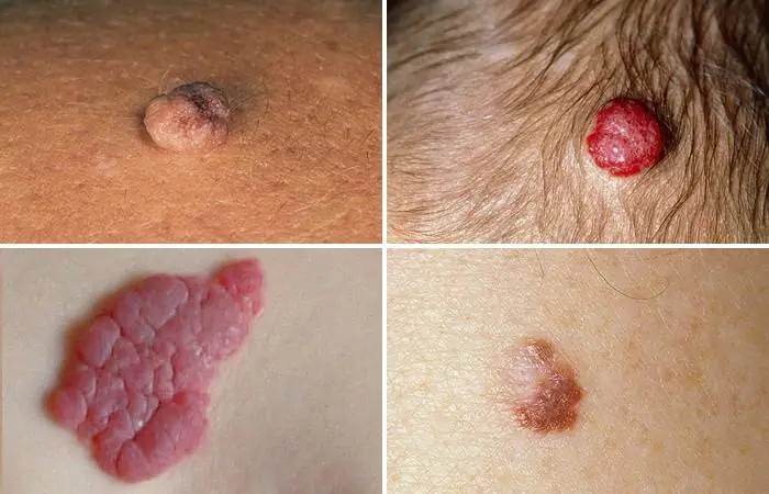
- pineal - a convex neoplasm that rises sharply above the skin;
- nodular - a small pinpoint formation that appears as a result of a blood vessel emerging onto the surface of the skin. This angioma does not have a capillary branch;
- branched or arachnid - a network of small blood vessels extends from the angioma;
- flat - a mole on the skin in the form of a compaction.
Depending on the vessels underlying the formation of red moles, they are called hemangiomas - moles formed due to abnormalities of blood vessels, and lymphangiomas - points that appear due to disruptions in the lymphatic system. Hemangiomas are impressive in size. Lymphangiomas are a rare occurrence. These are small nodules that are soft to the touch and easily compress when pressed. Formed on the neck, mouth, axillary and groin areas. Hemangiomas formed from vessels of the circulatory system are distinguished by external features and size:
- Capillary - formations that can be located anywhere in the body and look like a blue-purple or bright red spot. They are formed due to the expansion of a capillary vessel.
- Cavernous or cavernous are large formations containing several blood vessels that merge into large cavities. Such hemangiomas are located above the skin. A typical location is the face. Sometimes they can be located on internal organs - the uterus, spleen, liver.
- Branched - they are a pulsating, swollen formation that is filled with blood; they combine several simple moles.
- Punctate are the smallest hemangiomas, which appear as small dots.
Where are they located?
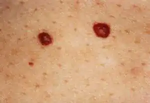
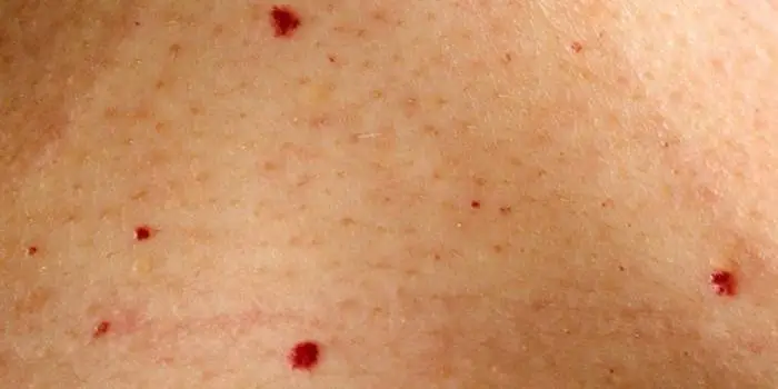
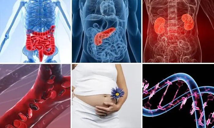
Angiomas are formations of different sizes and shapes. They are localized in tissues and organs: on the skin, in fatty tissue, bone tissue, liver, muscles, brain and kidneys. Such spots appear on the body in childhood and adolescence. Their appearance can also provoke pregnancy. Red dots can be single or multiple. If the former do not pose a danger, then the sudden appearance of multiple angiomas signals serious problems, which may include cancer.
Simple angiomas are located on the skin of the face and scalp. They are characterized by their small size, spherical shape, and purplish-blue color. In some cases, angiomas can occupy large areas on the eyelids, cheeks, nose, and external genitalia. Cavernous angiomas are localized in the subcutaneous area. Sometimes they grow into deep layers - into muscles and bones. This type of formation can develop in internal organs. 80% of angiomas are located in the upper part of the body: on the chest, back, head, face, neck, arms.
The most unfavorable location of angiomas is the head. This is explained by the possibility of damage during haircuts, combing or blow-drying. Red moles on the chest cause particular concern for women. Their single appearance does not pose any danger. However, if angiomas change color, begin to grow, itch or hurt, you should consult a doctor.
Causes of red moles on the skin
According to experts, in most cases such moles are congenital. The sources of their development are pathologies of the vessels of the lymphatic and circulatory systems. Angiomas can appear at any age. Red moles are often observed in children. From the point of view of doctors, this is explained by the transformation processes characteristic of this age.
In girls, angiomas form more often. Small formations go away on their own, without drug intervention. One of the reasons for the appearance of angiomas in newborns is considered to be infectious diseases suffered by the mother during pregnancy. In adults, the formation of angiomas occurs under the influence of hormonal changes and various disruptions in the functioning of the body. Among them:
- gastrointestinal diseases,
- pathology of the pancreas,
- exacerbation of chronic infections,
- pyelonephritis,
- deterioration of blood microcirculation,
- dysfunction of pigment cells,
- pregnancy,
- hereditary factor
- hormonal changes,
- lipid metabolism disorders,
- cardiovascular diseases.
Doctors believe that excessive sunbathing and solarium contribute to the appearance of red moles. It is assumed that angiomas can accompany diseases such as Henoch-Schönlein disease, rheumatoid arthritis, and lupus erythematosus. This happens because the immune system, due to emerging pathologies, sees the body’s cells as foreign and tries to suppress them. According to doctors, the appearance of angiomas may signal a blood clotting disorder.
How to remove red dots on the human body
In most cases, these formations do not require medical intervention. However, if the angioma bothers you - it itches, hurts or enlarges, it is recommended to remove it after an appropriate examination. Angiomas are removed if they negatively affect a person’s appearance, are located in places of contact with clothing, interfere with the performance of procedures to care for one’s appearance, or are often subject to accidental damage. Modern medicine offers several options for removing angiomas:

- Surgical excision. This method of removing a red mole is carried out only in a hospital setting. Performed by a surgeon without hospitalization. After excision, scars may remain on the surface of the skin, so the method is used to eliminate small moles located on the body. Surgical excision is not used for formations located on the face.
- Laser removal is the most popular way to get rid of angiomas. It is produced in layers, which ensures work with formations of any depth. Before the manipulation, the location of the angioma is anesthetized with an injection or application of an anesthetic gel. The procedure is short-term and lasts a few seconds. After excision, a crust forms at the site of the angioma within 24 hours, which disappears after 1-3 weeks. After complete healing, minor scars remain at the site of the mole.
- Cauterization is an effective and safe way to get rid of angiomas. Leaves no traces. Cauterization is performed using the coagulation method or using carbon dioxide and nitrogen. The first method can be used to remove large capillary formations. Before the procedure, local anesthesia is performed. Several coagulation methods are used - electrocoagulation, radio wave, infrared and light. Carbon dioxide is used to remove only small formations that are located above the surface of the skin.
Regardless of the chosen method of removing a red mole, it is recommended to make a preliminary diagnosis to exclude oncology. The specialist will be able to choose the best option for removing the angioma. After getting rid of a mole, it is not recommended to visit the sauna, solarium or expose the area where it was previously located to insolation for two months.
Treatment at home using traditional methods
Traditional methods of treating red dots can be used only when the mole is not inflamed, does not bleed, and is small in size. It is prohibited to treat large formations and angiomas that penetrate deep into the skin. According to reviews, the most popular and effective folk methods of getting rid of red moles are:

- Bee Honey. They lubricate the unwanted formation several times a day. After 10 days, the moles will begin to shrink.
- Castor oil. To reduce the size of a mole, apply it to its location overnight.
- Black radish is used to lighten moles. For this purpose, the root vegetable is grated on a fine grater, and the resulting pulp is applied to the problem area. The procedure should be carried out 2-3 times a day.
- Dandelion root. Clean, crushed root is applied to the mole every day for two hours.
- Onion. It is crushed and the juice is squeezed out of the resulting pulp. Lubricate the mole with it. After a month, the formation will dry out and disappear.
Are angiomas dangerous?
In most cases, angiomas are not dangerous. However, in some cases (very rarely), under the influence of unfavorable factors, they degenerate into malignant formations. Among these reasons, experts identify: damage to clothing, exposure to ultraviolet rays. In addition to the danger of degeneration, the cost of damage to the angioma's halo can be heavy bleeding.
Angiomas located in places of contact with clothing - in the abdomen, shoulders, neck, chest - require special attention. Hanging moles, which are easiest to pick off, and angiomas on the head are susceptible to increased trauma. Constant scratching, using a hairdryer, hairpins, headbands, and haircuts can damage the nevus. The danger is caused by angiomas located in the mouth, on the lips and near the organs of vision and hearing. In the oral cavity, moles can be subject to constant mechanical stress, which will contribute to their growth.
What to do if a red mole itches?
If a red mole begins to itch, this is a signal about the beginning of its growth, which may be associated with its degeneration into a malignant tumor. However, in some cases, angioma begins to itch when there is a disruption in the functioning of any body systems or a change in hormonal levels. Itching and flaking of red moles are often observed in pregnant women.
You will not be able to determine the reason for this behavior of a mole on your own, so to clarify the situation, you should consult a dermatologist or an oncology clinic. The hospital will order a series of tests, which include studying hormonal levels and determining the presence of malignant cells. Circular movements over the angioma or light pressure will help you get temporary relief from itching. You can't scratch a mole.
Is it possible to sunbathe and lead an active lifestyle?
The presence of red moles is not a contraindication to an active lifestyle. The only limitation may concern the possible methods of influencing angioma. If it interferes and is located in places of possible contact with surfaces and objects - on the arms, legs, it should be removed to avoid injury. As for sunbathing, as in the absence of red moles, doctors advise not to get carried away with sunbathing.
If there are any types of moles on the body, including red ones, being in the open sun is allowed before 10 a.m. and after 7 p.m. Due to increased activity of ultraviolet rays near bodies of water, you should stay away from water. To protect angiomas from sun rays, dermatologists recommend applying creams with a protection filter of 30 units.
There are a lot of moles on the skin of any person, which differ in different sizes and shapes.
Many of them are harmless, while others may pose a hidden threat.
Let's look at what red moles on the face are and why they appear, as well as what methods can be used to remove them safely.
What are red moles?
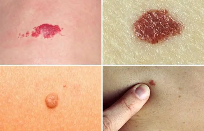
Science calls red moles angiomas. They are formed from blood vessels. If the neoplasm does not include blood vessels, but lymphatic vessels, then it is called lymphangioma. True angioma is divided into cavernous and simple varieties.
A simple capillary nevus of red color is located mostly on the face (on the cheeks or on the forehead); in size it can reach the dimensions of the palm.
Based on the characteristics of tissue composition, causes of appearance and placement in the layers of the epidermis red moles are divided into the following types:
- knotty – neoplasms in the form of dots, which indicate the emergence of a vessel on the surface of the epidermis. There are no branches in the form of capillaries around the mole;
- pineal – protrude sharply above the skin;
- branched – there are many blood vessels coming from the neoplasm;
- flat – the neoplasm looks like a plaque.
If a red mole appears, do not panic. Most often, it is not fraught with danger.
A common distinctive feature of red moles is that with slight pressure they become paler, and then return to their original shade.
Reasons for their appearance
Angiomas do not pose a health hazard, but if they begin to form in large quantities and frequently, this is a reason to consult a doctor. Enough The appearance of red moles is often associated with problems with the liver, its incorrect function. Other causes of red nevi are:
- improper diet, due to which excessive amounts of toxins are formed in the intestines;
- the occurrence is often provoked by abnormal proliferation of blood vessels;
- the size of such nevi ranges from 1-4 mm. They are placed on different parts of the body. The reason for the formation of moles on the face and other visible areas is often excessive exposure to the sun;
- The cause is also a hereditary factor;
- some changes in the hormonal background of the female body can also cause the formation of moles;
- age factor, when the body undergoes a change in the functioning of all systems and some malfunctions;
- disorders in lipid metabolism;
- diseases of the heart and blood vessels;
- pathological conditions of the liver and pancreas.
How can they be dangerous?
Representing a bundle of blood vessels, such moles can cause bleeding if there is mechanical damage. Particular attention should be paid to angiomas that are located in the area of excessive friction and contact with clothing.: on the neck, stomach, shoulders, chest. Moles on the scalp are subject to considerable trauma. And when located on the face, they can be accidentally touched during mechanical cleansing of the skin or scrubbing.
The appearance of one red mole or a scattering of such “surprises” on the skin is a message from the body about changes inside due to age, hormonal imbalances, and improper gastrointestinal function. If angiomas rapidly increase in size and occur in large numbers, it is important to consult a doctor.
Methods for removing angiomas
Cryodestruction
Similar method is based on freezing the tumor. It is used very successfully in medicine. The principle of influence is that liquid nitrogen with a temperature of minus 195 degrees Celsius is supplied to the tip of the device for the operation. He touches the nevus tissue. In this case, crystals immediately form in the cell and their functionality is stopped.
The procedure will take about two minutes. Over the course of a month, the mole disappears on its own, leaving a barely visible spot in its place.
Laser removal
This method differs in that angioma is burned out using a neodymium laser. It affects only the angioma cells, without damaging or heating the tissue next to it. So, after exposure, the mole acquires a dark burgundy color, and after a while it becomes lighter or disappears completely.
The laser affects the tumor gradually, layer by layer, causing a slight tingling sensation. Today this method of removal is considered the most progressive and successful.
Radio wave
Another non-traumatic and effective method of disposal is radio wave. The use of this method is a suitable choice for getting rid of red moles on the face and other visible areas of the skin, because nearby tissues are not injured in any way.
After regeneration, no trace remains. Using the radio wave method, you can eliminate red moles without a trace, because such a wave does not harm the tissues around the formation.
Drug treatment
The method has been used recently. Dynamic studies provide evidence that the medications used have a positive effect on the course of the disease. But absolute disappearance does not happen often. Thanks to the possibility of getting rid of angiomas with modern medications, patients are able to refuse surgery. Drugs for getting rid of red moles are as follows:
- Propranolol. It is able to influence capillary receptors, which causes their narrowing, reducing the rate of endothelial growth in blood vessels. The course of treatment reaches six months;
- Prednisolone. This is a hormonal agent that activates scar formation in the capillaries. They shrink into the mole, blood flow stops, and the angioma itself stops growing. The medicine is used for about 18 weeks;
- Vincristine. It is an antitumor drug that is used for large angiomas that grow into tissue. The process of cell division and enlargement in the mole is completed. But the medicine has many negative effects, so it is prescribed if it is impossible to use other methods. It is administered intravenously.
Patients with a large number of such nevi should visit a gastroenterologistto exclude disorders of the pancreas and liver. If they are present, treatment of skin defects is also accompanied by therapy for the main problem.
Folk remedies
Getting rid of red nevi at home is allowed for small formations if there is no tendency to actively increase. It is strictly prohibited to cauterize or lighten large tumors that affect the deep layers of the skin. Independent attempts to remove the tumor can cause the growth of moles, bleeding, and suppuration.
Any treatment method must be determined by a specialist! Self-medication in such a situation is unacceptable.
If you still decide to try traditional methods of treatment for yourself, here are the most popular recipes:
- after a month of use, results will be obtained by applying castor oil (castor oil) to the tumors every day;
- the use of ointment against viruses “acyclovir” also gives results after a month of daily use;
- Black radish gruel can lighten angiomas, which is applied to problem areas 2-3 times a day;
- Compresses from ground dandelion roots should be made every day for at least 120 minutes;
- You can dry the angioma using freshly squeezed onion juice;
- good results are achieved by applying honey to red nevi;
- grind the milkweed grass and apply a compress every day for a couple of hours for a week;
- You can also lubricate red moles with potato juice;
- chop the apple, add honey (in equal parts). The composition should be applied overnight. Cover the top with a cotton rag, and then insulate it with cling film. The course of treatment can reach three to four sessions;
- prepare a mixture of 50 ml of apple cider vinegar and three drops of lemon essential oil. Lubricate red growths twice a day;
- A lightening effect is achieved by treating angiomas alternately with lemon juice and then with garlic. Manipulations are carried out at least twice a day;
- lighten red nevi with pineapple juice, which is used as a lotion;
- Flax oil, castor oil, honey in equal proportions are applied to the angioma and left for half an hour.
Is it possible to squeeze out nevi on the face?
It is important to note that is strictly prohibited:
- expose the angioma to ultraviolet radiation;
- crush its contents;
- expose the mole to mechanical stress.
An attempt to squeeze out the contents of the angioma causes injury to the neoplasm itself and the surrounding epithelium. This is prohibited due to the possibility of infection and bleeding.
Prevention
Doctors claim that to prevent the formation of red nevi, systematic cleansing of the intestines and strengthening of the liver is required. In addition, there are other tips:
it is important to drink at least two liters of clean water per day; You should eat as many fresh vegetables and freshly squeezed juices as possible. Artichokes, carrots, and celery will bring a lot of benefits; spirulina is great at removing toxins that have accumulated in the intestines; It is important to include avocado and olive oil in the menu; You should drink fresh lemon juice in the morning; per day you should try to drink at least two glasses of pomegranate, pineapple or cranberry juice; Dandelion infusion, when taken every day, will wonderfully cleanse the body.To avoid the appearance of red nevi on the face and body, in addition to a proper and balanced diet, you should also protect yourself from the negative effects of the sun's rays. After all sun protection cream can prevent the occurrence of tumors on the skin of the face and neck.
It is also important to always maintain a good water balance of the skin, moisturize it, consume the amount of ascorbic acid necessary for the body, and avoid being in the sun from 12 o'clock to 4 o'clock in the evening.
Usually, red moles, once formed on the skin, do not disappear on their own. If they bother you too much, you can choose the appropriate remedy to get rid of them. But prevention is always the best medicine. Therefore, eat right and do not allow toxins to accumulate in the body.
In conclusion, it should be noted that most often specialists do not prescribe special treatment. If the tumor does not bother you in any way and does not grow, then it does not affect health and does not pose a danger.
If you decide to remove angiomas from your face, you should remember that this procedure is painless and will not cost much. After removal, a spot may remain, but it will soon go away.
