Modern people devote a sufficient amount of time to their appearance, especially the condition of their skin, because... prestigious work requires a beautiful and neat appearance. However, some elements of skin formations are not only difficult to treat, but are also very noticeable. Red moles are one of them.
They are called angiomas - nodules filled with overgrown blood vessels.
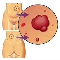
A red mole has appeared
Red moles are benign skin formations and appear as a result of improper functioning of the lymphatic and circulatory systems. They may look different and differ in the depth of location and type of vessel. By appearance they can be divided:
- Capillary hemangioma. It is the most common and represents a rupture of the vessel. Externally, it is bright red or purplish blue. It can be located anywhere on the face and body. In children they can be localized on the chest, neck, face, groin, arms.
- In a cavernous or cavernous hemangioma there are several vessels that connect into a large cavity. They are often located on the face, cause discomfort and disfigure the appearance. Covered with a thin layer of epidermis and located on the surface of the skin. Moles can also be on internal organs - in the uterus, liver, spleen and thus interfere with their functioning.
- Branched hemangioma looks like a blood-filled pulsating formation when several ordinary moles are combined with each other. If you press on it with your hand, you can see the blood flow out, then it fills again.
An angioma that looks like a red dot is called punctate. If small capillaries extend from it and it does not have clear contours, then it is an arachnid or stellate mole.
Why do red moles appear?
Doctors believe hormonal changes are one of the reasons for the appearance of angioma. Doctors believe that another cause of tumors is pancreatic disease or oncology. Disorders of the cardiovascular system and the function of pigment spots can also provoke the appearance of formations on the skin. Red moles sometimes appear during pregnancy. Some people believe that frequent visits to the solarium lead to the formation of angioma, but this is a misconception. In any case, if you find a red mole, you should consult a doctor.
Why is the mole red?
The red mole is well known and studied in medicine. It is filled with overgrown blood vessels, which explains its red color. Sometimes you can see small vessels extending from the base of the mole. This is a stellate or spider angioma. If several formations are located at a short distance, or one after another, then this is a cavernous or cavernous angioma. They are mostly congenital. A very large red mole that occurs due to active growth is called a hemangioma.
Bright red formations are located in different layers of the skin. This may be a capillary, venous or arterial site.
A distinctive feature of angioma is a change in color when pressed. This happens because blood is forced out of microscopic vessels under pressure. Then the color is restored again. Do not press, comb, rip off or scratch the mole.
Red moles reasons
Angiomas appear as a result of pathological changes in vascular disorders of all types. The most common is capillary, when transformation begins in the blood vessels and, as a result, a red bump appears on the surface of the skin - capillary angioma.
How to get rid of a red mole
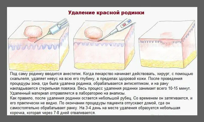
As a rule, red moles do not require removal or treatment, especially if they are not located on the face. Simple capillary hemangiomas are removed using the following methods:
- surgical excision;
- X-ray radiation;
- chemical sclerosis;
- cauterization with carbon dioxide;
- cryodestruction method;
- cauterization.
Treatment of red moles
Capillary and cavernous angiomas are treated with a course of hormonal therapy, after which they disappear.
Traditional medicine is rich in various recipes for cauterization and lightening of red moles. They can be used only when the formation is not large and does not affect the deep layers of the skin.
- At night, lubricate moles with castor oil. In a month their size will decrease.
- You can apply antiviral ointment every day.
- To lighten the formation, apply grated black radish three times a day.
- Apply a compress of pureed dandelion root every day for three hours.
- Cut a small potato into two parts and wipe with juice.
- Combine honey and grated apple in equal parts. Lubricate the mole and leave it under cellophane and a cotton bandage overnight.
- Twice a day, lubricate with a mixture of fifty milliliters of apple cider vinegar and three drops of lemon essential oil.
- Alternately apply garlic and lemon juice three times a day.
Removing red moles
- For x-ray removal of a red mole, the doctor prescribes a course of procedures, after which the angiomas will decrease in size or disappear completely. However, this type of treatment is the most dangerous.
- Small moles on the body can be removed at the clinic. Small scars may remain afterward.
- Small angiomas located above the surface of the skin are cauterized with carbon dioxide.
- Large hemangiomas are effectively treated using chemical sclerotherapy. A drug is injected around the mole, which blocks the blood vessels and separates the formation from the general blood flow. The mole decreases in size and disappears over time.
- Using the cryodestruction method, skin formations are frozen using liquid nitrogen. This is possible if the red mole is not large. The vessels that feed the mole become thinner, and over time they are destroyed.
- The angioma is cauterized using the coagulation method. This is the most effective and safest way. After the procedure there are no scars left. Coagulation can be light, infrared, radio wave and electrical. An anesthetic is applied to and around the mole. If the mole is large, local anesthesia is given.
- Branched and cavernous moles require surgical treatment in several stages. In small red moles, the vessels are ligated in the place where they are connected to healthy vessels or treated with radium application. At the same time, large trunks are removed.
- One of the most popular removal methods is the use of a vascular or carbon dioxide laser. Excisions are done in layers, which allows you to get rid of a formation located at any depth. The scars remain almost invisible. The procedure lasts a few seconds and is well tolerated. It is not recommended to visit the sauna and solarium for two months after the procedure.
Red moles in a child
Red moles are a common occurrence in children. They appear from the moment of birth and come in three groups:
- small diameter (from 0.5 to 1.5 cm);
- medium (from 1.5 to 10 cm);
- large – more than 10 cm.
Small moles are harmless and often resolve as they grow. They can be considered a cosmetic defect. If there are a lot of them and they are large, you need to see a specialist. Laser removal is carried out only for the following indications:
- peeling;
- bleeding;
- damage;
- change in size and color;
- itching
If your baby has a red mole, you must follow the same rules that apply to regular moles. Do not stay in the sun for a long time, do not allow the mole to be combed or picked off. If it begins to bleed and change shape, consult a doctor.
Other problems with moles
Red dot on a mole
Any changes in the mole itself, its pigmentation, or the appearance of any inclusions require consultation with an oncologist. This can be a dangerous degeneration into a malignant formation - melanoma. The disease is successfully treated only in the earliest stages.
Red around the mole
If redness appears around the mole, this indicates inflammation. The cause may be a scratch or exposure to sunlight. Be sure to contact an oncologist. Timely detection of a possible disease will prevent serious consequences. It is possible that the redness is caused by dysplasia; this is the initial stage of melanoma, which is easily treated, since the area of cancer cells is not yet large.
You cannot remove redness and swelling around a mole on your own.
It has been proven that ultraviolet radiation leads to changes in the skin, which can lead to the formation of tumors. People with fair skin are more susceptible to this effect.
Red raised moles
Convex red moles occur when a vessel is damaged. When you press on the mole, you feel pressure. Convex angiomas develop with hormonal disorders, diseases of the pancreas and with excess ultraviolet radiation. If such a mole causes inconvenience, discomfort or grows quickly, it is better to remove it under the supervision of a specialist. Large convex moles are dangerous for bleeding if they are damaged or infected.
Red mole growing
The degeneration of a mole from a benign state to a malignant formation occurs with various changes. The growth of a red mole is a sign of possible melanoma or skin cancer. If a mole enlarges on the face, it may be due to injury during shaving, friction or pressure. In women, this can happen during pregnancy due to hormonal changes or if they have thyroid disease. An increase in mole is observed in adolescence.
It happens that a person does not monitor changes in the body. The degeneration of formations can last from five to ten years. If you notice a change in the color of a mole, its rapid growth, or the appearance of itching, consult a doctor and, if necessary, conduct a histological examination.
Mole turned red
Often, redness of a mole is associated with injury. If you accidentally touch it, for example with a washcloth, while taking a shower, then contact not a cosmetologist, but an oncologist. Moles that are in areas of constant friction and contact with clothing may also turn red, and doctors often recommend removing them.
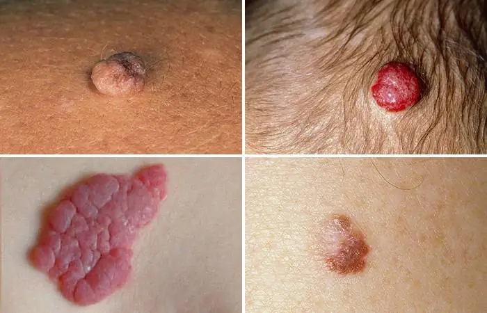
Red hanging mole
Convex hanging formations on a stalk of bright red color, in which there are many blood vessels, are called granuloma pyogenic or botryomycoma. A benign mole is characterized by:
- elevation above the skin;
- bleeding;
- sharp growth in two months;
- uneven papillary surface;
- diameter up to one centimeter.
In small children, such moles appear after a skin injury. In teenagers, they often appear on the hands and toes. If the formations are on the palm, the injury can lead to bleeding. In this case, the doctor will recommend removing it surgically or using a laser. Small convex moles on the stem are cauterized with silver nitrate or liquid nitrogen. Moles with a wide stalk with shallow excision give the most relapses.
Red mole itches
If a red mole itches, then you should not rip it off and injure it to alleviate the condition. Damage to an angioma is dangerous due to bleeding. If the itching is very difficult to bear, place your finger on the pad and gently massage it lightly in a circular motion. If you do damage the integrity of a mole, treat it with hydrogen peroxide and consult a doctor.
If itching is accompanied by pain and changes in shape and color, this may indicate the presence of cancer cells. Consultation with an oncologist and histology is necessary. Treatment is prescribed based on research results and your individual characteristics.
Angiomas, known as red moles, in medical practice are usually classified as benign formations that consist of lymphatic or blood vessels. Their appearance is explained by dysfunction of either the circulatory or lymphatic systems. They form throughout the entire period of a person’s life, but only in childhood up to 7 years are angiomas able to disappear on their own.
Red moles on the body - what are they?
What do red moles on the body mean? According to experts, they are an intermediate link between a malformation and a tumor. The medical literature provides little information about this phenomenon. This is due to the fact that red moles do not pose a particular danger to humans. They are believed to be innate.
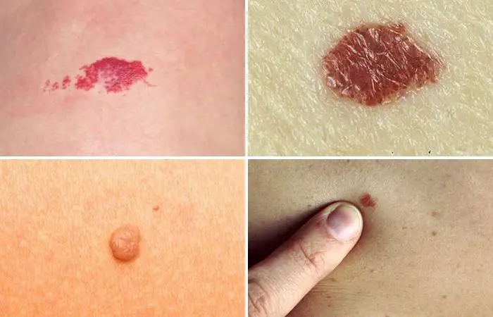
Types of red dots (angiomas) with photos
Red moles are usually divided into several varieties. Their classification is based on factors such as the cause of its appearance, location on the body, and the type of vessel that caused its formation. Depending on the cause of angioma and the composition of the tissue, there are red dots or moles of several varieties:
- pineal - a convex neoplasm that rises sharply above the skin;
- nodular - a small pinpoint formation that appears as a result of a blood vessel emerging onto the surface of the skin. This angioma does not have a capillary branch;
- branched or arachnid - a network of small blood vessels extends from the angioma;
- flat - a mole on the skin in the form of a compaction.
Depending on the vessels underlying the formation of red moles, they are called hemangiomas - moles formed due to abnormalities of blood vessels, and lymphangiomas - points that appear due to disruptions in the lymphatic system. Hemangiomas are impressive in size. Lymphangiomas are a rare occurrence. These are small nodules that are soft to the touch and easily compress when pressed. Formed on the neck, mouth, axillary and groin areas. Hemangiomas formed from vessels of the circulatory system are distinguished by external features and size:
- Capillary - formations that can be located anywhere in the body and look like a blue-purple or bright red spot. They are formed due to the expansion of a capillary vessel.
- Cavernous or cavernous are large formations containing several blood vessels that merge into large cavities. Such hemangiomas are located above the skin. A typical location is the face. Sometimes they can be located on internal organs - the uterus, spleen, liver.
- Branched - they are a pulsating, swollen formation that is filled with blood; they combine several simple moles.
- Punctate are the smallest hemangiomas, which appear as small dots.
Where are they located?
Angiomas are formations of different sizes and shapes. They are localized in tissues and organs: on the skin, in fatty tissue, bone tissue, liver, muscles, brain and kidneys. Such spots appear on the body in childhood and adolescence. Their appearance can also provoke pregnancy. Red dots can be single or multiple. If the former do not pose a danger, then the sudden appearance of multiple angiomas signals serious problems, which may include cancer.
Simple angiomas are located on the skin of the face and scalp. They are characterized by their small size, spherical shape, and purplish-blue color. In some cases, angiomas can occupy large areas on the eyelids, cheeks, nose, and external genitalia. Cavernous angiomas are localized in the subcutaneous area. Sometimes they grow into deep layers - into muscles and bones. This type of formation can develop in internal organs. 80% of angiomas are located in the upper part of the body: on the chest, back, head, face, neck, arms.
The most unfavorable location of angiomas is the head. This is explained by the possibility of damage during haircuts, combing or blow-drying. Red moles on the chest cause particular concern for women. Their single appearance does not pose any danger. However, if angiomas change color, begin to grow, itch or hurt, you should consult a doctor.
Causes of red moles on the skin
According to experts, in most cases such moles are congenital. The sources of their development are pathologies of the vessels of the lymphatic and circulatory systems. Angiomas can appear at any age. Red moles are often observed in children. From the point of view of doctors, this is explained by the transformation processes characteristic of this age.
In girls, angiomas form more often. Small formations go away on their own, without drug intervention. One of the reasons for the appearance of angiomas in newborns is considered to be infectious diseases suffered by the mother during pregnancy. In adults, the formation of angiomas occurs under the influence of hormonal changes and various disruptions in the functioning of the body. Among them:

- gastrointestinal diseases,
- pathology of the pancreas,
- exacerbation of chronic infections,
- pyelonephritis,
- deterioration of blood microcirculation,
- dysfunction of pigment cells,
- pregnancy,
- hereditary factor
- hormonal changes,
- lipid metabolism disorders,
- cardiovascular diseases.
Doctors believe that excessive sunbathing and solarium contribute to the appearance of red moles. It is assumed that angiomas can accompany diseases such as Henoch-Schönlein disease, rheumatoid arthritis, and lupus erythematosus. This happens because the immune system, due to emerging pathologies, sees the body’s cells as foreign and tries to suppress them. According to doctors, the appearance of angiomas may signal a blood clotting disorder.
How to remove red dots on the human body
In most cases, these formations do not require medical intervention. However, if the angioma bothers you - it itches, hurts or enlarges, it is recommended to remove it after an appropriate examination. Angiomas are removed if they negatively affect a person’s appearance, are located in places of contact with clothing, interfere with the performance of procedures to care for one’s appearance, or are often subject to accidental damage. Modern medicine offers several options for removing angiomas:
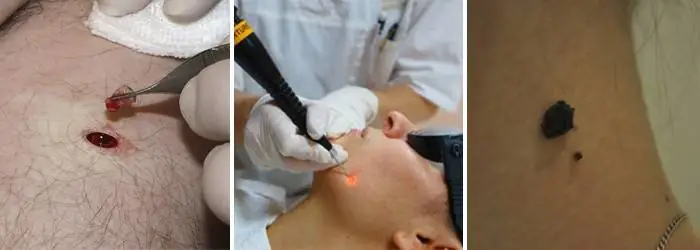
- Surgical excision. This method of removing a red mole is carried out only in a hospital setting. Performed by a surgeon without hospitalization. After excision, scars may remain on the surface of the skin, so the method is used to eliminate small moles located on the body. Surgical excision is not used for formations located on the face.
- Laser removal is the most popular way to get rid of angiomas. It is produced in layers, which ensures work with formations of any depth. Before the manipulation, the location of the angioma is anesthetized with an injection or application of an anesthetic gel. The procedure is short-term and lasts a few seconds. After excision, a crust forms at the site of the angioma within 24 hours, which disappears after 1-3 weeks. After complete healing, minor scars remain at the site of the mole.
- Cauterization is an effective and safe way to get rid of angiomas. Leaves no traces. Cauterization is performed using the coagulation method or using carbon dioxide and nitrogen. The first method can be used to remove large capillary formations. Before the procedure, local anesthesia is performed. Several coagulation methods are used - electrocoagulation, radio wave, infrared and light. Carbon dioxide is used to remove only small formations that are located above the surface of the skin.
Regardless of the chosen method of removing a red mole, it is recommended to make a preliminary diagnosis to exclude oncology. The specialist will be able to choose the best option for removing the angioma. After getting rid of a mole, it is not recommended to visit the sauna, solarium or expose the area where it was previously located to insolation for two months.
Treatment at home using traditional methods
Traditional methods of treating red dots can be used only when the mole is not inflamed, does not bleed, and is small in size. It is prohibited to treat large formations and angiomas that penetrate deep into the skin. According to reviews, the most popular and effective folk methods of getting rid of red moles are:

- Bee Honey. They lubricate the unwanted formation several times a day. After 10 days, the moles will begin to shrink.
- Castor oil. To reduce the size of a mole, apply it to its location overnight.
- Black radish is used to lighten moles. For this purpose, the root vegetable is grated on a fine grater, and the resulting pulp is applied to the problem area. The procedure should be carried out 2-3 times a day.
- Dandelion root. Clean, crushed root is applied to the mole every day for two hours.
- Onion. It is crushed and the juice is squeezed out of the resulting pulp. Lubricate the mole with it. After a month, the formation will dry out and disappear.
Are angiomas dangerous?
In most cases, angiomas are not dangerous. However, in some cases (very rarely), under the influence of unfavorable factors, they degenerate into malignant formations. Among these reasons, experts identify: damage to clothing, exposure to ultraviolet rays. In addition to the danger of degeneration, the cost of damage to the angioma's halo can be heavy bleeding.
Angiomas located in places of contact with clothing - in the abdomen, shoulders, neck, chest - require special attention. Hanging moles, which are easiest to pick off, and angiomas on the head are susceptible to increased trauma. Constant scratching, using a hairdryer, hairpins, headbands, and haircuts can damage the nevus. The danger is caused by angiomas located in the mouth, on the lips and near the organs of vision and hearing. In the oral cavity, moles can be subject to constant mechanical stress, which will contribute to their growth.
What to do if a red mole itches?
If a red mole begins to itch, this is a signal about the beginning of its growth, which may be associated with its degeneration into a malignant tumor. However, in some cases, angioma begins to itch when there is a disruption in the functioning of any body systems or a change in hormonal levels. Itching and flaking of red moles are often observed in pregnant women.
You will not be able to determine the reason for this behavior of a mole on your own, so to clarify the situation, you should consult a dermatologist or an oncology clinic. The hospital will order a series of tests, which include studying hormonal levels and determining the presence of malignant cells. Circular movements over the angioma or light pressure will help you get temporary relief from itching. You can't scratch a mole.
Is it possible to sunbathe and lead an active lifestyle?
The presence of red moles is not a contraindication to an active lifestyle. The only limitation may concern the possible methods of influencing angioma. If it interferes and is located in places of possible contact with surfaces and objects - on the arms, legs, it should be removed to avoid injury. As for sunbathing, as in the absence of red moles, doctors advise not to get carried away with sunbathing.
If there are any types of moles on the body, including red ones, being in the open sun is allowed before 10 a.m. and after 7 p.m. Due to increased activity of ultraviolet rays near bodies of water, you should stay away from water. To protect angiomas from sun rays, dermatologists recommend applying creams with a protection filter of 30 units.
Red moles are one of the types of benign neoplasms that can occur on human skin. They can form in people of different ages, including newborns. From this article you will learn whether red moles on the body are dangerous, why they appear, and how such growths can be removed.
What are moles?
There are many classifications of moles, and all of their components have their own medical term or name. But still, experts distinguish the two most common groups - pigmented moles and vascular ones. The former are formed due to an excess of melanin, the latter – due to problems with blood vessels (in particular with capillaries). Moles can also be congenital or acquired. Moreover, congenital ones are not always visible immediately; their pigmentation appears only after some time.
Large moles are the most dangerous, so you need to monitor them constantly and consult a doctor if there is the slightest change. Large formations can quickly transform into malignant tumors, which can negatively affect not only the skin, but also internal organs, and also contribute to the development of various diseases.
What are angiomas or red moles
Red moles, or angiomas, are benign tumor growths that develop from blood vessels. Red moles are quite common. Approximately 22% of all benign tumors are hemangiomas.
Red moles and red dots arise as a result of a congenital malformation of blood vessels and are detected from the moment the child is born.
What do red moles look like?
There are convex red moles and flat ones like a spot. They can range in size from a small red dot to a very large spot that occupies the entire limb.
Diagnostics
If the red mole is located on the surface of the skin, then examination and histological examination are sufficient.
If there is a suspicion of the presence of hemangiomas with a different localization, then angiography and radiography are indicated.
Causes of red moles on the body
By its nature, a red mole is a cluster of vessels and capillaries, which normally perform the function of oxygen and nutritional supply to the epidermal structures. With the development of any internal pathological process or under the influence of exogenous factors, small vessels can connect, forming a kind of bundle, which in medicine is often called an angioma.
When asked by a patient regarding small red moles, “What is this?”, the specialist will most often answer – angioma. Typically, these pigmented formations are formed during the period of active growth of the body, that is, in early childhood, which is associated with a serious transformation of the human vascular system at this age.
A distinctive feature of red pigmented nevi is the loss of color intensity of the formation when pressing on it, which is associated with a vascular reaction. In addition, red moles normally do not hurt or itch, and do not change their shape and size over time.
The following etiotropic factors may be the causes of angiomas in adult patients::
- Excessive sun exposure (excessive ultraviolet radiation);
- Hormonal imbalance (puberty, pregnancy, menopause, etc.);
- Pathologies of the gastrointestinal tract, especially the pancreas;
- Injuries and mechanical damage to the epidermal integument;
- Diseases of the cardiovascular system;
- Violation of the formation or destruction of pigmented cells.
Today, doctors do not give a clear answer regarding the causes of red moles on the body, however, the factors listed above can become a provoking condition for their appearance.
Types of red moles
The appearance of red moles on the body can be accompanied by various visual characteristics of the defect. Such epidermal formations can be either flat, forming in the deep dermal layers, or have a superficial nature, protruding above the skin. Moreover, according to the nature of the visual picture, all angiomas are usually divided into two groups:
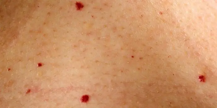
- Spot. The pigmented area has clear red borders and is a point at which a collection of vessels and capillaries is concentrated. Often, punctate angiomas are multiple in nature, manifesting themselves in the form of specific “rashes” on the patient’s skin.
- Star-shaped. Such moles are a collection of tiny thin vessels that are visible through the epidermis and converge at a central point, forming a kind of star. These structures are also associated with diseases such as rosacea.
In addition, a separate group includes especially large red moles on the body - hemangiomas, which usually significantly spoil the patient’s appearance and require cosmetic correction.
Red moles in newborns
During the neonatal period and infancy, red moles in children are very noticeable, but they often disappear by the age of 5.
Such benign tumors do not pose a danger if they:
- They do not itch, do not hurt, do not cause any concern;
- They do not become larger, for example, in a few months they do not increase in size by 3 times or more;
- Not located under the eye, on the nose, on the face or on the genitals.
The fact is that as the mole grows, it will destroy nearby tissues, causing defects not only cosmetic, but also functional. This is relevant in the case when hemangiomas are located on the ears, under the eyes, etc. Normal functioning of organs becomes impossible due to compression by the overgrown formation.
Features of red moles in adults
Red moles, as primary formations, do not appear in adulthood. They arise from previously undetected vascular proliferations. Most often, they are treated before the child enters school, so if a hemangioma is present in adulthood, this means that it was either not treated, or the tumor is located on internal organs.
Prevalence and localization
According to available statistics, hemangiomas are detected immediately after the birth of a child. This happens in 87% of cases. Girls are at risk; they account for about 70% of all diagnosed red moles. By the way, it is hemangiomas that make up up to 48% of all soft tissue formations that are detected in childhood.
A red mole can be found in any part of the body, but 80% of all such defects are localized in the upper part of the body. It is also possible to detect hemangiomas, although extremely rarely, on the bones, in the brain, lungs, and liver.
- 95% of all hemangiomas are simple formations;
- 3% – cavernous formations;
- 2% – mixed type formations.
The most common are red moles on the skin. But angiomas can also be located on the mucous membranes and even in internal organs. Red moles are especially common on the face and neck.
Red dots on the body are often detected at birth or in early childhood, and they increase as the body grows. Small red dots can appear at any age throughout life.
Red moles, or so-called port-wine stains, can be significant in size and cover most of the face, leading to pronounced cosmetic imperfections. But more often there are small formations in the form of small red dots.
Red moles on the chest appear more often in women. And in men, red dots most often appear on or near the nose.
Why are red moles dangerous?
Many doctors believe that red moles do not pose any danger, despite the fact that they are benign tumors. For many people, it is simply a matter of beauty and external aesthetics. Of course, red moles cause some concern, but often their owners simply do not notice angiomas on their body and do not attach much importance to their appearance. However, there is an opinion that red moles can directly affect the health of internal organs.
In particular, with diseases of the pancreas and liver, red dots are localized only on the upper part of the body, mostly on the arms. The more intense the localization of red moles, the more the disease worsens. Red moles can also indicate the presence of rheumatic diseases, such as lupus, dermatomyositis or arthritis. But in this case there is no clear location of the angiomas. They can be of various shapes and sizes.
If a red mole does not bother its owner and does not change its external characteristics, it can be argued that such a formation does not pose a significant threat to the health and life of the patient. At the same time, you should get professional medical advice and resort to modern therapeutic methods if you have the following manifestations:
- The red mole grows or changes its shape;
- The patient experiences pain, itching or burning in the area where the pigmented formation is located;
- The mole is bleeding;
- Superficial structures or ulcerations appear;
- There are more than 6 small red dots in one area of the body.
All these symptoms may indicate the development of an oncological process. In addition, the category of dangerous moles includes angiomas located in places where they can be easily injured when wearing clothes, shoes, jewelry, etc. Mechanical damage to such a formation can provoke the appearance of new red moles on the body, activate malignant transformation and leave scars or scars on the skin.
What to do if a red mole appears on the body
Since red moles tend to go away on their own, specialists usually do not prescribe a special course of treatment. If an angioma does not grow and does not bother a person in any way, then it does not pose a danger. It is not recommended to touch neoplasms in closed areas of the body. Removal may be approved by a specialist if the mole is located, for example, on the face, i.e. for cosmetic purposes. An indication for immediate consultation with a doctor is the growth of angioma.
There are several treatment options for red moles, and it should only be chosen by a medical specialist after conducting a series of studies. Among the most popular and effective methods of treating angiomas is laser surgery: vascular sclerosis, X-ray therapy, infrared or light coagulation. Flat red moles are treated much faster and easier than raised ones. The operations are performed without anesthesia, although they are quite unpleasant in sensation. Sometimes after the procedure it is necessary to use a numbing cream. After removal of the angioma, it is recommended to avoid visits to the solarium and prolonged exposure to sunlight for several months.
Complications
Hemangiomas (red moles) can disrupt the functioning of organs that are located next to them. This is especially dangerous if they grow in the liver, near the eyes, or in the brain.
- Inflammation and the appearance of ulcers on the mole are possible. As the pathological process fades, spontaneous disappearance of the mole is not excluded.
- Bleeding from a mole as a result of its traumatization. Bleeding is dangerous if there is a large mole, if it is a cavernous or combined formation localized on the internal organs.
- Infection.
How to get rid of red moles
Removal of small red moles is usually carried out for aesthetic reasons. If red dots on the hands rarely bother you, then a red dot on the face does not look aesthetically pleasing and attracts attention. Treatment for red moles involves removing them.
And, most importantly, the removal of the tumor should be carried out by a dermato-oncologist. He will be able not only to correctly diagnose, but also to carefully rid you of red moles and dots.
Medical correction of red moles
The doctor, after conducting an initial examination and the necessary diagnostic measures, will make a conclusion regarding the likelihood of malignancy of the red mole. Based on this diagnosis, a strategy for further therapeutic actions is developed. If a red mole does not pose an oncological threat and is located in a closed area of the body, its removal is not necessary.
In cases where the red nevus causes aesthetic or physiological discomfort to the patient, it can be removed using modern hardware techniques, the priority among which is laser destruction.
This method of removing red moles guarantees painlessness and safety for the patient, and also has a low level of trauma.
So, how to remove a red mole
The least traumatic and most effective method is removal using the Surgitron radio wave device. Using the radio wave method is the best choice for removing red spots and moles on the face and other open areas of the body, since the surrounding tissues are not injured. After healing, as a rule, no trace remains.
Using the Surgitron radio wave device (made in the USA), it is possible to remove red dots without leaving a trace, since the radio wave does not damage surrounding tissues.
If you are deciding where to remove a red mole in Moscow, pay attention to the method that the clinic offers and the qualifications of the doctors.
Treatment of red moles
Most red moles do not require treatment and go away on their own. If a mole does not interfere, does not increase in size and does not bother its owner in any way, it does not have a negative effect on the human body and is completely harmless. Experts do not recommend removing red moles that appear on closed areas of the body. The situation is different with those moles that are localized, for example, on the face and spoil a person’s appearance. Rapid growth and change in color of a mole requires immediate contact with a specialist.
A mole is most often located in the deep layers of the skin; only its upper part is on the surface. If, after removing a mole, its roots remain in the skin, it may reappear in the same place. The optimal treatment method is prescribed by the doctor after examining the mole for the presence of benign cells. Most often, laser surgery is used to get rid of a red mole.
The most modern methods for removing red moles are light or infrared coagulation of blood vessels, sclerosis of the vascular bed and radiotherapy treatment. Flat moles can be treated faster than those that rise above the skin. During the removal procedure, a numbing cream may be used if necessary. Anesthesia is usually not required in most cases.
It should be remembered that the procedure for removing a mole is very unpleasant and requires some investment. After it is carried out, small reddish spots appear at the site of the removed moles. After some time they disappear completely. After removing a red mole, it is recommended to refrain from visiting a solarium and prolonged exposure to the sun for at least one month.
Spontaneous problem resolution
Red moles go away on their own if they are superficial or simple. This is possible in no more than 15% of cases. Most often, formations that are localized on closed parts of the body regress.
At first, the tumor becomes less bright and decreases in size. After six months, the mole becomes a pale pink spot located at skin level. In this case, the skin over it atrophies. After another 3-4 years, only a small depigmented area remains on the skin.
