Many people are born with birthmarks on their body and face. Often these formations are small in size, but in some cases they can be located in the most visible places. In addition, they can be dangerous to human health. However, only a doctor can understand what a red birthmark means, when it should be removed and when it can be left. The sooner diagnosis is made and treatment is prescribed, the easier it will be to prevent such a serious disease as melanoma.
Characteristics of the disease
Birthmarks are spots on the skin that are discovered at birth or appear later. All moles and marks that have a color that does not match the main skin tone are called nevi. According to statistics from neonatologists, red birthmarks occur in newborns in 30% of cases. These cutaneous vascular marks in infants can be raised or smooth, have different sizes and locations. Moreover, their shade can range from burgundy red to pink. Once a red birthmark is discovered in a child or adult, it is recommended to constantly monitor its condition, noting any changes.
Varieties
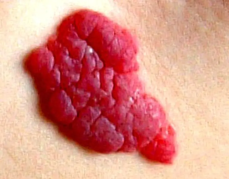
The most common type of red birthmark is a hemangioma. It is a vascular formation. Typically, hemangioma does not cause pain and is safe for the body. Doctors involved in research into the causes of birthmarks still cannot determine what exactly influences the occurrence of this type of nevus. The color of a hemangioma ranges from bright scarlet to pale red and depends on how developed the blood vessels are in this area of the skin.
- Strawberry hemangioma is a common type of red birthmark in a newborn. In most cases, it disappears when the child is 9-10 years old. This subspecies is characterized by a bright red hue and slight wrinkling. This sign may remain on the skin even after the formation disappears.
Additional classification
Based on the degree of bulging above the surface of the skin, dermatologists distinguish the following types:
- flat;
- knotty;
- branched;
- pineal.
To classify red birthmarks on the legs, arms or body, the shape of the formation means a lot. This parameter is divided into:
- Arachnids - capillary arrows move in different directions, becoming more noticeable over time.
- Dotted - do not have pronounced vascular branches, more like red dots.
- Multiple - look like small formations located close to each other.
If a red birthmark appears on the body or face, this should alert you. To diagnose the pathology and properly treat it, it is best to contact a medical institution so that the doctor can determine the type of disease and talk about treatment options.
Causes
In most cases, doctors cannot determine the factors that influence the appearance of red birthmarks. As a rule, they are not formed due to genetic predisposition; this happens extremely rarely. Much more often, red birthmarks on the face or other parts of the body are associated with various disorders in the body. The main reasons include:
- hormonal disorders;
- liver diseases;
- avitaminosis;
- frequent exposure to the sun;
- deterioration of lipid metabolism;
- skin injury;
- heart and vascular diseases.
If it is determined that red nevi appeared due to a lack of nutrients, then an additional examination is carried out to determine the level of vitamins K and C. The lack of these components leads to a weakening of the vascular walls and the appearance of various formations. They can appear even after slight pressure on the surface of the skin.
After statistical research in this area, it has been proven that fair-skinned people are more prone to developing red birthmarks. Therefore, dermatologists recommend that they refrain from prolonged exposure to direct sunlight or choose the first half of the day for summer walks. If you follow this advice, the likelihood of various formations appearing can decrease by 30-40%.
As for red pigment spots in newborns, they most often occur due to excess melanin. This pigment is responsible for the color of the skin. The level of melanin in each person is individual, so it is impossible to predict the appearance of formations. Among the causes of red birthmarks in babies, doctors name fetal hypoxia, various infections of the mother suffered during pregnancy, and multiple pregnancies. According to statistics, such nevi are observed much more often in female infants.
Location
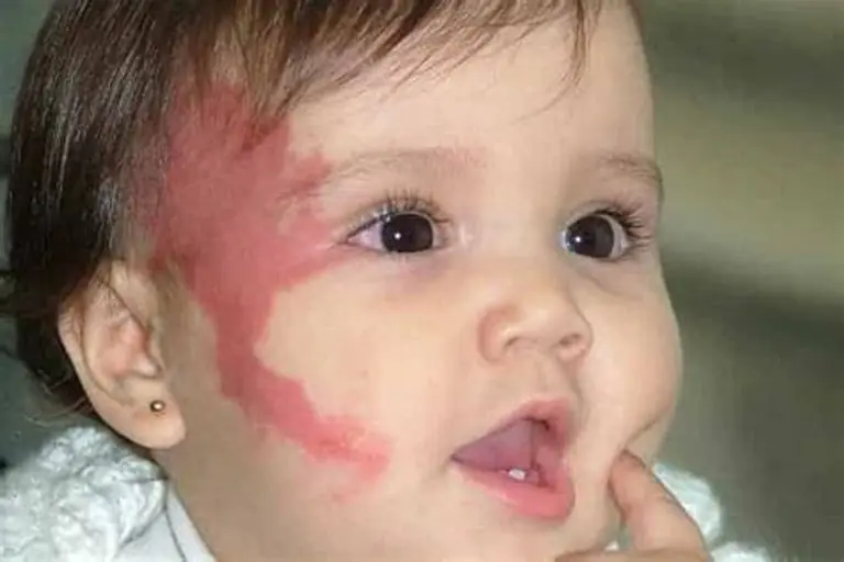
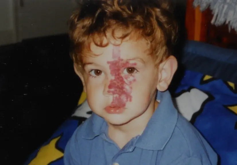
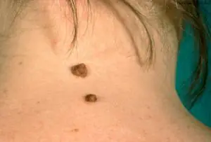
As a rule, this type of nevus can have different localizations. They are distinguished by their location around natural physiological openings on the face - mouth, ear, eye, cheeks, bridge of the nose. This is how the “stork’s kiss” is localized. Much less common are red birthmarks on the arms, legs or near the genitals. It is very important that the formations do not come into contact with tight clothing, because this increases the likelihood of damage.
Strawberry hemangiomas can be located on the scalp, and these red birthmarks also often appear on the back or stomach. It is very important to immediately determine where the marks are located in a child or adult. This will help to carry out further diagnostics and determine whether treatment is required in this case.
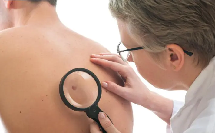
Diagnostics
If a red birthmark is found on the skin, you should immediately bring it to the attention of your doctor. After this, the doctor must determine the cause of the disease using modern diagnostic methods. Initially, a visual examination is carried out, which helps to record data such as color, shape of capillaries, elevation above the surface of the skin, and localization of the red birthmark.
A comprehensive examination may be ordered, including laboratory tests of blood and urine. But, according to dermatologists, the most effective is a full examination of the blood vessels adjacent directly to the formation.
Among the most accurate diagnostic methods are computer capillaroscopy. During this procedure, the relationship of capillaries to the skin surface can be determined. They can be located superficially, have a tortuous or expanded structure. In order to exclude such a serious disease as melanoma, you will have to perform a biopsy of the red birthmark. To do this, the doctor takes a small piece of the skin lesion for examination. Laboratory analysis will confirm or deny the presence of cancer cells.
Treatment
If nothing bothers the patient, the formation does not increase in size and does not interfere with wearing everyday clothes, then no treatment is required. In cases where a red birthmark has increased in shape over a short period of time, you should consult a doctor for advice. The type of treatment will depend on the cause of the pathology, which is determined by the doctor. Sometimes all therapy comes down to taking drugs that improve vascular circulation. If the reason lies in a lack of microelements and nutrients, then a special balanced diet is prescribed that will help compensate for the lack of vitamins K, A and C.
Special formulations will help you get rid of a red birthmark, allowing you to reduce the intensity of the color after repeated treatment of the skin. For example, the pharmaceutical drug "Tsindol" successfully fights different types of pigmented nevi, if they are located on the surface of the skin and the capillaries do not lie too deep. For children under 9-10 years of age, physical therapy is additionally prescribed, which involves the use of special medical devices that improve microcirculation of the skin. Such procedures include magnetic therapy.
Folk recipes
Since ancient times, there have been folk techniques with which healers removed small red birthmarks. To do this, they recommended using effective recipes and lubricating the formations:
- bee propolis;
- castor oil;
- juice of garlic or onions;
- potato mush;
- dandelion leaf juice.

Some methods actually helped reduce the size of the birthmark or even get rid of it completely. The main effect was achieved due to the whitening effect of these ingredients. However, if the growth continues to increase in size, it is necessary to seek medical help and not self-medicate the red birthmark. The value of professional therapy is much more important than witchcraft knowledge.
When Removal is Required
Red birthmarks that are located on closed parts of the body usually cannot be removed. In this case, the doctor advises conservative treatment. However, if the formation is affected by negative factors, it can degenerate into malignant. This often happens if the birthmark becomes damaged after coming into contact with clothing. It is dangerous to leave growths that appear in the eye area, because as they increase in size, they can significantly impair vision.
Reasons to remove red birthmarks include:
- frequent injury;
- cracking and peeling;
- bleeding;
- color change;
- rapid growth;
- the appearance of asymmetry;
- severe itching.
These signs may indicate degeneration into melanoma, a disease that belongs to the category of cancer. If there is the slightest doubt, you need to do a biopsy and use the prescribed treatment to prevent the growth of cancer cells.
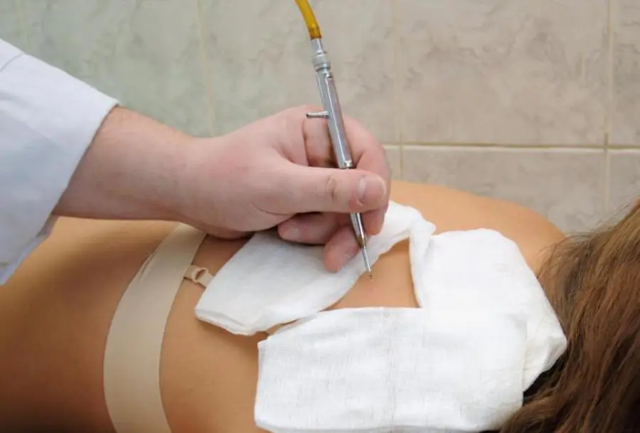
Removal methods
Excision is not always performed due to pathological complications. Sometimes it is necessary to remove the formation if the mark is located on a visible part of the body and leads to the fact that the patient begins to feel complex about his appearance.
Currently, the following types of red birthmark removal are distinguished:
- X-ray irradiation is a course of treatment consisting of several procedures. If the dosage is prescribed correctly, the formation will decrease and darken, and soon disappear altogether. However, this method has many contraindications due to the high health risks, so it is used extremely rarely.
- Surgical excision – often used for red birthmarks that protrude above the surface of the skin, also suitable for cavernous and branched nevi. The main disadvantage is the long healing time of wounds. In addition, after excision with a surgical scalpel, unaesthetic scars remain.
- The carbon dioxide method is suitable only for surface formations. Before carrying out the procedures, capillaroscopy will be needed to determine the depth of the pathological vessels.
- Chemical sclerosis - for this removal method, special drugs are introduced that block the vessels and prevent them from participating in the general blood flow. Not suitable for small patients.
- Cryodestruction involves the use of liquid nitrogen. This substance successfully copes with red birthmarks that have a convex structure. But, for successful removal, they should not be located too deep, otherwise you will have to repeat the procedure or choose an alternative treatment method.
- Electrocoagulation is another modern method that works well with red nevi. During treatment, small doses of electric current are applied to the birthmark. The procedure can be painful and requires appropriate anesthesia.
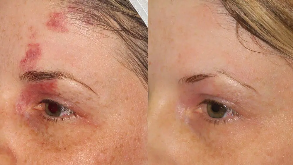
- Laser exposure has numerous advantages over other methods. During the micro-operation, which takes no more than 10 minutes, no anesthesia is required. The patient will not feel any discomfort.
Among the described methods, laser coagulation is the most preferable. This procedure is even used to treat red birthmarks on the back of the head, face and body in children. However, this method also has contraindications, so the decision to carry out therapy must be made directly by the doctor who conducted the examination.
Prevention of new formations
After the red birthmark is removed, you need to change your life taking into account harmful factors so that similar formations no longer appear on other areas of the skin. Doctors recommend following some rules for prevention:
- Stay in the sun less, avoiding direct rays.
- Avoid injury to the areas where removal was performed.
- Monitor your hormonal levels, taking any medications only as prescribed by your doctor.
- Conduct an independent preventive examination and, at the slightest suspicion of deterioration of the skin condition, contact a dermatologist.
If you regularly conduct a medical examination and examine your skin, and also avoid injuries and cuts, you can forever forget about such a pathology as red birthmarks.
It's rare to see a person without small dark marks on their body. Is it worth paying attention to these points? Only a doctor will distinguish between dangerous and normal moles - malignant melanoma or harmless nevus - and give recommendations on what to do with them. Is it worth worrying about the appearance of new formations, when immediate contact with specialists is required, what are the signs of cancer development - the answers to these questions remain to be found out. No one is immune from disaster, and early diagnosis will protect you from severe consequences.
What is a mole
The first tiny spots may appear in children in infancy. A mole is a small formation on the skin - a nevus - that is considered benign and harmless. The basis for their appearance is melanocyte cells that accumulate the natural pigment melanin. Depending on its quantity, a difference in color is observed. Available colors:
The shape of the tumors depends on the location and concentration of melanin. They may have a stalk or be located under the skin, be flat and convex. The most common type is round, but there are exceptions. The development of neoplasms is provoked by ultraviolet radiation - natural from the sun, in a solarium. Hereditary factors cannot be excluded. A common cause of growth is hormonal imbalance, characteristic of periods:
- puberty;
- pregnancy;
- menopause.
What types of moles are there?
One person may discover very different tumors. Types of moles are classified according to several criteria. This helps in correct diagnosis in case of changes. They differ in:
- origin– congenital, newly acquired;
- structure– pigment, vascular;
- place of education – in depth, on the surface, in the boundary layer;
- raised above the skin – flat – even, protruding as a hemisphere, pedunculated, larger birthmarks;
- potential threats – dangerous, degenerating into melanoma, non-dangerous.
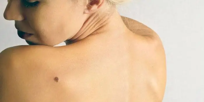
Safe moles
Those who have dark spots on their skin should be wary of their changes. In time, detected signs of degeneration into melanoma contribute to the timely removal of the formation and preservation of health. Safe moles are different:
- the presence of a stalk – it cannot be formed by malignant cells that grow randomly;
- long-term condition without changes.
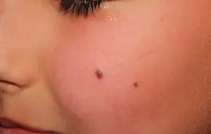
Spots that appear shortly after birth are not considered dangerous. It is important that they are small in size. Good – non-dangerous – signs of neoplasms include:
- flesh tone;
- unchanged pattern of the skin of the nevus and adjacent tissues;
- soft consistency;
- hair on the surface of the neoplasm - growing from the skin, indicates the absence of pathologies;
- diameter no more than 5 mm;
- symmetry;
- nevus in the form of a spot.
Which moles are dangerous?
Why do people with nevi on their bodies need to monitor their changes? There is always a threat of degeneration of non-dangerous tumors into a cancerous tumor. What moles are dangerous to health? Key signs you need to know:
- change in shades towards the dark side, the appearance of multi-color;
- rapid increase in size - exceeds two millimeters per year;
- occurrence of cracks;
- the formation of asymmetry due to uneven growth;
- lack of elasticity;
- the appearance of itching, burning;
- presence of discomfort.
The appearance of dangerous moles requires an immediate visit to a specialist to clarify the nature of the changes and the likelihood of developing skin cancer. Pathological transformations provoke:
- injury to the nevus due to negligence;
- self-removal;
- abuse of exposure to the sun, use of a solarium;
- location of the formation in places of frequent contact with clothing - on the neck, head, genitals, legs;
- placement in the hair, on the face, palms - where there is a high probability of injury;
- previously removed melanoma.
Why are moles dangerous?
Not a single person is protected from the sudden proliferation of cells of a harmless mole. Melanoma is an extremely serious disease. Changes not detected at the initial stage can result in death. The provoking factor is unsuccessful independent removal of tumors. Moles are dangerous because of their ability to:
- transform into an atypical – precancerous form;
- grow to large sizes;
- turn into cancerous;
- with minor external changes, metastases actively spread throughout the body through the circulatory and lymphatic channels.
How quickly does melanoma develop from a mole?
The transformation of a nevus into a cancerous formation can occur in different ways. The process depends on the stage of the disease and the type of tumor. Instant metastases are dangerous. Begins:
- growth of cancer (oncological) cells in the deep layers of the epidermis;
- their entry into the blood and lymph;
- penetration into the lungs, liver, kidneys;
- growth in these organs;
- complete damage to the body;
- death.
The growth phases of pigment cells are observed, along which melanoma develops from a mole. There are varieties:
- horizontal– damage to the upper layers of the skin occurs, lasting up to 10 years, but metastases do not appear;
- vertical– accompanied by the spread of cancer cells throughout the organs, can last two years, has an unfavorable prognosis;
- nodal – especially dangerous – characterized by deep spread within two months.
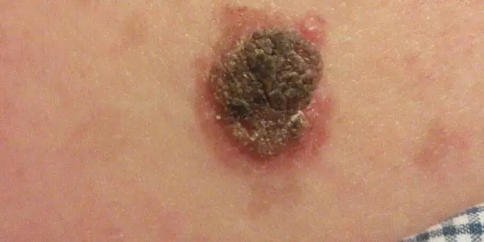
The first signs of melanoma
The patient can be assisted only when suspicious changes begin to be identified. The diagnosis, research, and referral for surgical treatment save a person’s life. The first signs of melanoma:
- increase in the height of the tumor;
- bleeding;
- the appearance of discharge;
- redness;
- burning, itching;
- swelling of tissues;
- softening of the nevus;
- the appearance of a crust;
- thickening;
- hair loss;
- expansion of pigmentation around the lesion.
With the further development of dangerous melanoma, the following are observed:
- significant change in size;
- the appearance of pain;
- enlarged lymph nodes;
- surface ulceration;
- formation of new foci;
- bleeding from places of pigmentation;
- liquid separation;
- skin thickening;
- the appearance of an earthy tint;
- signs of metastases are chronic cough, weight loss, cramps, headaches.
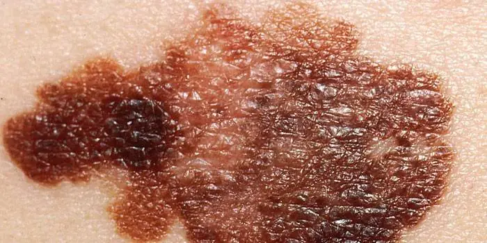
How to distinguish a mole from melanoma
To recognize which moles are dangerous and which are not dangerous, you need to know what they look like. A person with nevi, in order to avoid dire consequences, must constantly monitor the appearance of new formations and changes that occur. You can distinguish a mole from melanoma by its signs. Non-dangerous neoplasm:
- symmetrical;
- with smooth edges;
- uniform in color;
- with dimensions not exceeding 6 millimeters.
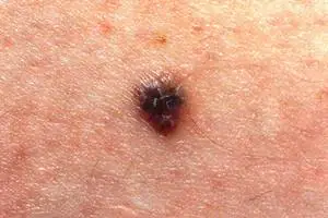
Features of dangerous melanoma that require seeking help from dermatologists:
- growth in a short time;
- pronounced asymmetry of shape;
- heterogeneity in color - the presence of inclusions of several shades;
- lack of clear boundaries - the contour line is blurred, jagged, and looks like a coastline on a geographical map;
- increased diameter over six millimeters;
- variability of any parameters - color, size, shape.
What dangerous moles look like
What do nevi that are subject to pathological changes look like? Only a doctor can correctly distinguish between non-dangerous tumors. Dangerous formations look like this:
- blue– compactions under the skin with clear boundaries, with dimensions no more than 10 mm;
- nodal– round, flat in shape, color – brown, black;
- cutaneous– often pale, convex;
- halo nevus – pigment surrounded by a light or white rim;
- spitz- looks like a dome-shaped tumor of pink shades, with the possible presence of a hole through which blood and liquid leak;
- connecting- connect individual entities into a whole.
Mole with jagged edges
One of the signs of a non-hazardous formation turning into a dangerous one is a change in contours. It often has blurred edges and scalloped borders. There are non-dangerous types of nevi - dysplastic. Only a specialist can make a correct diagnosis. A mole with uneven edges can be dangerous if there are additional signs of melanoma:
- accelerated changes in size;
- the presence of clearly defined asymmetry;
- the appearance of highly indented boundaries.
Rough mole
Such a neoplasm is harmless if its diameter is no more than 5 mm and remains constant in size. Often its appearance signals a lack of vitamins and nutritional disorders. Doctors advise coming for a consultation if it is discovered that:
- the smooth nevus turned into a rough one;
- bothered by burning, itching, tingling;
- irregularities and compactions appeared in the middle;
- areas with different shades formed;
- diameter has increased significantly.
A dangerous rough mole requires immediate examination if:
- the appearance of bleeding;
- development of the inflammatory process;
- rapid change in size;
- formation of asymmetry;
- formation of purulent discharge;
- the occurrence of painful sensations when touched;
- the emergence of an irregular shape, blurred boundaries, along the edges of the neoplasm.
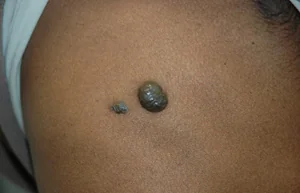
Large moles
Large formations on the skin are pigment spots. When they remain unchanged and do not cause inconvenience, this is not a dangerous phenomenon. It is important to constantly monitor their appearance, color, and size. To eliminate worries, you need to consult a dermatologist. During the visit, the specialist will conduct a diagnosis and give a forecast of the risk of developing a malignant neoplasm. Large moles become dangerous if they:
- injured;
- thickened;
- started to itch;
- were unsuccessfully removed independently;
- changed in size, shape;
- are bleeding.
What moles can be removed
Often nevi cause trouble for women when they are in a visible place - the face, neck. Even if they do not bother you, using removal will be the right decision - the appearance will improve significantly. After the procedure, the doctor must necessarily send the tissue for histological analysis to decide whether the mole is malignant or not. If the neoplasm is not dangerous, does not bother you, and does not change in size, surgery is not required. What moles cannot be removed? Experts believe:
- there are no contraindications;
- It is important to choose the right excision technique.
You should be careful about skin growths; it is unacceptable to remove them yourself. Only the doctor will determine whether a nevus is dangerous or not and decide what to do with it. You can delete it if:
- injured from clothing - on the neck, in the groin area, under the armpits;
- cause pain when touched;
- are located under the hair on the head and can be damaged when combing or cutting;
- change color, shape, outline;
- significantly increase in size;
- characterized by the presence of burning, itching;
- accompanied by inflammation and bleeding.
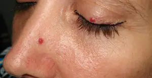
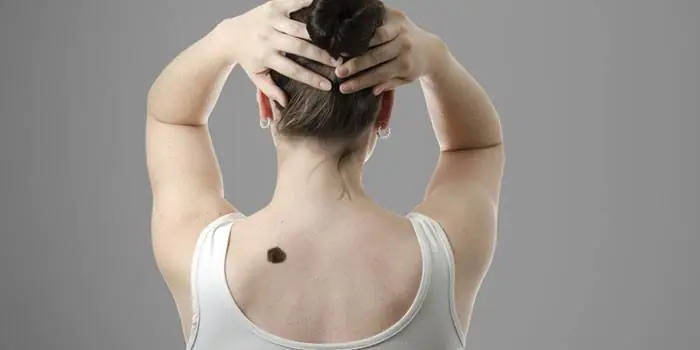
A phenomenon familiar to most people, which many do not notice or do not attach due importance to, is moles. But due to the connection of most of these types of birthmarks to oncology, they deserve close attention. Moles on the body can have different types, sizes, surface texture, color and shape.
Moles, acquired and congenital
All moles that appear on the body can be divided into congenital and acquired. The first, in turn, can be classified by size:
- Small ones reach a size of one and a half centimeters in diameter.
- Average moles can reach ten centimeters.
- Large spots exceed ten centimeters in diameter.
- Giant birthmarks are localized over the entire anatomical region, that is, they cover the entire face or chest.
Small moles, which very rarely can develop into cancer, are considered harmless. The most dangerous are considered to be giant moles, which develop into cancer in half of the cases. Owners of such neoplasms should visit a dermatologist regularly for consultation. Many experts insist on removing particularly large birthmarks in order to avoid unwanted consequences.
Due to individual characteristics of a person at the genetic level, acquired moles may appear. Most often, they form in early childhood during the period of intense pigment spots appearing on the surface of the skin. Dividing moles by location, they can be classified into the following types:
- Epidermal birthmarks characterize the accumulation of melanocytes in the upper layers of the skin.
- Intradermal moles are accumulations of melanocytes in the deep layers of the skin.
- Clusters of melanocytes at the border of the dermis and epidermis are borderline moles.
Many doctors insist on removing almost all types of acquired birthmarks.
Moles - types of moles on the body with photos
In my own way location and form moles can be of the following types:
Hemangiomas are vascular formations. The vessel from which they are formed determines their main color: red, pink or with a hint of blue. They reach different sizes and may have uneven edges. They, in turn, can take the form of capillary moles, which are flat in structure and located on the surface of the skin, or cavernous, located in the thickness of the skin, lumpy and nodular.- Non-vascular moles, similar to pigmentation of the skin, can also rise above the skin in the form of warts. According to their characteristics, these can be multiple or single elements with a keratinized surface of different shapes and colors from gray to brown. In some cases, they can be covered with hair and reach a black tint in color.
- It can be a light or dark formation in the upper layers of the epidermis from melancites and have the familiar name of a mole or a flat birthmark, which is familiar to many. Over time, their number and size do not change.
- Pigment spots of a brown or brown hue, which increase in intensity in old age or adolescence, are called lentigo. They can develop into a so-called disease, lentiginosis.
- In the deep layers of the epidermis, convex spots can form, which are bumpy or smooth formations with a diameter not exceeding ten millimeters and in most cases having a hair in the center of the growth. The color scheme can vary from black to yellow. Locations in intimate areas, on the palms or soles of the feet.
- A mole that looks like a smooth hemisphere with a diameter of up to two millimeters or a small dense growth with an elevation above the surface of the skin is called a blue nevus or blue mole. It can be located on the face, buttocks or limbs and range in color from light blue to dark blue.
- The congenital formation is a giant pigmented nevus, which is represented by colors of gray, black or brown. This pigmented spot has its own peculiarity: it increases in size along with the growth of the human body.
- Formations of moles with different shapes and sizes exceeding one centimeter are called dysplastic nevus. They are birthmarks with blurry outlines that have genetic inheritance.
Description of melanoma-dangerous moles on the body
Blue nevus, localized on the buttocks, limbs or face. Represents dense nodule, without hair, does not exceed five millimeters in diameter. The color can vary from light blue to dark blue.
Nevus of Ota is a large dark brown pigmented spot on the face and is a type of blue nevus. The location can be any part of the face, which creates the effect of dirty skin.
A borderline pigmented nevus is a nodule with a flat, dry and smooth surface, not exceeding ten millimeters in diameter. The color ranges from dark brown to black with localization in the intimate area, nail beds, palms or soles of the feet.
Giant pigmented nevus resembles a wart with a cracked, bumpy surface and has a gray to black color. This species is capable of increasing along with the growth of the human body.
Dubreuil melanosis is a single pigmented spot of pre-melanoma skin lesion with slow growth. From a light brown color, the spot acquires a dark brown tint as it grows, even black. It is localized in open areas of the skin, but is most often located on the face in patches with a diameter of up to thirty millimeters.
Melanomic types of moles
Nevus of Setton is a red or brown nodule reaching a size of five millimeters in diameter. It rises slightly above the surface of the skin. A round or oval nodule, usually surrounded by a white ring of depigmented skin. Such a nodule is capable of spontaneous disappearance and is located mainly on the arms and torso.
Nevus fibroepithelial It is a slowly growing formation with a soft elastic surface and a spherical shape. The dimensions do not exceed a diameter of ten millimeters. May contain vellus or bristly hair and have a natural color. The color scheme can also have a bluish, pinkish or dark brown tint. It can be located singly or in dozens throughout the body or on the face. It differs from the borderline pigmented nevus in that it has a less saturated color, regular outline and the presence of hair.
An intradermal pigmented nevus is a simple brown birthmark that can be located in any part of the body.
Mongolian spot located in the lumbosacral area. A mole is a round, irregularly shaped spot of blue or brown color. Appears in children from birth and goes away on its own by adolescence.
A papillomatous nevus is a formation with irregular outlines and an uneven surface. The color is usually natural or dark brown with dimensions up to several centimeters. Typically, a mole is riddled with hairs and is most often localized on the scalp.
Nevus verrucous is a type of papillomatous nevus. The difference is the greater pigmentation and roughness of the surface, often cut by deep cracks.
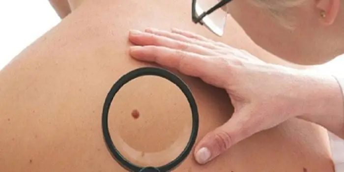
Differences between good types of moles and bad ones
Many owners of such formations on the skin are concerned with the question of how to distinguish a benign mole from a malignant one. This is quite simple to do; you just need to carefully examine the mole. Can be considered non-hazardous clearly defined birthmarks small size. They have a uniform structure and do not protrude much above the skin. The color range varies from light yellow to black. But if in doubt, it is better to undergo an examination by a dermatologist using special studies.
Bad birthmarks include the appearance of intense pain of various types in their area. If within two months the mole has increased significantly or itching has appeared in its area, then this is a reason to consult a doctor. Additional concern should be caused by the appearance of additional crusts, bulges or ulcers on the surface.
Moles on the body and the reasons for their appearance
The appearance of these benign formations can be influenced by various factors that stimulate excessive division of skin cells in a limited area of the skin. Modern doctors the following factors are identified, as the main ones, when moles appear:

Ultraviolet radiation.- Hormonal imbalances and related diseases.
- Genetic changes in the body contribute to the hereditary transmission of birthmarks to children, identical to the birthmarks of the parents, appearing in certain places of the body.
- Various defects in skin development that lead to the appearance of congenital moles in children starting from the age of two months.
- The possibility of the appearance of moles with long-term use of drugs containing homones.
- Long-term and untreated viral and bacterial infections.
What moles should be removed and methods for their elimination
Only those birthmarks that are pose a potential threat for humans and can turn into cancer. Removing a mole cannot provoke the development of this dangerous disease. Surgeries to remove birthmarks are completely safe and can have side effects only in the event of an allergic reaction to certain medications.
Modern medicine welcomes several methods for getting rid of birthmarks:
- Surgical operations.
- Laser exposure.
- Removal using liquid nitrogen.
- Exposure to electric current.
- Removal by radio waves.
The method of treatment for removing a birthmark is selected at an appointment with a dermatologist strictly on an individual basis. Removal using laser or liquid nitrogen refers to more gentle methods of combating unwanted tumors. All manipulations are carried out without the appearance of blood and are as gentle as possible on the skin around the mole.
