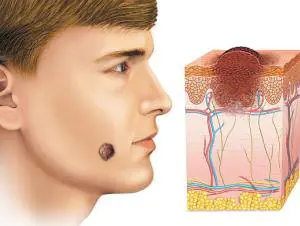
Moles in medical terminology they are called nevi.
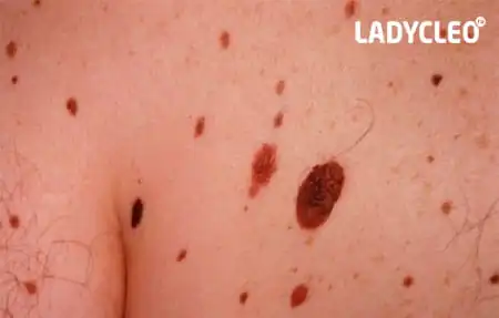
They can be found on the body of any person in the form of small round dots, small spherical formations that rise above the surface of the body, and large pigmented areas of the skin.
What it is?
Moles consist of specialized cells of the epidermis - melanocytes, responsible for the production of melanin in the body (a pigment that colors the skin in various shades of brown). Under the influence of external or internal factors, an excess amount of them accumulates in certain places of the skin - this is why moles appear on the body of an adult, child or elderly person.

In some people, nevi remain unchanged on the skin all their lives, in others they disappear, in others they grow or change their shape and color. The latter become dangerous and can degenerate into malignant formations.
- Common moles have a rounded symmetrical shape, depending on the concentration of melanin in them, nevi are colored beige, brown or black.
- Meet and red dots on the body as moles, for the most part this phenomenon is associated with impaired skin pigmentation due to hepatic or endocrine pathology, as well as natural age-related changes (i.e. as a result of aging).
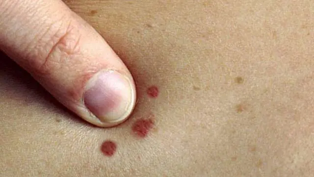
The difference between these spots and moles is that they usually appear in groups on a limited area of the skin. When metabolic processes are restored, the red spots disappear.
In adults, the appearance of nevi is associated with hormonal crises due to illness, menopause or nervous exhaustion. The appearance of moles in them can be observed constantly or appear spontaneously, in response to a certain stimulus. In childhood, science has noted the wavy growth of nevi.
Periods when moles appear in children:
- 6 months - six months, at this moment the child’s endocrine system adapts to external conditions
- 5-7 years, the stage of active growth of the skeletal system and skeletal muscles, requiring rapid metabolic reactions
- 12-16 years, puberty with significant changes in the functions of the whole organism.
Why do they appear?
Nevi are essentially benign skin formations.
Some people get scared when they see many moles on their body. What does this mean for a specialist? Only that the patient’s body is prone to accumulation (accumulation) of melanin in the surface layers of the epidermis.
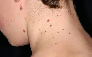
The reasons for the appearance of numerous or single nevi are varied.:
- exposure to ultraviolet radiation, one of the most common factors in the formation of moles due to increased melanin levels during tanning
- traumatic damage to the epidermis, systematic violations of the integrity of the skin contribute to the appearance of pathological changes in it
- exposure to radiation, which rapidly changes normal skin cells
- consumption of harmful products (GMOs, fast food, alcohol) and smoking, these habits negatively affect metabolic processes in the body.
- endocrine disorders and diseases, any changes in hormonal levels can cause the appearance of skin pathologies, pigmentation, moles
- hereditary predisposition, the presence of various nevi in the family.
Classification and photo
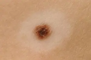
1. A flat nevus or birthmark is a pigmented island of skin with clearly defined boundaries. It can take the form of lentigo - multiple brown or brownish formations in the upper layers of the epidermis.
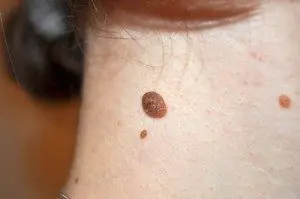
2. Convex nevus or mole. It has a diameter of up to a centimeter, a smooth or lumpy surface and rises above the level of the skin. Its color varies from beige to black, and a hair is usually located in the center of such a formation.
3. Blue nevus or blue mole. It looks like a smooth hemisphere, slightly raised above the skin, sometimes reaching a size of 2 cm. The color of this benign formation ranges from blue to dark blue.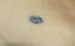
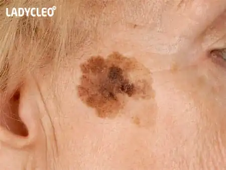
4. Giant nevus. It is a large spot on the body, gray, bright beige (sometimes brick), black or brown.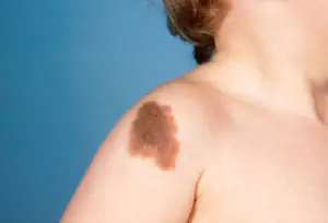
Dangerous and non-dangerous moles
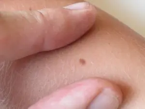
Ordinary nevi do not cause any discomfort to their carriers sometimes they even disappear without a trace. Their shape is stable, size and color remain unchanged.
But benign moles are sometimes removed to prevent their degeneration if they are of rather large size (pedunculated) and are located in areas of the body where a person constantly injures them (during vigorous activity or parts of clothing).
Remember dangerous moles in the photo and be vigilant!
If nevi degenerate into cancerous moles, this is dangerous, since such neoplasms quickly metastasize to other organs.
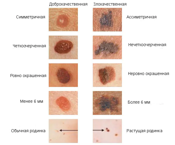
Therefore, it is very important to see signs of malignancy in time.:
1. The shape of the mole changes, it loses its symmetry and begins to grow in one direction.
2. The edges of the nevus become uneven (“cut up”, “torn”).
3. The color of the mole is uneven and contains yellow, red or black inclusions.
4. The nevus grows or “shrinks”, its size changes rapidly.
5. The texture of the mole becomes different, smooth becomes rough, bumpy becomes flat, etc.
6. Loss of hair growing from the nevus.
7. Itching, peeling and burning in the mole area.
There are several reasons why a nevus itches:
– pathological cells multiply
– there are active processes of death of healthy tissues
– the area around the formation becomes inflamed and swollen.
8. The appearance of microcracks and ulcerations.
9. Bleeding and soreness of the mole.
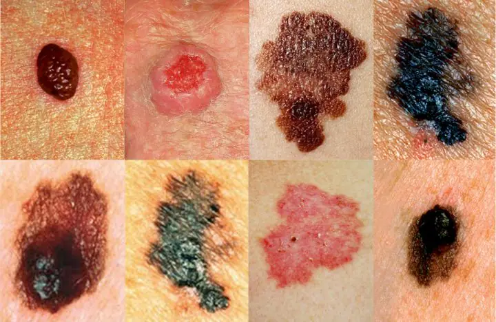
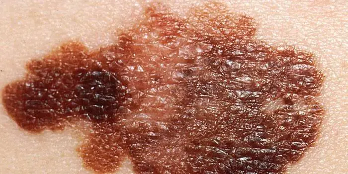
Cancerous moles (melanomas): photo
What happens if you rip off a mole?
You cannot remove a nevus yourself.
Firstly, it is dangerous, and secondly, it is simply ineffective. If the mole is located close to blood vessels, prolonged bleeding may develop. Often such self-medication leads to re-formation of the nevus, its growth or malignancy.
Therefore, it is difficult to predict what will happen if a mole is torn off. It may have no consequences, or it may cause serious complications for the health and life of patients.
Doctors warn that any trauma to the nevus is extremely undesirable, but pedunculated moles or small convex formations can be accidentally removed with nails or hard items of clothing.
What to do if you rip off a mole:
- cauterize the wound with an alcohol solution
- stop the bleeding by applying a gauze bandage
- come to see a specialist.
In cases of partial removal of a mole, do not touch the remaining formation, do not cut it off or tear it off.
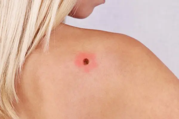
Sometimes such pigmentation appears before the disappearance of a mole (as a sign of depigmentation), and in other cases it may signal its degeneration.
White spots around the mole appear as a characteristic sign of Setton's nevus. This formation is considered harmless in terms of degeneration into a more malignant form, however, melonomas (aggressive cancerous formations) can also have such a white rim, so the appearance of white spots is a reason to contact a medical specialist.
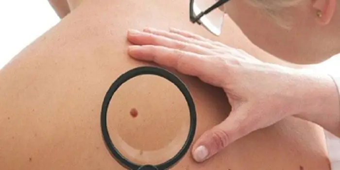
Diagnostics
Dermatologists and oncologists are involved in determining the type of nevus.
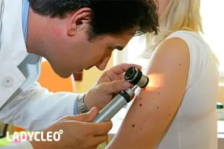
Using a dermatoscope, the doctor examines the formation and determines its nature (benign or malignant). Sometimes a histological examination (scraping method) is required.
Biopsy (tissue sampling) for nevi is not used due to their trauma during this procedure. And as you know, it’s better not to touch moles again!
Removal
Many people want to get rid of birthmarks and nevi not only in cases of malignancy (pathological change), but also to correct a cosmetic defect.
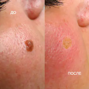
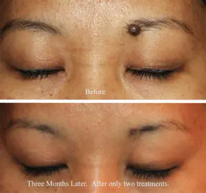
However, removal is carried out at the oncology center for people with malignant tumors and with a high probability of degeneration of nevi.
Often the indication for surgery is the localization of moles on the body: on the scalp, on the neck, on the chin, in the area of the shoulder blades.
Methods for removing nevi (with information on how much it costs to remove them):
- surgical, using local anesthesia and a scalpel (in municipal surgical hospitals, according to indications - free of charge, in medical centers from 300-500 rubles)
- cryofreezing with liquid nitrogen (1000-1500 rubles)
- electrocoagulation - cauterization with electric current (600-1300 rubles)
- photodynamic - ultraviolet irradiation (1000-1200 rubles)
- laser - removal of moles with a beam (800-2000 rubles)
- radio wave, destruction of nevi by shock radio wave (RUB 700-1400)
The choice of method for removing moles is made by the doctor prices for these procedures depend on the size and type of nevus.
Prevention of malignancy
Preventive methods for reducing the risk of malignancy of moles include:
- limited sunbathing, cancellation of visits to the solarium
- minimizing skin trauma
- maintaining a healthy lifestyle (healthy eating and avoiding bad habits).
It's rare to see a person without small dark marks on their body. Is it worth paying attention to these points? Only a doctor will distinguish between dangerous and normal moles - malignant melanoma or harmless nevus - and give recommendations on what to do with them. Is it worth worrying about the appearance of new formations, when immediate contact with specialists is required, what are the signs of cancer development - the answers to these questions remain to be found out. No one is immune from disaster, and early diagnosis will protect you from severe consequences.
What is a mole
The first tiny spots may appear in children in infancy. A mole is a small formation on the skin - a nevus - that is considered benign and harmless. The basis for their appearance is melanocyte cells that accumulate the natural pigment melanin. Depending on its quantity, a difference in color is observed. Available colors:
The shape of the tumors depends on the location and concentration of melanin. They may have a stalk or be located under the skin, be flat and convex. The most common type is round, but there are exceptions. The development of neoplasms is provoked by ultraviolet radiation - natural from the sun, in a solarium. Hereditary factors cannot be excluded. A common cause of growth is hormonal imbalance, characteristic of periods:
- puberty
- pregnancy
- menopause.
What types of moles are there?
One person may discover very different tumors. Types of moles are classified according to several criteria. This helps in correct diagnosis in case of changes. They differ in:
- origin– congenital, newly acquired
- structure– pigment, vascular
- place of education – in depth, on the surface, in the boundary layer
- raised above the skin – flat – even, protruding as a hemisphere, pedunculated, larger birthmarks
- potential threats – dangerous, degenerating into melanoma, non-dangerous.

Safe moles
Those who have dark spots on their skin should be wary of their changes. In time, detected signs of degeneration into melanoma contribute to the timely removal of the formation and preservation of health. Safe moles are different:
- the presence of a stalk – it cannot be formed by malignant cells that grow randomly
- long-term condition without changes.
Spots that appear soon after birth are not considered dangerous. It is important that they are small in size. Good – non-dangerous – signs of neoplasms include:
- flesh tone
- unchanged pattern of the skin of the nevus and adjacent tissues
- soft consistency
- hair on the surface of the neoplasm - growing from the skin, indicates the absence of pathologies
- diameter no more than 5 mm
- symmetry
- nevus in the form of a spot.
Which moles are dangerous?
Why do people with nevi on their bodies need to monitor their changes? There is always a threat of degeneration of non-dangerous tumors into a cancerous tumor. What moles are dangerous to health? Key signs you need to know:
- change in shades towards the dark side, the appearance of multi-color
- rapid increase in size - exceeds two millimeters per year
- occurrence of cracks
- the formation of asymmetry due to uneven growth
- lack of elasticity
- the appearance of itching, burning
- presence of discomfort.
The appearance of dangerous moles requires an immediate visit to a specialist to clarify the nature of the changes and the likelihood of developing skin cancer. Pathological transformations provoke:
- injury to the nevus due to negligence
- self-removal
- abuse of exposure to the sun, use of a solarium
- location of the formation in places of frequent contact with clothing - on the neck, head, genitals, legs
- placement in the hair, on the face, palms - where there is a high probability of injury
- previously removed melanoma.
Why are moles dangerous?
Not a single person is protected from the sudden proliferation of cells of a harmless mole. Melanoma is an extremely serious disease. Changes not detected at the initial stage can result in death. The provoking factor is unsuccessful independent removal of tumors. Moles are dangerous because of their ability to:
- transform into an atypical – precancerous form
- grow to large sizes
- turn into cancerous
- with minor external changes, metastases actively spread throughout the body through the circulatory and lymphatic channels.
How quickly does melanoma develop from a mole?
The transformation of a nevus into a cancerous formation can occur in different ways. The process depends on the stage of the disease and the type of tumor. Instant metastases are dangerous. Begins:
- growth of cancer (oncological) cells in the deep layers of the epidermis
- their entry into the blood and lymph
- penetration into the lungs, liver, kidneys
- growth in these organs
- complete damage to the body
- death.
The growth phases of pigment cells are observed, along which melanoma develops from a mole. There are varieties:
- horizontal– damage to the upper layers of the skin occurs, lasting up to 10 years, but metastases do not appear
- vertical– accompanied by the spread of cancer cells throughout the organs, can last two years, has an unfavorable prognosis
- nodal – especially dangerous – characterized by deep spread within two months.
The first signs of melanoma
The patient can be assisted only when suspicious changes begin to be identified. The diagnosis, research, and referral for surgical treatment save a person’s life. The first signs of melanoma:
- increase in the height of the tumor
- bleeding
- the appearance of discharge
- redness
- burning, itching
- swelling of tissues
- softening of the nevus
- the appearance of a crust
- thickening
- hair loss
- expansion of pigmentation around the lesion.
With the further development of dangerous melanoma, the following are observed:
- significant change in size
- the appearance of pain
- enlarged lymph nodes
- surface ulceration
- formation of new foci
- bleeding from places of pigmentation
- liquid separation
- skin thickening
- the appearance of an earthy tint
- signs of metastases are chronic cough, weight loss, cramps, headaches.
How to distinguish a mole from melanoma
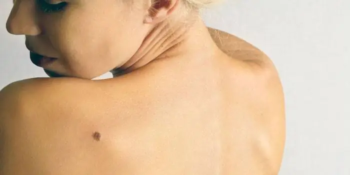
To recognize which moles are dangerous and which are not dangerous, you need to know what they look like. A person with nevi, in order to avoid dire consequences, must constantly monitor the appearance of new formations and changes that occur. You can distinguish a mole from melanoma by its signs. Non-dangerous neoplasm:
- symmetrical
- with smooth edges
- uniform in color
- with dimensions not exceeding 6 millimeters.
Features of dangerous melanoma that require seeking help from dermatologists:
- growth in a short time
- pronounced asymmetry of shape
- heterogeneity in color - the presence of inclusions of several shades
- lack of clear boundaries - the contour line is blurred, jagged, and looks like a coastline on a geographical map
- increased diameter over six millimeters
- variability of any parameters - color, size, shape.
What dangerous moles look like
What do nevi that are subject to pathological changes look like? Only a doctor can correctly distinguish between non-dangerous tumors. Dangerous formations look like this:
- blue– compactions under the skin with clear boundaries, with dimensions no more than 10 mm
- nodal– round, flat in shape, color – brown, black
- cutaneous– often pale, convex
- halo nevus – pigment surrounded by a light or white rim
- spitz- looks like a dome-shaped tumor of pink shades, with the possible presence of a hole through which blood and liquid leak
- connecting- connect individual entities into a whole.
Mole with jagged edges
One of the signs of a non-hazardous formation turning into a dangerous one is a change in contours. It often has blurred edges and scalloped borders. There are non-dangerous types of nevi - dysplastic. Only a specialist can make a correct diagnosis. A mole with uneven edges can be dangerous if there are additional signs of melanoma:
- accelerated changes in size
- the presence of clearly defined asymmetry
- the appearance of highly indented boundaries.
Rough mole
Such a neoplasm is harmless if its diameter is no more than 5 mm and remains constant in size. Often its appearance signals a lack of vitamins and nutritional disorders. Doctors advise coming for a consultation if it is discovered that:
- the smooth nevus turned into a rough one
- bothered by burning, itching, tingling
- irregularities and compactions appeared in the middle
- areas with different shades formed
- diameter has increased significantly.
A dangerous rough mole requires immediate examination if:
- the appearance of bleeding
- development of the inflammatory process
- rapid change in size
- formation of asymmetry
- formation of purulent discharge
- the occurrence of painful sensations when touched
- the emergence of an irregular shape, blurred boundaries, along the edges of the neoplasm.
Large moles
Large formations on the skin are pigment spots. When they remain unchanged and do not cause inconvenience, this is not a dangerous phenomenon. It is important to constantly monitor their appearance, color, and size. To eliminate worries, you need to consult a dermatologist. During the visit, the specialist will conduct a diagnosis and give a forecast of the risk of developing a malignant neoplasm. Large moles become dangerous if they:
- injured
- thickened
- started to itch
- were unsuccessfully removed independently
- changed in size, shape
- are bleeding.
What moles can be removed
Often nevi cause trouble for women when they are in a visible place - the face, neck. Even if they do not bother you, using removal will be the right decision - the appearance will improve significantly. After the procedure, the doctor must necessarily send the tissue for histological analysis to decide whether the mole is malignant or not. If the neoplasm is not dangerous, does not bother you, and does not change in size, surgery is not required. What moles cannot be removed? Experts believe:
- there are no contraindications
- It is important to choose the right excision technique.
You should be careful about skin growths it is unacceptable to remove them yourself. Only the doctor will determine whether a nevus is dangerous or not and decide what to do with it. You can delete it if:
- injured from clothing - on the neck, in the groin area, under the armpits
- cause pain when touched
- are located under the hair on the head and can be damaged when combing or cutting
- change color, shape, outline
- significantly increase in size
- characterized by the presence of burning, itching
- accompanied by inflammation and bleeding.
Almost every person has moles. Some people have only a few of them, while some people can count dozens or even hundreds of such marks. Most moles are harmless. But among them there are specks that can cause a lot of trouble. What types of moles are distinguished and when do they require our close attention?

Morphological classification of moles
A mole in medical terminology is called a “nevus”. This is a cluster of cells - melanocytes, containing a special pigment - melanin. The color of the mole is determined by the concentration, quantity and depth of melanin in the nevus.
Moles are classified according to a number of parameters. They may vary in color, shape and size.
The color range of spots is varied. It depends not only on the characteristics of the cells that form the mole, but also on the color type of the carrier’s skin. Available palette:
- From light brown to almost black.
- Pink - red - crimson.
- Blue - purple - dirty blue.
Form
Flat and convex, round and oblong, nodular and “pedunculated”, smooth and rough - the dimorphism of spots is impressive! As a rule, even on the body of one person, moles of various shapes can “get along” quite peacefully.
Size
From 1 mm to extensive nevi covering a significant area. As a rule, size is not directly related to the risk of nevus degeneration into an oncogenic form.
Attention! Any change in the shape, color, size and structure of the nevus is a sufficient reason to contact a dermato-oncologist for timely diagnosis!
Moles that have:
- smooth edges
- no more than 0.5 cm in diameter
- evenly colored.
Even the subjective feeling that a mole is different from the rest, and even more so that it is itchy, growing and changing color, should alert you. According to some data, the 5-year survival rate after treatment of malignant tumors detected at an early stage reaches more than 90%.
Types of moles based on benignity
Morphological features of moles provide primary information about their structure and character. The full picture can only be revealed by histological examination. Thus, there are three conditional groups of skin neoplasms:
- Benign neoplasms - nevi.
- Precancerous or borderline - basilioma.
- Malignant - melanoma, skin cancer.
Benign nevi
Widespread. We can say that upon closer examination they can be found in the majority. Such plaques have smooth and clear edges, they can be of different solid colors. They are capable of increasing in size, but this process occurs slowly and often goes unnoticed. Their growth is not associated with discomfort - it does not cause itching or inflammation.
Borderline neoplasms
These include potentially dangerous pigmented formations - atypical moles and basiliomas. When certain conditions are created (trauma, excess sun), they can turn into a malignant form. There is a simple formula - the abbreviation AKORD. With its help, you can try to independently determine the presence of an atypical mole.
An asymmetrical mole with uneven edges of uneven color, changing its size and changing its appearance is atypical.
Malignant structures
Melanoma, or skin cancer, occurs less frequently. According to some reports, only one mole in a thousand is dangerous. However, in some countries, doctors began to remove all pigmented formations on the skin without waiting for their degeneration. In Russia, this method of combating melanoma is not practiced. And there is a good reason for this - the removal operation itself can also cause a malignant change.
Unfortunately, it is impossible to visually determine the degree of malignancy of a mole. And the line between them is so thin that in case of any suspicion it is better to consult a doctor. The presence of atypical spots requires careful observation and examination every three months.
Structural classification
Based on the structures involved in the body of the mole, dermatologists distinguish between pigmented, vascular and warty varieties.
Pigmented moles
Appearance: smooth or rough, may have hairs coming from inside. The color varies, but in all cases it is darker than that of freckles. The color of pigmented nevi is determined by a combination of black eumelanin and brown pheomelanin.
Pigmented nevi consist of two types of cells - epidermal and nerve cell sheaths, located in the upper layers of the skin.
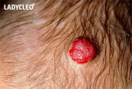
Vascular moles
Nevi of this type lie deeper, localized in the basal layer of cells between the epidermis and dermis. They are more convex and include blood vessels in their structure. The color of such formations ranges from pink to bright red.
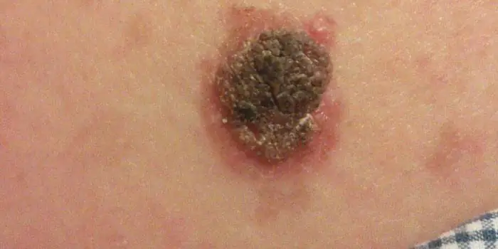
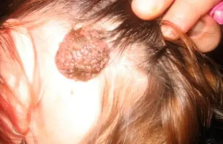
Warty moles
A collection of bubbles that are dirty gray or brown in color. The surface is granular, keratinized. The external resemblance to a wart determines the name. Mostly localized on the head, neck, and behind the ears.
More often, a warty nevus is found in the fair sex. Unfortunately, these moles do not give them any charm, since they are large in size, very noticeable in the photo and have an unpleasant appearance. From 2 to 10% of such formations can degenerate into cancer, and therefore require careful attention and observation by a dermatologist.
Medical classification
Nevi are diverse and changeable - the variety of their shapes, structures and degree of danger requires different approaches to classification. Let us describe the types of moles that are identified by practicing dermatologists when making a diagnosis.
Lentigo
Somewhat reminiscent of freckles. The main difference is a more saturated color. In addition, under the influence of ultraviolet radiation, the number of such spots and the intensity of their color do not change. Border formations located in the basal layer between the epidermis and dermis.
Epidermo-dermal
They are non-convex nevi of small size up to 1 cm. Like lentigo, they are based in the border area. Color from flesh to black. Localized on the skin of the feet, palms and genital area.
Intradermal nevi
The melanocytes that form this type of nevus are located in the thickness of the dermis. The deeper the melanocytes lie, the more convex the nevus. In this case, the plaques necessarily protrude above the skin. Color - from beige to dark brown.
Complex nevi
Melanocytes of such a nevus are located in both the dermis and epidermis. These moles always protrude above the skin and are very dark in color.
Sutton's nevi
A characteristic feature of this type is the ability to spontaneously disappear and appear. They are easily distinguished from others by the presence of a ring of unpigmented skin around the nevus.
Dysplastic nevi
They have a number of characteristic features:
- They first appear in people over 35 years of age.
- Diameter - up to 12 mm.
- They are located in areas hidden from sunlight (buttocks, chest, scalp).
- Often these are numerous clusters.
- Passed on by inheritance.
- Paradoxically, these irregularly shaped plaques with blurred edges and uneven coloring, although they contain signs of cancer, extremely rarely turn into malignant forms.
Blue nevi
The color range of such nevi is varied - from gray-blue to blue and dark blue. A distinctive feature is color variations within the blue palette. These are towering formations with a smooth surface and a clear boundary. The size does not exceed 2 cm, hair does not grow in their area. Located on the face, limbs and buttocks.
Cellular blue nevus
Visually, this type of nevus is indistinguishable from a simple blue nevus. Histological examination shows that this type of mole is distinguished by its ability to rapidly divide melanocytes. This is an unfavorable sign indicating the possibility of developing melanoma.
Giant pigmented nevus
It is a flat spot, the color of which can be either brown or dark gray. It can reach impressive sizes, since it is a congenital formation that tends to grow with the child.
Nevi of early childhood
In addition to the giant pigmented nevus, there are other congenital pigmentation disorders. As a rule, these are vascular types of moles, namely hemangiomas, port-wine stains and pigmentation at the base of the skull. The latter goes away on its own within 1 - 1.5 years.
Hemangioma
Benign formations that developed as a result of abnormal formation of a blood vessel. They appear during the first week of life and look like a pink-red spot the size of a needle point. For some time they may increase slightly in size, but in most cases they turn pale and disappear by 3 years.
Wine stains
Another name for them is flaming nevi. They occur due to dilation of blood vessels on the face and scalp. With age, they do not disappear or fade, but grow with the child. If such a spot appears, it is better to immediately contact a dermatologist and begin treatment as early as possible in order to prevent it from growing and turning into a serious cosmetic defect.
Be attentive to yourself and your loved ones! Visit a dermatologist, study your moles - this will give you peace of mind and confidence in your health.
