Birthmarks can be found on the body of almost any person. There was always close attention and interest in them. Now their popularity has waned, but any formation on the skin should not be ignored. What kinds of moles there are, how they can be dangerous, whether you need to see a doctor and in what cases, we will consider in this article.
In medicine, a mole on the body is called a nevus. It is understood as a congenital or acquired transformation of the skin, expressed in the growth or change in color of its individual areas. Congenital formation can be seen only several months after birth. Acquired moles are often associated with hormonal imbalances, skin injuries and ultraviolet radiation.
Types of moles and their descriptions
The types of moles are determined based on their color, shape and size. In shape, the neoplasm can appear flat, oblong, round, smooth or with a rough structure. The skin of a mole can be light brown, have all shades of the red spectrum, black and even purple, which directly depends on the color type of a particular person. Its minimum size is usually 1 mm, and its maximum is difficult to predict; sometimes it covers a significant area.
Depending on the danger, moles are of the following types:
1. Nevus is a benign neoplasm. It does not cause discomfort, its shape has clear outlines, and does not change its original color. Most moles belong to this type.
2. Basalioma is a type of precancerous condition of a birthmark.
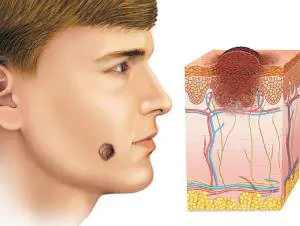
3. Melanoma. All malignant moles have this name in medicine. To identify it, a thorough examination by an oncodermatologist and diagnostics are necessary.
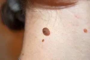
In the medical literature, structural types of moles and photos with descriptions are often found. Let's look at them in more detail. The pigmented neoplasm is smooth to the touch, sometimes it can have a rough appearance due to a small amount of hair. The color is usually dark.
Vascular moles are distinguished by different shades of red, because... their structure includes vessels of the circulatory system and have a convex appearance. The last type of structural classification is warty moles. They are similar in appearance and color to ordinary warts. The difference lies in the nature of occurrence. Warts are basically viral in nature. The skin of women is more susceptible to the appearance of warty nevus on it than that of men. About 10% of them may carry a risk of developing cancer.
Let's consider a description of all types depending on the diagnoses made by dermatologists.
- Lentigines are classified as borderline formations. They are small in shape and size, resembling freckles. A distinctive feature is a darker and more saturated shade.
- The epidermodemal type is most often located in the intimate area, on the palms and soles. The color may vary (from flesh-colored to bright black).
- Sutton's nevi. The skin around the mole does not contain pigment, which is how this variety is easily distinguished from other formations. Also, such a mole on the body can disappear on its own, and after a while appear again.
- Dysplastic moles select the body of a mature person after 35 years of age and are hereditary in nature. They are often located on parts of the body protected from sunlight. The diameter can reach 12 mm.
- Nevi are blue in color. Their color varies from light blue to deep blue. The most dangerous moles of this type are cellular nevi.
- Pigmented giant nevus is congenital. Considering that this neoplasm grows with a person, it sometimes has an impressive size. Characterized by a flat shape.
The human body can contain several types of nevi at the same time. Many of them are harmless, but in order to notice malignant moles in time, you should treat them carefully.
How to recognize them?
A safe mole on the skin should have a clear contour with smooth edges, uniform color and its diameter should not exceed 0.5 cm. There are certain risk factors, the presence of which in a person increases the risk of a simple mole transforming into melanoma. These include:
- Light skin with freckles.
- Frequent sunburn.
- Men, especially older ones.
- Relatives who have a history of malignant moles.
- If a person’s body is practically covered with moles.
A great danger lies in the presence of moles such as blue and borderline nevus.
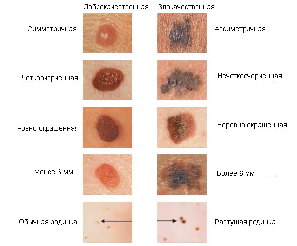
In order to notice dangerous moles in time, you should carefully and independently examine the body for changes. The simple formula for self-diagnosis proposed by oncodermatologists - ACORD - will help eliminate the danger. This abbreviation includes all the main signs of a malignant formation: asymmetry, edges, color, size and dynamics.
A harmless mole will have symmetrical halves if an imaginary line is drawn along its center. For greater clarity, use a ruler. The appearance of a malignant mole does not have clear boundaries, the edges are blurred. Her color is distributed unevenly. If, after examining the body, you find similar moles, then you should track their dynamics. This type of education changes frequently. You can measure its size; if after a few days the nevus increases, then this is a dangerous sign. The detection of a mole with a diameter of more than 6 mm should also alert you.
The considered signs are considered the most important, but there are also secondary ones, which can also indicate a dangerous type of birthmark. These include:
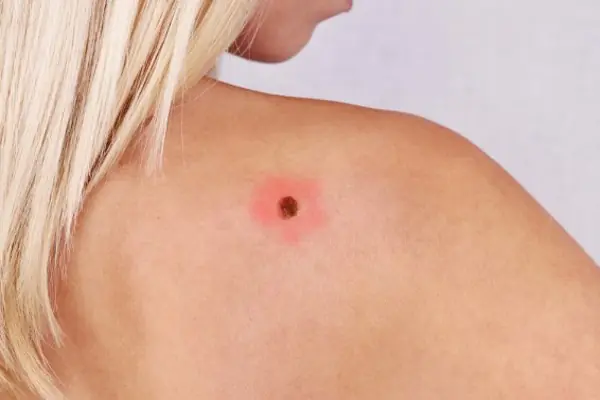
- The presence of inflammation, in which the skin around the mole becomes reddish and painful.
- Bleeding, wetness, or crusting appears. Also, such a formation may become covered with small ulcers.
- The mole increases not in width, but in length.
- The skin of the mole is covered with satellites (multiple pink dots).
- Detection of the “leg” of the nevus. The nodular variety of moles is usually found on the head, back, neck and limbs.
- Change in the matte surface of the birthmark and the appearance of a slight shine.
An external examination provides only tentative conclusions. Only an oncodermatologist can determine an accurate diagnosis by performing a biopsy.
Cases when specialist consultation is necessary
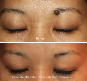
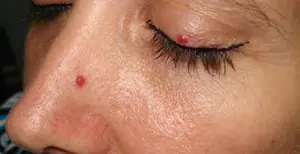
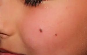
You should know that detecting melanoma at an early stage increases the likelihood of a complete cure. Detection of any change, even the most insignificant, is a reason to visit a specialist’s office for a more complete examination. If a mole on the face begins to bleed, itching appears and is constantly increasing, then this is an extremely unfavorable signal. The situation may worsen if you begin to feel pulsation in the birthmark. Perhaps the mole is a type of blue nevus, which quite often changes into melanoma.
The moles have increased in size so much that they begin to cause discomfort and pain when they come into contact with clothing. Sometimes the pain and redness of moles is temporary, but don't let your body fool you. After a while, all the unpleasant symptoms will return again, but now they will become stronger.
Light skin, which has numerous birthmarks, requires a more careful and serious attitude. There are frequent cases of death with late detection of melanoma. Its danger lies in the fact that metastases occur almost immediately, affecting internal organs.
Now modern diagnostics include completely safe and painless methods (biopsy, X-ray, computed tomography and laboratory tests). The largest birthmarks should be removed to eliminate the risk of developing cancer. At the moment, they are removed surgically using radiation therapy.
The meaning of moles depending on their location on the body
At many times, people tried to unravel the reason for this or that location of birthmarks. A body with moles may well suggest the answer to any question, if you can correctly interpret the meaning of their location.
1. A mole on a man’s face will help determine his suitability for family life. If you find a birthmark in the corner of your left eye, then this indicates a jealous character. But a man with a nevus near his right eye will turn out to be a wonderful family man. Finding it in the center of the forehead will tell about love and frivolity. A nevus on the eyelid indicates a high level of intelligence.
2. Skin on the back with moles will indicate a grouchy, but honest and open character. Nevi on the legs will indicate indecision. However, if they are found on the feet, then their meaning is associated with travel.
3. A woman who has a body or face with moles is also able to see some meaning in them. The location near the lips often reveals a passionate nature, the skin of the cheeks indicates hot temper, but easy reconciliation. The owner of a mole between the eyebrows is endowed with good intuition and intelligence.
4. Having thoroughly examined her own body, a woman can even find out how many children she will have. Count the moles on your waist, their number will be the answer. Skin on the shoulders and arms with birthmarks speaks of good luck, success and a happy life.
5. The meaning of a nevus depends not only on the part of the body where it is located, but also on the shape and type. For example, there is an opinion that triangle-shaped moles can only be found in a person with strong energetic and extrasensory abilities. According to another version, the skin of indigo people often has this sign. If this type of mole is on the inside of the palm, then this indicates success in activities related to communication. Finding it on your head promises a successful scientific career.
In conclusion, we should recall preventive measures that will significantly reduce the risk of a mole degenerating into a malignant tumor. You should not injure birthmarks; if they are located in a place that is constantly in contact with clothing, it is better to remove such a nevus. When showering, use soft sponges.
Never remove moles that have a nodule or a “leg” yourself, even if it seems to you that the nodule is very thin. Protect skin, especially those with a lot of moles, from direct sunlight. You should also avoid frequent visits to the solarium. According to the results of studies conducted in California, it turned out that on average 5-10 years pass from the first visit to a solarium by people at risk until the discovery of melanoma.
Remember that monitoring your birthmarks and regular thorough self-diagnosis will help you avoid serious health problems. Let moles become for their owner only a symbol of aesthetic beauty.
Moles in medical terminology they are called nevi.
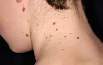
They can be found on the body of any person in the form of small round dots, small spherical formations that rise above the surface of the body, and large pigmented areas of the skin.
What it is?
Moles consist of specialized cells of the epidermis - melanocytes, responsible for the production of melanin in the body (a pigment that colors the skin in various shades of brown). Under the influence of external or internal factors, an excess amount of them accumulates in certain places of the skin - this is why moles appear on the body of an adult, child or elderly person.

In some people, nevi remain unchanged on the skin all their lives, in others they disappear, in others they grow or change their shape and color. The latter become dangerous and can degenerate into malignant formations.
- Common moles have a rounded symmetrical shape, depending on the concentration of melanin in them, nevi are colored beige, brown or black.
- Meet and red dots on the body as moles, for the most part this phenomenon is associated with impaired skin pigmentation due to hepatic or endocrine pathology, as well as natural age-related changes (i.e. as a result of aging).
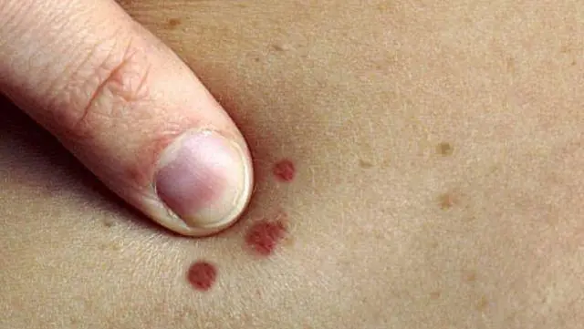
The difference between these spots and moles is that they usually appear in groups on a limited area of the skin. When metabolic processes are restored, the red spots disappear.
In adults, the appearance of nevi is associated with hormonal crises due to illness, menopause or nervous exhaustion. The appearance of moles in them can be observed constantly or appear spontaneously, in response to a certain stimulus. In childhood, science has noted the wavy growth of nevi.
Periods when moles appear in children:
- 6 months - six months, at this moment the child’s endocrine system adapts to external conditions;
- 5-7 years, the stage of active growth of the skeletal system and skeletal muscles, requiring rapid metabolic reactions;
- 12-16 years, puberty with significant changes in the functions of the whole organism.
Why do they appear?
Nevi are essentially benign skin formations.
Some people get scared when they see many moles on their body. What does this mean for a specialist? Only that the patient’s body is prone to accumulation (accumulation) of melanin in the superficial layers of the epidermis.
The reasons for the appearance of numerous or single nevi are varied.:
- exposure to ultraviolet radiation, one of the most common factors in the formation of moles due to increased melanin levels during tanning;
- traumatic damage to the epidermis, systematic violations of the integrity of the skin contribute to the appearance of pathological changes in it;
- exposure to radiation, which rapidly changes normal skin cells;
- consumption of harmful products (GMOs, fast food, alcohol) and smoking, these habits negatively affect metabolic processes in the body.
- endocrine disorders and diseases, any changes in hormonal levels can cause the appearance of skin pathologies, pigmentation, moles;
- hereditary predisposition, the presence of various nevi in the family.
Classification and photo
1. A flat nevus or birthmark is a pigmented island of skin with clearly defined boundaries. It can take the form of lentigo - multiple brown or brownish formations in the upper layers of the epidermis.
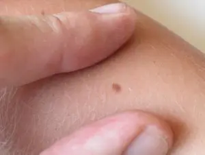
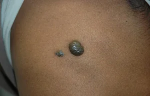
2. Convex nevus or mole. It has a diameter of up to a centimeter, a smooth or lumpy surface and rises above the skin level. Its color varies from beige to black, and a hair is usually located in the center of such a formation.
3. Blue nevus or blue mole. It looks like a smooth hemisphere, slightly raised above the skin, sometimes reaching a size of 2 cm. The color of this benign formation ranges from blue to dark blue.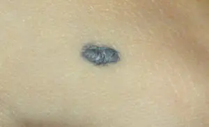
4. Giant nevus. It is a large spot on the body, gray, bright beige (sometimes brick), black or brown.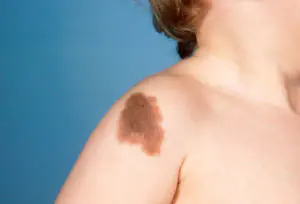
Dangerous and non-dangerous moles
Ordinary nevi do not cause any discomfort to their carriers; sometimes they even disappear without a trace. Their shape is stable, size and color remain unchanged.
But benign moles are sometimes removed to prevent their degeneration if they are of rather large size (pedunculated) and are located in areas of the body where a person constantly injures them (during vigorous activity or parts of clothing).
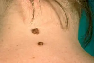
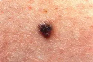
Remember dangerous moles in the photo and be vigilant!
If nevi degenerate into cancerous moles, this is dangerous, since such neoplasms quickly metastasize to other organs.
Therefore, it is very important to see signs of malignancy in time.:
1. The shape of the mole changes, it loses its symmetry and begins to grow in one direction.
2. The edges of the nevus become uneven (“cut up”, “torn”).
3. The color of the mole is uneven and contains yellow, red or black inclusions.
4. The nevus grows or “shrinks”, its size changes quickly.
5. The texture of the mole becomes different, smooth becomes rough, bumpy becomes flat, etc.
6. Loss of hair growing from the nevus.
7. Itching, peeling and burning in the mole area.
There are several reasons why a nevus itches:
– pathological cells multiply;
– there are active processes of death of healthy tissues;
– the area around the formation becomes inflamed and swollen.
8. The appearance of microcracks and ulcerations.
9. Bleeding and soreness of the mole.
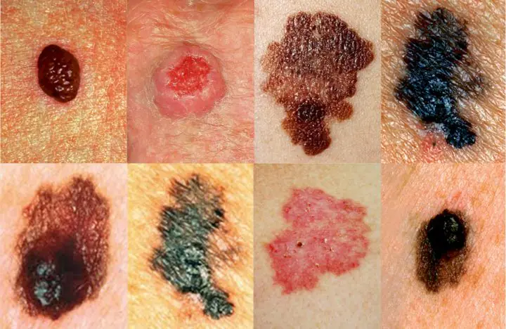
Cancerous moles (melanomas): photo
What happens if you rip off a mole?
You cannot remove a nevus yourself.
Firstly, it is dangerous, and secondly, it is simply ineffective. If the mole is located close to blood vessels, prolonged bleeding may develop. Often such self-medication leads to re-formation of the nevus, its growth or malignancy.
Therefore, it is difficult to predict what will happen if a mole is torn off. It may have no consequences, or it may cause serious complications for the health and life of patients.
Doctors warn that any trauma to the nevus is extremely undesirable, but pedunculated moles or small convex formations can be accidentally removed with nails or hard items of clothing.
What to do if you rip off a mole:
- cauterize the wound with an alcohol solution;
- stop the bleeding by applying a gauze bandage;
- come to see a specialist.
In cases of partial removal of a mole, do not touch the remaining formation, do not cut it off or tear it off.
Sometimes such pigmentation appears before the disappearance of a mole (as a sign of depigmentation), and in other cases it may signal its degeneration.
White spots around the mole appear as a characteristic sign of Setton's nevus. This formation is considered harmless in terms of degeneration into a more malignant form, however, melonomas (aggressive cancerous formations) can also have such a white rim, so the appearance of white spots is a reason to contact a medical specialist.
Diagnostics
Dermatologists and oncologists are involved in determining the type of nevus.
Using a dermatoscope, the doctor examines the formation and determines its nature (benign or malignant). Sometimes a histological examination (scraping method) is required.
Biopsy (tissue sampling) for nevi is not used due to their trauma during this procedure. And as you know, it’s better not to touch moles again!
Removal
Many people want to get rid of birthmarks and nevi not only in cases of malignancy (pathological change), but also to correct a cosmetic defect.
However, removal is carried out at the oncology center for people with malignant tumors and with a high probability of degeneration of nevi.
Often the indication for surgery is the localization of moles on the body: on the scalp, on the neck, on the chin, in the area of the shoulder blades.
Methods for removing nevi (with information on how much it costs to remove them):
- surgical, using local anesthesia and a scalpel (in municipal surgical hospitals, according to indications - free of charge, in medical centers from 300-500 rubles)
- cryofreezing with liquid nitrogen (1000-1500 rubles);
- electrocoagulation - cauterization with electric current (600-1300 rubles);
- photodynamic - ultraviolet irradiation (1000-1200 rubles);
- laser - removal of moles with a beam (800-2000 rubles);
- radio wave, destruction of nevi by shock radio wave (RUB 700-1400)
The choice of method for removing moles is made by the doctor; prices for these procedures depend on the size and type of nevus.
Prevention of malignancy
Preventive methods for reducing the risk of malignancy of moles include:
- limited sunbathing, cancellation of visits to the solarium;
- minimizing skin trauma;
- maintaining a healthy lifestyle (healthy eating and avoiding bad habits).
A common phenomenon for most people, which many do not notice or do not attach due importance to, is moles. But due to the connection of most of these types of birthmarks to oncology, they deserve close attention. Moles on the body can have different types, sizes, surface texture, color and shape.
Moles, acquired and congenital
All moles that appear on the body can be divided into congenital and acquired. The first, in turn, can be classified by size:
- Small ones reach a size of one and a half centimeters in diameter.
- Average moles can reach ten centimeters.
- Large spots exceed ten centimeters in diameter.
- Giant birthmarks are localized over the entire anatomical region, that is, they cover the entire face or chest.
Small moles, which very rarely can develop into cancer, are considered harmless. The most dangerous are considered to be giant moles, which develop into cancer in half of the cases. Owners of such neoplasms should visit a dermatologist regularly for consultation. Many experts insist on removing particularly large birthmarks in order to avoid unwanted consequences.
Due to individual characteristics of a person at the genetic level, acquired moles may appear. Most often, they form in early childhood during the period of intense pigment spots appearing on the surface of the skin. Dividing moles by location, they can be classified into the following types:
- Epidermal birthmarks characterize the accumulation of melanocytes in the upper layers of the skin.
- Intradermal moles are accumulations of melanocytes in the deep layers of the skin.
- Clusters of melanocytes at the border of the dermis and epidermis are borderline moles.
Many doctors insist on removing almost all types of acquired birthmarks.
Moles - types of moles on the body with photos
In my own way location and form moles can be of the following types:
Hemangiomas are vascular formations. The vessel from which they are formed determines their main color: red, pink or with a hint of blue. They reach different sizes and may have uneven edges. They, in turn, can take the form of capillary moles, which are flat in structure and located on the surface of the skin, or cavernous, located in the thickness of the skin, lumpy and nodular.- Non-vascular moles, similar to pigmentation of the skin, can also rise above the skin in the form of warts. According to their characteristics, these can be multiple or single elements with a keratinized surface of different shapes and colors from gray to brown. In some cases, they can be covered with hair and reach a black tint in color.
- It can be a light or dark formation in the upper layers of the epidermis from melancites and have the familiar name of a mole or a flat birthmark, which is familiar to many. Over time, their number and size do not change.
- Pigment spots of a brown or brown hue, which increase in intensity in old age or adolescence, are called lentigo. They can develop into a so-called disease, lentiginosis.
- In the deep layers of the epidermis, convex spots can form, which are bumpy or smooth formations with a diameter not exceeding ten millimeters and in most cases having a hair in the center of the growth. The color scheme can vary from black to yellow. Locations in intimate areas, on the palms or soles of the feet.
- A mole that looks like a smooth hemisphere with a diameter of up to two millimeters or a small dense growth with an elevation above the surface of the skin is called a blue nevus or blue mole. It can be located on the face, buttocks or limbs and range in color from light blue to dark blue.
- The congenital formation is a giant pigmented nevus, which is represented by colors of gray, black or brown. This pigmented spot has its own peculiarity: it increases in size along with the growth of the human body.
- Formations of moles with different shapes and sizes exceeding one centimeter are called dysplastic nevus. They are birthmarks with blurry outlines that have genetic inheritance.
Description of melanoma-dangerous moles on the body
Blue nevus, localized on the buttocks, limbs or face. Represents dense nodule, without hair, does not exceed five millimeters in diameter. The color can vary from light blue to dark blue.
Nevus of Ota is a large dark brown pigmented spot on the face and is a type of blue nevus. The location can be any part of the face, which creates the effect of dirty skin.
A borderline pigmented nevus is a nodule with a flat, dry and smooth surface, not exceeding ten millimeters in diameter. The color ranges from dark brown to black with localization in the intimate area, nail beds, palms or soles of the feet.
Giant pigmented nevus resembles a wart with a cracked, bumpy surface and has a gray to black color. This species is capable of increasing along with the growth of the human body.
Dubreuil melanosis is a single pigmented spot of pre-melanoma skin lesion with slow growth. From a light brown color, the spot acquires a dark brown tint as it grows, even black. It is localized in open areas of the skin, but is most often located on the face in patches with a diameter of up to thirty millimeters.
Melanomic types of moles
Nevus of Setton is a red or brown nodule reaching a size of five millimeters in diameter. It rises slightly above the surface of the skin. A round or oval nodule, usually surrounded by a white ring of depigmented skin. Such a nodule is capable of spontaneous disappearance and is located mainly on the arms and torso.
Nevus fibroepithelial It is a slowly growing formation with a soft elastic surface and a spherical shape. The dimensions do not exceed a diameter of ten millimeters. May contain vellus or bristly hair and have a natural color. The color scheme can also have a bluish, pinkish or dark brown tint. It can be located singly or in dozens throughout the body or on the face. It differs from the borderline pigmented nevus in that it has a less saturated color, regular outline and the presence of hair.
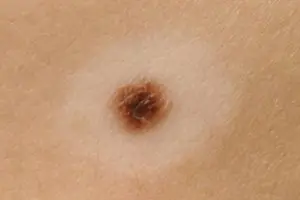
An intradermal pigmented nevus is a simple brown birthmark that can be located in any part of the body.
Mongolian spot located in the lumbosacral area. A mole is a round, irregularly shaped spot of blue or brown color. Appears in children from birth and goes away on its own by adolescence.
A papillomatous nevus is a formation with irregular outlines and an uneven surface. The color is usually natural or dark brown with dimensions up to several centimeters. Typically, a mole is riddled with hairs and is most often localized on the scalp.
Nevus verrucous is a type of papillomatous nevus. The difference is the greater pigmentation and roughness of the surface, often cut by deep cracks.
Differences between good types of moles and bad ones
Many owners of such formations on the skin are concerned with the question of how to distinguish a benign mole from a malignant one. This is quite simple to do; you just need to carefully examine the mole. Can be considered non-hazardous clearly defined birthmarks small size. They have a uniform structure and do not protrude much above the skin. The color range varies from light yellow to black. But if in doubt, it is better to undergo an examination by a dermatologist using special studies.
Bad birthmarks include the appearance of intense pain of various types in their area. If within two months the mole has increased significantly or itching has appeared in its area, then this is a reason to consult a doctor. Additional concern should be caused by the appearance of additional crusts, bulges or ulcers on the surface.
Moles on the body and the reasons for their appearance
The appearance of these benign formations can be influenced by various factors that stimulate excessive division of skin cells in a limited area of the skin. Modern doctors the following factors are identified, as the main ones, when moles appear:

Ultraviolet radiation.- Hormonal imbalances and related diseases.
- Genetic changes in the body contribute to the hereditary transmission of birthmarks to children, identical to the birthmarks of the parents, appearing in certain places of the body.
- Various defects in skin development that lead to the appearance of congenital moles in children starting from the age of two months.
- The possibility of the appearance of moles with long-term use of drugs containing homones.
- Long-term and untreated viral and bacterial infections.
What moles should be removed and methods for their elimination
Only those birthmarks that are pose a potential threat for humans and can turn into cancer. Removing a mole cannot provoke the development of this dangerous disease. Surgeries to remove birthmarks are completely safe and can have side effects only in the event of an allergic reaction to certain medications.
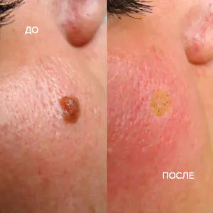
Modern medicine welcomes several methods for getting rid of birthmarks:
- Surgical operations.
- Laser exposure.
- Removal using liquid nitrogen.
- Exposure to electric current.
- Removal by radio waves.
The method of treatment for removing a birthmark is selected at an appointment with a dermatologist strictly on an individual basis. Removal using laser or liquid nitrogen refers to more gentle methods of combating unwanted tumors. All manipulations are carried out without the appearance of blood and are as gentle as possible on the skin around the mole.
