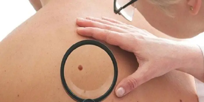
It's rare to see a person without small dark marks on their body. Is it worth paying attention to these points? Only a doctor will distinguish between dangerous and normal moles - malignant melanoma or harmless nevus - and give recommendations on what to do with them. Is it worth worrying about the appearance of new formations, when immediate contact with specialists is required, what are the signs of cancer development - the answers to these questions remain to be found out. No one is immune from disaster, and early diagnosis will protect you from severe consequences.
What is a mole
The first tiny spots may appear in children in infancy. A mole is a small formation on the skin - a nevus - that is considered benign and harmless. The basis for their appearance is melanocyte cells that accumulate the natural pigment melanin. Depending on its quantity, a difference in color is observed. Available colors:
The shape of the tumors depends on the location and concentration of melanin. They may have a stalk or be located under the skin, be flat and convex. The most common type is round, but there are exceptions. The development of neoplasms is provoked by ultraviolet radiation - natural from the sun, in a solarium. Hereditary factors cannot be excluded. A common cause of growth is hormonal imbalance, characteristic of periods:
- puberty;
- pregnancy;
- menopause.
What types of moles are there?
One person may discover very different tumors. Types of moles are classified according to several criteria. This helps in correct diagnosis in case of changes. They differ in:
- origin– congenital, newly acquired;
- structure– pigment, vascular;
- place of education – in depth, on the surface, in the boundary layer;
- raised above the skin – flat – even, protruding as a hemisphere, pedunculated, larger birthmarks;
- potential threats – dangerous, degenerating into melanoma, non-dangerous.

Safe moles
Those who have dark spots on their skin should be wary of their changes. In time, detected signs of degeneration into melanoma contribute to the timely removal of the formation and preservation of health. Safe moles are different:
- the presence of a stalk – it cannot be formed by malignant cells that grow randomly;
- long-term condition without changes.
Spots that appear shortly after birth are not considered dangerous. It is important that they are small in size. Good – non-dangerous – signs of neoplasms include:
- flesh tone;
- unchanged pattern of the skin of the nevus and adjacent tissues;
- soft consistency;
- hair on the surface of the neoplasm - growing from the skin, indicates the absence of pathologies;
- diameter no more than 5 mm;
- symmetry;
- nevus in the form of a spot.
Which moles are dangerous?
Why do people with nevi on their bodies need to monitor their changes? There is always a threat of degeneration of non-dangerous tumors into a cancerous tumor. What moles are dangerous to health? Key signs you need to know:
- change in shades towards the dark side, the appearance of multi-color;
- rapid increase in size - exceeds two millimeters per year;
- occurrence of cracks;
- the formation of asymmetry due to uneven growth;
- lack of elasticity;
- the appearance of itching, burning;
- presence of discomfort.
The appearance of dangerous moles requires an immediate visit to a specialist to clarify the nature of the changes and the likelihood of developing skin cancer. Pathological transformations provoke:
- injury to the nevus due to negligence;
- self-removal;
- abuse of exposure to the sun, use of a solarium;
- location of the formation in places of frequent contact with clothing - on the neck, head, genitals, legs;
- placement in the hair, on the face, palms - where there is a high probability of injury;
- previously removed melanoma.
Why are moles dangerous?
Not a single person is protected from the sudden proliferation of cells of a harmless mole. Melanoma is an extremely serious disease. Changes not detected at the initial stage can result in death. The provoking factor is unsuccessful independent removal of tumors. Moles are dangerous because of their ability to:
- transform into an atypical – precancerous form;
- grow to large sizes;
- turn into cancerous;
- with minor external changes, metastases actively spread throughout the body through the circulatory and lymphatic channels.
How quickly does melanoma develop from a mole?
The transformation of a nevus into a cancerous formation can occur in different ways. The process depends on the stage of the disease and the type of tumor. Instant metastases are dangerous. Begins:
- growth of cancer (oncological) cells in the deep layers of the epidermis;
- their entry into the blood and lymph;
- penetration into the lungs, liver, kidneys;
- growth in these organs;
- complete damage to the body;
- death.
The growth phases of pigment cells are observed, along which melanoma develops from a mole. There are varieties:
- horizontal– damage to the upper layers of the skin occurs, lasting up to 10 years, but metastases do not appear;
- vertical– accompanied by the spread of cancer cells throughout the organs, can last two years, has an unfavorable prognosis;
- nodal – especially dangerous – characterized by deep spread within two months.
The first signs of melanoma
The patient can be assisted only when suspicious changes begin to be identified. The diagnosis, research, and referral for surgical treatment save a person’s life. The first signs of melanoma:
- increase in the height of the tumor;
- bleeding;
- the appearance of discharge;
- redness;
- burning, itching;
- swelling of tissues;
- softening of the nevus;
- the appearance of a crust;
- thickening;
- hair loss;
- expansion of pigmentation around the lesion.
With the further development of dangerous melanoma, the following are observed:
- significant change in size;
- the appearance of pain;
- enlarged lymph nodes;
- surface ulceration;
- formation of new foci;
- bleeding from places of pigmentation;
- liquid separation;
- skin thickening;
- the appearance of an earthy tint;
- signs of metastases are chronic cough, weight loss, cramps, headaches.
How to distinguish a mole from melanoma
To recognize which moles are dangerous and which are not dangerous, you need to know what they look like. A person with nevi, in order to eliminate terrible consequences, must constantly monitor the appearance of new formations and changes that occur. You can distinguish a mole from melanoma by its signs. Non-dangerous neoplasm:
- symmetrical;
- with smooth edges;
- uniform in color;
- with dimensions not exceeding 6 millimeters.
Features of dangerous melanoma that require seeking help from dermatologists:
- growth in a short time;
- pronounced asymmetry of shape;
- heterogeneity in color - the presence of inclusions of several shades;
- lack of clear boundaries - the contour line is blurred, jagged, and looks like a coastline on a geographical map;
- increased diameter over six millimeters;
- variability of any parameters - color, size, shape.
What dangerous moles look like
What do nevi that are subject to pathological changes look like? Only a doctor can correctly distinguish between non-dangerous tumors. Dangerous formations look like this:
- blue– compactions under the skin with clear boundaries, with dimensions no more than 10 mm;
- nodal– round, flat in shape, color – brown, black;
- cutaneous– often pale, convex;
- halo nevus – pigment surrounded by a light or white rim;
- spitz- looks like a dome-shaped tumor of pink shades, with the possible presence of a hole through which blood and liquid leak;
- connecting- connect individual entities into a whole.
Mole with jagged edges
One of the signs of a non-hazardous formation turning into a dangerous one is a change in contours. It often has blurred edges and scalloped borders. There are non-dangerous types of nevi - dysplastic. Only a specialist can make a correct diagnosis. A mole with uneven edges can be dangerous if there are additional signs of melanoma:
- accelerated changes in size;
- the presence of clearly defined asymmetry;
- the appearance of highly indented boundaries.
Rough mole
Such a neoplasm is harmless if its diameter is no more than 5 mm and remains constant in size. Often its appearance signals a lack of vitamins and nutritional disorders. Doctors advise coming for a consultation if it is discovered that:
- the smooth nevus turned into a rough one;
- bothered by burning, itching, tingling;
- irregularities and compactions appeared in the middle;
- areas with different shades formed;
- diameter has increased significantly.
A dangerous rough mole requires immediate examination if:
- the appearance of bleeding;
- development of the inflammatory process;
- rapid change in size;
- formation of asymmetry;
- formation of purulent discharge;
- the occurrence of painful sensations when touched;
- the emergence of an irregular shape, blurred boundaries, along the edges of the neoplasm.
Large moles
Large formations on the skin are pigment spots. When they remain unchanged and do not cause inconvenience, this is a harmless phenomenon. It is important to constantly monitor their appearance, color, and size. To eliminate worries, you need to consult a dermatologist. During the visit, the specialist will conduct a diagnosis and give a forecast of the risk of developing a malignant neoplasm. Large moles become dangerous if they:
- injured;
- thickened;
- started to itch;
- were unsuccessfully removed independently;
- changed in size, shape;
- are bleeding.
What moles can be removed
Often nevi cause trouble for women when they are in a visible place - the face, neck. Even if they do not bother you, using removal will be the right decision - the appearance will improve significantly. After the procedure, the doctor must send the tissue for histological analysis to decide whether the mole is malignant or not. If the neoplasm is not dangerous, does not bother you, and does not change in size, surgery is not required. What moles cannot be removed? Experts believe:
- there are no contraindications;
- It is important to choose the right excision technique.
You should be careful about skin growths; it is unacceptable to remove them yourself. Only the doctor will determine whether a nevus is dangerous or not and decide what to do with it. You can delete it if:
- injured from clothing - on the neck, in the groin area, under the armpits;
- cause pain when touched;
- are located under the hair on the head and can be damaged when combing or cutting;
- change color, shape, outline;
- significantly increase in size;
- characterized by the presence of burning, itching;
- accompanied by inflammation and bleeding.
We are all familiar with small red dots, very similar to moles, that can appear anywhere on our body out of the blue. Often, we simply do not pay special attention to them, however, these spots indicate certain changes occurring in our body. Although knowledgeable people advise that you should not worry about the appearance of red current on the body, for some reason it appeared! Therefore, at least for your own peace of mind, it would not hurt to contact a specialist if the appearance of a red dot worries you. Extra examination will not be a hindrance.
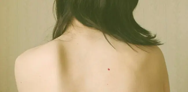
Red dots on the body - what are they?
Red dots on the body, like moles, what are they? These are benign formations, pink or scarlet, that appear on any part of our body at any age, even in young children. Most often it is believed that the reasons why red specks appear, called capillary angiomas, are senile changes in the skin. In most cases, this is true, however, these points can also indicate that some kind of malfunction has occurred in our body. Often, such points indicate hidden diseases of the internal organs.
If you think carefully, what age-related changes can a child or a very young person have who have red moles? Here there is a clear answer - it is necessary to examine such a person for the presence of diseases of the internal organs. It is better to prevent an impending problem than to deal with it later.
If suddenly one or more red dots appear on the skin of the chest, back or abdomen, quickly disappearing or, on the contrary, increasing in size, then you should pay attention to the pancreas and other organs of the gastrointestinal tract, which may be in danger. Also, there is a possibility that the cardiovascular system is sick, or the hormonal balance is disturbed. It is necessary to undergo an appropriate examination, which will reveal what is wrong with our body.
In a small child, the appearance of red moles indicates the presence of benign vascular formations of the skin, the cause of which lies in the mother’s illness in the first months of pregnancy with viral respiratory ailments.
The dots can either appear suddenly or disappear, however, if their number increases sharply, this should not be ignored, you should quickly run to the doctor.
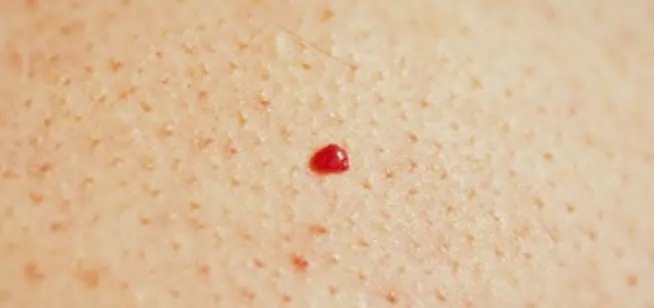
Types of red dots - angiomas
Typically, red dots on the body do not cause pain and do not exceed a diameter of 2 millimeters. They don't grow any further. Cases when they begin to hurt, itch, or cause discomfort in general well-being are very rare. Moles can appear anywhere on the body. If, when such spots appear, unpleasant, painful sensations appear, or the temperature rises, you should immediately run to the clinic.
Red dots differ from each other in the reasons for their appearance and shape; they usually consist of small capillaries and are pink or red in color. According to their form they are divided into:
They can also be divided according to their structure and the results of histological examination. If an ordinary angioma grows, then a large mole appears, which is called a hemangioma.
Causes of appearance on the skin
The reasons for the appearance of angiomas on the body are not fully understood, however, heredity is considered the main reason. There is a possibility that their appearance is also facilitated by hormonal and age-related changes in the skin, in the presence of certain diseases of the internal organs, mainly digestion, skin trauma, pigmentation disorders or vascular malfunction. With prolonged exposure to the sun, the appearance of small red dots has also been noticed, which go away on their own. In any case, if you have such a problem, for your own peace of mind, it is best to consult a specialist.
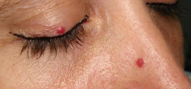
How to remove red dots on the human body
Red dots often disappear on their own, without any intervention. If angiomas do not cause concern, do not grow and are located on closed parts of the body, then it is not recommended to touch them. You can remove such a point if it is on an exposed part of the body, such as the neck or face. The sign when an angioma begins to enlarge is also an indication for its removal.
Removal by medical methods
There are several medical methods for removing red moles:
- Surgical removal, in which the doctor cuts out the angioma and then stitches it together. This method has been considered the most common since time immemorial, however, it is a painful procedure, and after removal a scar remains;
- removal by electric current - electrocoagulation is also a painful method and leaves traces;
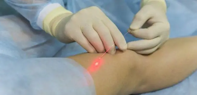
- The laser treatment method is the most effective and does not leave scars, but it can only be used to remove flat angiomas; the laser may not treat very convex angiomas, since it only affects the upper layers of the skin. After the procedure, there is a possibility that the red dot will reappear;
- X-ray therapy, and vascular sclerosis.
The intervention option is chosen by the attending physician in accordance with the diagnosis, the cause of occurrence and the studies conducted at the medical institution.
Treatment at home using traditional methods
To avoid surgical intervention, you can use traditional medicine methods to remove formations, which are often even more effective than the above methods. However, it is easier to prevent the appearance of angiomas than to treat them later. And to do this, you should stay in the sun as little as possible, and not get carried away with the solarium. If you have red hair and fair skin, you are most susceptible to the appearance of pink spots.
Cauterization of moles will not lead to proper results, since in most cases they are located in the inner layers of the skin, and on top there is only a small part, which may disappear when cauterized, but later, the angioma will appear again.
The most common medications for treating red spots grow on the windowsill. This is aloe and euphorbia.
An aloe leaf or its juice is applied directly to the affected area three times a day until the bulge disappears. You can make a bandage with aloe juice and wear it constantly until the mole disappears.
Euphorbia is used in the same way as aloe - either juice or crushed pulp from the leaves of the plant is applied.

One popular treatment for red moles is apple cider vinegar. Before using it, do a test - apply a little liquid to the skin; if there is no reaction, you can lubricate the affected areas by applying cotton swabs soaked in vinegar to them. If you use such lotions twice a day, the skin problem will disappear within a week.
Garlic will also help you in treating this scourge. To do this, you need to mash two cloves to a paste, lightly add salt, and apply this mixture to the red dot for five days until it falls off.
Honey is a good way to soften moles. If you smear the bulge with honey for a week, it will become soft and soon fall off.
Use a paste made from soda to remove angiomas. It is prepared like this: mix one tablespoon of powder with four tablespoons of water, and apply the finished mixture to the diseased surface. Leave it on for about an hour, then wash it off. The procedure is carried out twice a day.
You can use iodine purchased at a pharmacy. Use the drug diluted with water (1:5), which is applied to the mole with a cotton swab and left for 5 minutes.
A mixture of aspirin and water will help greatly. It will dry out the mole and it will fall off. It is prepared like this: three tablets of acetylsalicylic acid are crushed and mixed with two teaspoons of water. If you use this remedy for a couple of weeks, the angioma will dry out and disappear.

A good remedy for treating angiomas is dandelion root, the juice of which is applied to the sore spot three times a day for a week.
You can whiten a mole with lemon, grapefruit or pomegranate juice. The fresh juice of these fruits is applied with a cotton swab directly to the area where the sore is located, which will discolor over time.
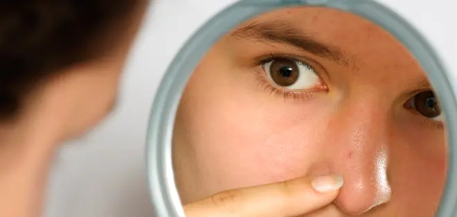
How to make sure it's not cancer
Angiomas, in most cases, do not pose any danger in themselves, however, in order to exclude the occurrence of skin cancer, you should still monitor the emerging mole. If the tumor begins to change color, size and shape, then you need to consult a doctor to make an accurate diagnosis. This will help prevent serious illness and begin treatment in a timely manner.

If a mole is asymmetrical and has uneven edges, is it worth paying attention to this? Malignant formations, for the most part, have jagged, uneven borders.
A darker color may also indicate that it may be a malignant tumor, in which case you should also consult a doctor.
Another criterion is increasing the size. Simple angiomas do not have large diameters and do not tend to grow on their own. If a mole begins to grow rapidly, you should also immediately run to the doctor.
What to do if the red dot itches
It often happens that angiomas begin to change both in size and shape. Then, they will itch, itch and hurt. Of course, in such cases, you must immediately contact a skin specialist or oncologist to make a correct diagnosis.
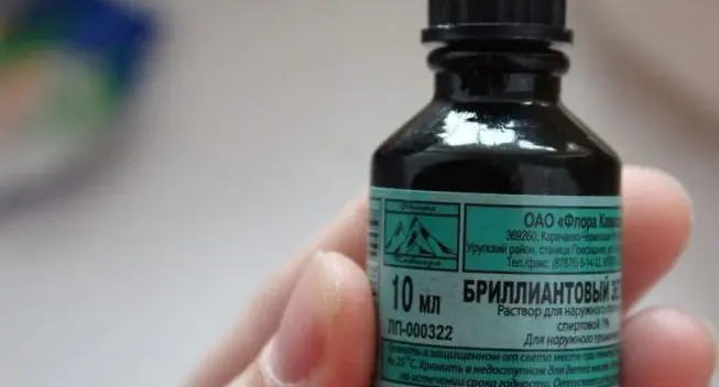
You can scratch the angioma, but you need to do it very carefully - you don’t need to use improvised means for this, especially if you don’t do it with your hands. You can use the same vinegar, which is used to moisten a piece of sterile bandage and wipe the disturbing area, or use alcohol if the red dot is damaged. If it’s not there, then green stuff will do.
Remember, for any changes in the angioma, and if itching or pain occurs, consult a doctor immediately!
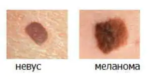
Every person has birthmarks of different types, textures, colors, and shapes on their body. These harmless formations arise in the epidermis from melanocytes and grow in clusters. The scientific name of a mole is nevus. This medical term applies to all skin abnormalities.
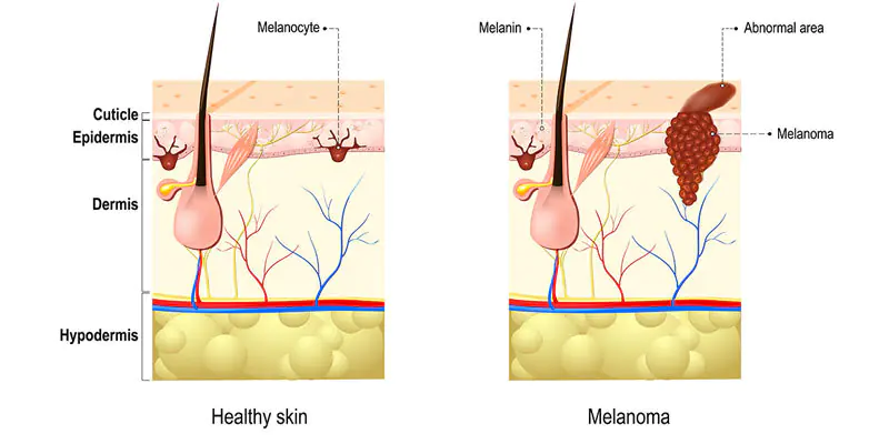
However, these so-called “floaters” can hide the most aggressive malignant tumor – melanoma. Therefore, you should know what dangerous moles are and be able to recognize the main differences between benign and malignant ones. Cancerous transformations most often occur on the basis of pigmented skin tissues.
What moles are dangerous?
Cancerous moles, like regular ones, consist of melanocytes. But this is an aggressive form of the tumor, prone to rapid spread and damage to other organs. In this regard, it is recommended to be careful with such pigmented skin formations as:
Atypical nevi
This type does not look like a regular birthmark because its size is larger than a pencil eraser, its shape is unclear, and its color is uneven. Moreover, the potential danger is posed by congenital formations, not acquired ones. Most of them are inherited and are larger than 1 cm.
Hutchinson's melanotic freckles (lentigo)
Appear as a flat spot containing two or more shades of darkening. They are quite common after the age of 50 and are localized particularly on the face. Gradually they become larger and darker, transforming into skin cancer.
Skin neoplasms of unknown etiology
Neoplasms that appear suddenly develop very quickly, are aggressive in appearance and do not at all resemble an ordinary “fly”. In 60% of all cases of melanoma, this type of pigmentation functions.
Dangerous moles: signs
Color Changes
A mole that has begun to change color is potentially cancerous. For example, one-color pigmentation has acquired some other spots around or in the middle.
Height change
An important feature is a change in the height of the previously flat spot and density (thickening).
Painful sensations
The mole hurts, the surface becomes larger, erosions appear, the release of fluid, purulent masses or blood appears.
Satellite pigmentations
The skin around the formation is also distinguished by redness, swelling or the presence of new color spots, the so-called. satellite pigmentations.
Itching and burning
There are sensations such as tingling, burning, and the mole itches.
Consistency changes
For example, a mole softens, is divided into small pieces that break off easily, or resemble scratches that do not heal.
Which moles are potentially dangerous?
There are certain categories of birthmarks that are prone to transform into a malignant form. All of them refer to abnormal skin lumps.
1. Nodular pigmented nevi: usually brown or black moles, round and flat.
2. Skin pigmented nevi: they have a raised appearance, pale color, and sometimes a hairy surface.
3. Connecting nevi combine elements of different formations.
4. Halo nevus is a pigmented area of skin surrounded by a discolored white ring.
5. Dysplastic nevus (otherwise known as Clark) is a specific neoplasm.
6. Spitz nevus: looks like a tumor-like growth on the skin. This spot is pink (but a combination of different colors is possible), dome-shaped, prone to bleeding. May have a hole through which liquid leaks.
7. Blue nevus has one of the shades of blue, shows well-defined borders, any size (but most often does not exceed 1 cm), looks like a lump under the skin.
THE MAIN DIFFERENCES OF BENIGN MOLES FROM MALIGNANT MOLES
A number of characteristics allow you to accurately determine which moles are dangerous. The benign formation is not asymmetrical. If you draw a line through the middle, then both sides will correspond to each other.
A cancerous lump does not meet these requirements.
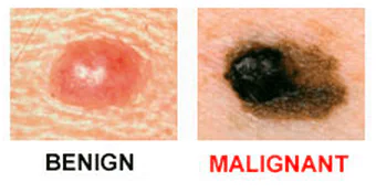
Unlike melanoma, a common pigmented spot has smooth, rather than jagged, borders.
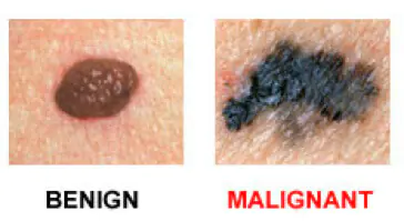
The presence of colors and brightness is another exciting symptom.
The formation changes size over time and becomes larger than 6 mm. Noncancerous nevi look the same. You need to be alert if a mole begins to grow or gives other unusual signals regarding its general condition.
The only way to accurately establish a diagnosis and confirm or refute suspicions of cancer is to conduct a histological examination of cells using a biopsy.
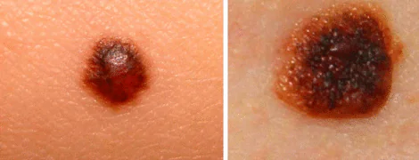
Melanoma symptoms
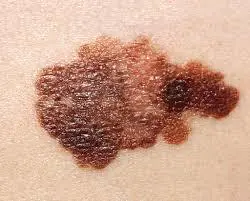
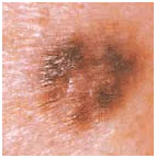
Cancerous pigmentation can vary greatly in its symptoms. Sometimes a person is able to adequately assess only some of the features. You should pay attention to what a dangerous mole looks like. Uneven edges, but a fairly clear border with healthy tissue. Diameter – 10 mm.
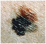
Blue-black newly formed melanoma that has irregular borders. It arose from a dysplastic nevus (pink-brown area in the upper left corner). Size about 12 mm.
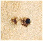
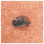
Oncological dysplastic nevus with black distant malignant extension, which was previously absent. It is only about 3 mm.
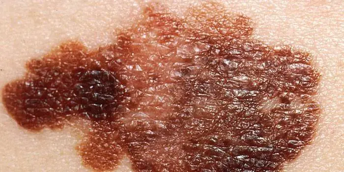
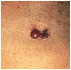
A malignant skin tumor consisting of three parts: a dark brown area on the left, a red area on the right and a light area on top. Size – about 15 mm.
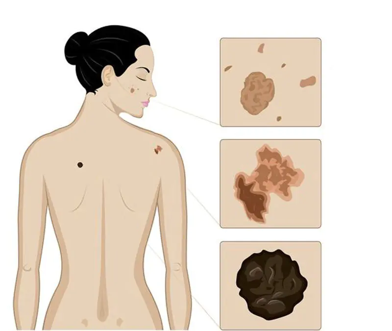
Photos of dangerous moles
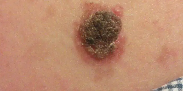
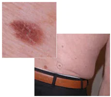
Melanoma in a dysplastic nevus: irregular contours, bright color in a relatively small size (1/3 inch).
Transformation of a single atypical pigmentation with the presence of black, brown and pink colors (1/2 inch).
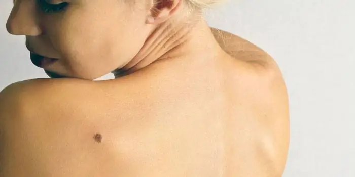
Tumor formation on the lower back demonstrates asymmetry, color saturation and changes in the zone bordering healthy skin.
Every person should be attentive to the condition of their skin in order to diagnose dangerous moles in time and prevent possible consequences that are detrimental to health and life.
Remember that if detected early, skin cancer can be successfully treated. Published by econet.ru.
If you have any questions, ask them here
P.S. And remember, just by changing your consumption, we are changing the world together! © econet
Did you like the article? Then support us press:
