There is an opinion that moles on the head in the hair are dangerous, although there are also moles whose existence their owner is not even aware of.
Moles or nevi can appear on different parts of the human body. These are neoplasms that stand out on the skin with their color and structure.
It is not uncommon for moles to appear on the head. Such moles can cause a feeling of discomfort and are easily damaged as a result of external influences.
Causes of moles on the head
The factor that causes the appearance of nevi on the head is the increased content of melanin in the upper part of the skin.
This feature can arise as a result of several reasons:
- weakened immune system;
- genetic predisposition;
- strong susceptibility to ultraviolet radiation and sun exposure;
- birth defect;
- skin diseases;
- hormonal imbalance of the body (occurs during the period of teenage changes in the body or pregnancy);
- skin damage.
Such factors that cause the appearance of nevi can occur throughout a person’s life., even in old age, so you should be careful about the occurrence of neoplasms.

To find out which moles on the head in the hair are dangerous, you should visit a dermatologist.
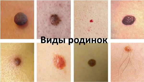
Types of moles that appear on the head in the hair
Nevi can be divided into several different groups depending on different characteristics.
Based on their origin, such neoplasms can be divided into 2 types:
- congenital – arise as a result of disruption of melanocyte division even at the stage of fetal development in utero;
- acquired - appear as a result of external exposure to the skin of an excessive amount of ultraviolet rays.
If we talk about the structure of moles, the following subgroups can be distinguished:
- vascular – arise as a result of the growth and close proximity of several vessels, usually have a reddish or bluish tint;
- non-vascular – appear due to a significant number of melanocytes, have a dark color;
- flat – the most common nevi, they are not damaged as a result of external influences and, as a rule, do not cause any discomfort to the owner;
- convex – rise slightly above the surface of the skin and look like a hemisphere; having similar moles on the head in the hair is dangerous, as they are very easy to injure, so experts usually advise removing them;
- hanging – rarely appear in the head area, they represent a head connected to the skin with a kind of leg; due to the high likelihood of damage, the best option is to remove them.
Based on their size, nevi are divided into:
- small – with a diameter of no more than 1.5 cm;
- average – 1.5-10 cm;
- large – with a diameter of more than 10 cm;
- gigantic – occupy more than half of the scalp.
Which moles on the head are safe?
In most cases, moles on the head are benign neoplasms that do not pose any danger to the owner.
The safest in this regard are flat non-vascular nevi, with a diameter not exceeding 0.5 cm. Such moles do not require any special care or special attention.
Any moles on the head in the hair can be dangerous if they begin to cause discomfort to their owner.
Moles in the hair on the head that require specialist consultation
Nevi on the head are not just an external defect, but a phenomenon that requires careful attention and careful handling.
A direct indication for seeking medical advice from a specialist is the presence of large convex, and especially hanging, moles in the hair. Such formations can very easily be injured when combing or washing your hair.
The main problem with such moles is that they can easily degenerate into malignant neoplasms or melanomas., successful treatment of which is possible mainly in the initial stages.

Such neoplasms develop quickly and metastasize to other organs, which can lead to death within a few months.
It is important to know! Often the cause of nevus degeneration is various injuries that provoke negative processes.
That is why moles that rise above the surface of the skin and are large in size require contacting a specialist and making a decision about their removal.
Dangerous resort to traditional methods of removing moles on the head in the hair, this can cause damage and degeneration.
Also, you should not cover such formations with a band-aid, since the greenhouse effect can also provoke unwanted processes.

How to properly care for a mole on your head
Moles in the hair require careful handling.
There are several simple rules that, if followed, can minimize the risk of damage to tumors on the head:
- You should not use cosmetics containing solid substances that can cause mechanical damage to the skin (scrubs, peelings), as well as shampoos and masks that contain strong chemicals;
- Avoid blow-drying this area of the skin;
- Do not scratch or scratch your head with sharp objects, use hairpins and hairpins carefully;
- If the mole is still damaged, it is necessary to treat it with hydrogen peroxide, stop the bleeding, in case of severe damage, seek medical help;
- Carefully monitor the condition of the mole and consult a doctor if any changes occur.
Be careful! Microtrauma to a mole in itself is not dangerous; it is important to prevent infection and inflammation.
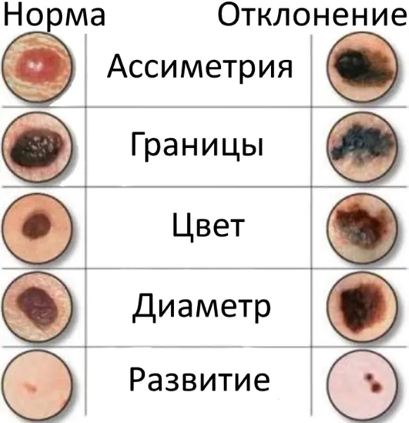
Signs of degeneration of a mole
There are 5 main signs of degeneration of a mole, the appearance of which requires urgent medical intervention:
- The emergence of asymmetry – in a normal state, if you mentally divide the mole in half, both parts should be the same;
- Color change – darkening or redness of the nevus is an alarming symptom. Normally, the color of a mole should be uniform and unchanged;
- Jagged edges – benign neoplasms have clear, even boundaries;
- Resizing – if a mole increases in size or becomes convex from flat, then you should immediately contact a specialist;
- The appearance of unpleasant sensations – pain, itching, peeling, bleeding. Under normal circumstances, a nevus should not cause discomfort.
Important to remember! Even the appearance of one of the above signs is a reason to seek medical advice.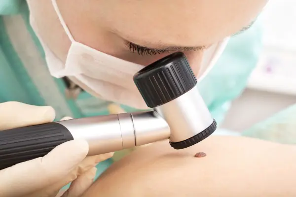
Is it necessary to remove moles on the head in the hair, when is it dangerous?
Very often the question arises: to remove moles or not. The decision should be made only after consultation with a specialist.
There are several indications for removing nevi:
- the emergence of processes of degeneration of a mole into a malignant neoplasm;
- convex or hanging shape of the mole, as it is easy to injure in everyday life;
- finding a mole in a place where it is constantly exposed to mechanical influence;
- a cosmetic defect that causes discomfort to the owner of the mole;
- the appearance of large moles in a child, since children cannot take the necessary precautions.
| Methods for removing moles on the head in hair | Peculiarities |
| Surgical | Allows you to send tissue for histological examination and find out whether these formations are dangerous or not. |
| Laser | The most gentle method, leaves no traces. |
| Using liquid nitrogen | It is carried out in several passes and leaves no traces. |
| Electrocoagulation |
Produced using high-frequency current, it can cause discomfort after the procedure. Radio wave A gentle, non-contact method using exposure to radio waves.
Any new growths on the head require careful and careful treatment.
It is important to follow simple rules to reduce the risk of injury to moles and the development of undesirable consequences. Any change in the nevus or the occurrence of unpleasant sensations is a reason for an immediate visit to the doctor.
This video will tell you about moles on the head, in the hair, on the body, and how dangerous they can be:
In this video you will see and hear which moles can be hazardous to health:
A mole (nevus) is a widespread benign pigmented formation consisting of melanocyte cells that produce melanin. Due to the excess amount of pigment, which is concentrated in certain areas of the skin, a birthmark is formed.
It can form in a child at birth or occur after some time. The bulk of moles appear during puberty or during pregnancy due to hormonal changes in the body.
Nevi are localized on any part of the skin and mucous membranes (mainly in women). In 20% of people, neoplasms occur on the head in the hair (on the back of the head, crown) or on the temples.
Moles in hair, what is the danger
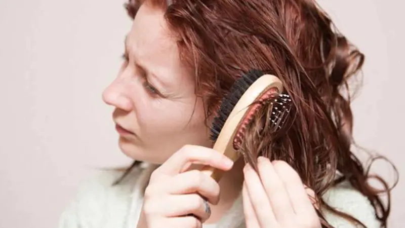
Due to this location, a mole on the head (especially hidden in the hair) goes unnoticed for a long time. It is more susceptible to injury than other areas of the body. It can be damaged when wearing an uncomfortable headdress, when combing, cutting, styling and even washing your hair. Regular exposure of the birthmark to hair care products can cause irritation, swelling, and itching.
It is believed that not all moles have the ability to degenerate into a malignant tumor, but only a few under the influence of certain factors.
Causes of moles
The mechanism of the appearance of birthmarks has not been fully studied, but it is known for sure that such formations are formed as a result of excess content of the melanin pigment.
Also, the appearance of moles is the result of uncontrolled cell division, which the immune system cannot cope with on its own.
There are also some additional factors that provoke the appearance of birthmarks:
- excessive sun exposure;
- frequent visits to the solarium;
- hormonal disorders;
- X-ray and radiation exposure;
- genetic predisposition;
- pathological changes in the functioning of internal organs and systems;
- skin aging;
- repeated injury to the epidermis.
Nevi can change throughout life: grow, change color, shape, sometimes disappear completely or appear in a new place. And it is important not to miss the appearance of dangerous signs of their degeneration into a malignant tumor.
What are the types of moles on the head?
Formations can be different in their structure, shape, and size. There are the following types of moles on the head:
- Large - is a congenital skin defect, has a brown color. It grows with a person and can reach significant sizes.
- Convex - a formation of red, brown or burgundy color that occurs in the inner layer of the skin and rises above it. Often the mole is covered with hairs. This type of mole appears in adulthood and there is a significant risk of damage.
- Flat is the most common type of birthmark on the head. It is located at the same level with the skin and does not cause discomfort to the owner of such a mark.
- Blue is very rare, but this does not mean that it is more dangerous than others. This formation is cone-shaped, bluish in color and protrudes slightly above the level of the epidermis.
- Hanging (in the form of a wart) are rarely found on the head. They are easily damaged and therefore require removal.
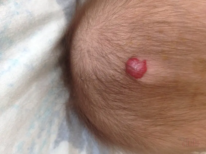
- A red - vascular mole is formed at birth; as the child grows older, it can disappear on its own.
How to understand that a neoplasm is not malignant
A benign formation must have the correct shape, small size, symmetry, clear contours, even color, uniform structure, and absence of discharge.
Quite often, hair grows from a mole, which is considered normal and should not be a cause for concern.
If the tumor meets these criteria, then everything is in order.
Differential diagnosis of a mole and skin cancer
All people with birthmarks should closely monitor them, especially those who have a family history of cancer. At the first suspicious symptoms, it is recommended to urgently visit a doctor, who, through diagnostic measures, will find out the cause of this transformation and determine the degree of danger to the person.
The appearance of a malignant tumor is the result of uncontrolled cell division.
To make a diagnosis confirming or refuting the nature of the tumor, a large number of techniques are used:
- visual examination of the mole and medical history;
- blood test - helps to identify a tumor marker characteristic of melanoma;
- dermatoscopy – allows, by magnifying the skin tenfold (using a dermatoscope), to examine not only the upper layer of the epidermis, but also the deeper layers of the skin;
- biopsy - to remove the affected tissue to determine the nature of the formation;
- ultrasound examination - allows you to assess the condition of the lymph nodes, measure the thickness of melanoma and determine the extent of its spread;
- computed tomography – helps to identify metastasis in various organs;
- magnetic resonance imaging – allows you to obtain images in transverse and longitudinal sections;
- radiothermometry – for early diagnosis of malignant tumors by measuring the temperature of internal tissues.
The most effective diagnostic method is positron emission tomography. It is carried out using a special radiopharmaceutical, which is introduced into the body and accumulates in the affected cells. When scanned, it appears as luminous spots.
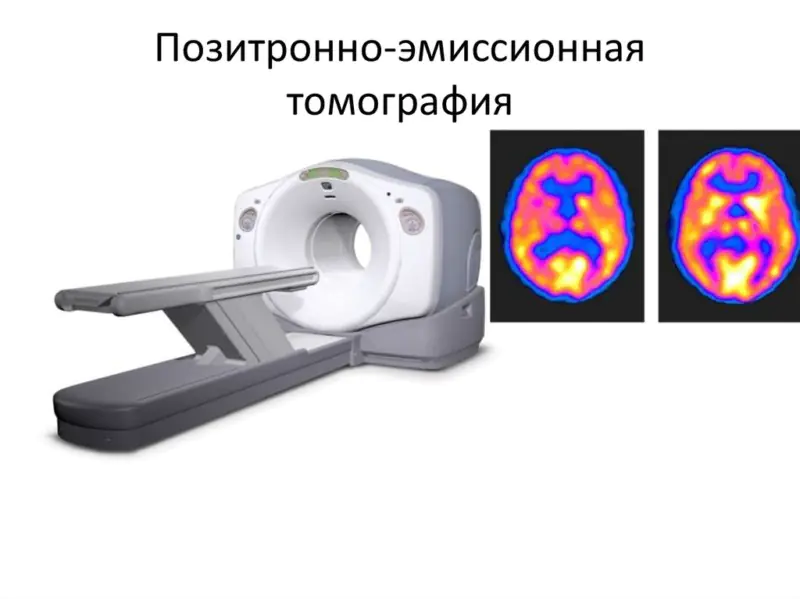
A correct diagnosis allows you to more accurately select the necessary treatment method in each specific case.
When does a tumor need to be removed?
Melanoma is the most dangerous and aggressive malignant tumor, which is formed as a result of the degeneration of melanocyte cells at the site of the mole. It progresses very quickly in the early stages of the disease with multiple metastases (in the lungs, brain, bones).
Once metastasis occurs, the disease is considered incurable and can cause death, so it is very important to closely monitor your moles in order to identify the approaching danger in time.
Signs to pay close attention to:
- a rapid increase in old or new marks begins (size reaches more than 6 mm), while uneven edges of the growth are observed;
- the color of the neoplasm changes (to darker or lighter);
- the presence of individual inclusions, crusts, cracks, and flakes is observed;
- serous fluid is released or bleeding appears;
- there is an increase in lymph nodes;
- a feeling of soreness, burning and itching at the site of formation appears.
People at risk for moles turning into melanoma include:
- with fair skin with a lot of freckles and other growths;
- having increased sensitivity to solar radiation;
- working under the sun;
- sunburned at any age;
- living in hot climates.
If the growth is removed at the initial stage of transformation into a malignant tumor, there is a significant chance of complete recovery.
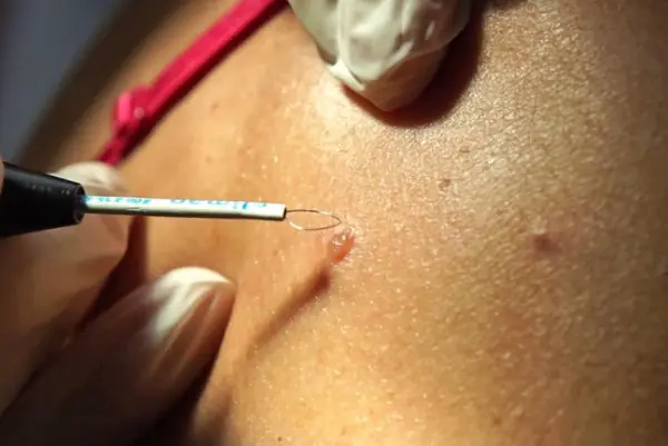
Ways to combat nevi
The following methods for removing moles on the head are known:
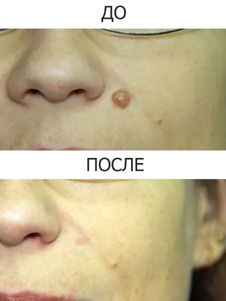
- Laser therapy – removal of a birthmark using a high-precision laser beam. It destroys the cells of formation without leaving any traces.
- Cryodestruction - freezing the nevus using liquid nitrogen. A mole on the head becomes covered with a white crust, which subsequently falls off, leaving behind healthy skin.
- Radio wave therapy - using a radio wave knife, resembles surgical removal, only less traumatic for the head.
- Electrocoagulation is the cauterization of a growth on the head using a high-frequency current, which promotes its peeling and death.
If dangerous symptoms are observed and there is a suspicion that a mole on the head is turning into a malignant formation, immediate surgical intervention is necessary. It is performed by excision of the tumor and adjacent skin with a scalpel. The operation is dangerous due to bleeding and the formation of scars. In some cases, if the formation occupies a large area, skin grafting may be necessary.
Preventing skin cancer
To reduce the risk of melanoma, you must:
- avoid sun exposure to the skin, especially during peak sun activity;
- protect exposed areas of the body (if it is necessary to stay outside for a long time on sunny days);
- use sunscreen;
- exclude visiting the solarium;
- comb and wash hair with care;
- inspect moles daily, in hard-to-reach places, use the help of relatives or friends;
- choose hats that do not cause squeezing and rubbing of moles;
- use non-aggressive hair care products;
- warn the hairdresser about a mole on the head when visiting a beauty salon.
Under no circumstances should you remove the tumor yourself - this will lead to irreversible consequences.
The main protection against an incurable disease is early consultation with a doctor if negative signs appear.
It should be remembered that melanoma can be defeated only with timely treatment.
Moles (nevi) and birthmarks are benign neoplasms that can appear on any part of the body and in any quantity. In most cases, they are not dangerous to human health and life. A mole on the head can be dangerous when it is often injured or changes its color, shape, or size. In this case, you need to consult a doctor, undergo an examination and, possibly, remove the formation.
What does it look like
The appearance of the formation in the photo may vary. All of the following options are normal:
- The color is most often brown, but can be black, red and even purple. A white rim may appear around.
- They rise (convex) or do not rise (flat) above the skin level.
- The mole can be located on any part of the head: the scalp (back of the head, crown), face.
- The size ranges from a pinpoint to 5 cm or even more. Birthmarks are often larger in size.
- There are no additional symptoms. The patient should not be bothered by pain, numbness, or burning. The appearance of these symptoms most likely means that we are talking about another pathology.
Red mole on the head: is it dangerous?
In most cases, moles on the head are not dangerous and do not require diagnosis or treatment. However, there is caution associated with the likelihood of malignancy (malignancy) of such formations. Under the influence of various factors (damage, ultraviolet radiation), a mole can degenerate into melanoma. That is why every person should be able to distinguish a dangerous mole using special criteria.
Hazard criteria
Moles on the scalp require special attention, as they are often injured when combing or other manipulations. There are 5 criteria that help assess the danger of a nevus: asymmetry, edge, color, size, dynamics (abbreviated as ACORD).
If you draw a conditional line along the center of the formation, both halves should be identical, even, and symmetrical. Asymmetric halves of a nevus are one of the signs of danger.
Normally, the edge of the nevus is smooth and clear. Blurry edges, as well as jagged edges and deformation are a sign of danger.
The formation can have different colors: brown, black, red (all of these options are the norm). It is not the color itself that is important, but its uniformity. The presence of any inclusions, darkening, or blanching should alert the patient.
The larger the nevus, the higher the risk of malignancy. The diameter of a formation greater than 6 mm is an alarming sign.
It is very important to monitor the nevus over time. You should be wary of the appearance of roughness, ulceration, as well as the rapid growth of formation.
If the formation is localized on the head, it is not always possible to conduct a self-examination. It is better to ask someone close to you to examine hard-to-reach places (the back of the head, the crown of the head).
What should a patient do if the criteria exist?
If you identify at least one of the above criteria, you should definitely contact a dermatologist. This does not always indicate malignancy of the nevus, but it is better to be safe.
Who is at risk
There is a group of people who are especially at risk of nevus degeneration into melanoma. The risk group includes:
- men over 50 years old;
- persons with a family history (melanoma in relatives);
- persons with more than 100 moles;
- persons susceptible to sunburn;
- persons with fair skin, red hair, freckles.
Anyone who is at risk for melanoma should be regularly monitored by a dermatologist. The frequency of preventive examinations is once a year (even without dangerous signs).
How does an appointment with a doctor work?
The doctor will carefully examine the nevus, determine the presence of signs of malignancy, and collect an anamnesis of life and disease. An additional examination is prescribed, usually the consultation is limited to dermatoscopy. Dermatoscopy is a method of visual assessment of skin tumors. For the study, a dermatoscope is used, with which you can obtain an image magnified 10 times.
Dermatoscopy is a preliminary diagnostic method. It will not be possible to make a final diagnosis (benign or malignant formation, what type), this requires a histological examination.
Mole on the head in the hair: what causes the appearance
A mole is a collection of melanocytes that are located between the lower (dermis) and upper layers of the skin (epidermis). Melanocytes are skin cells that produce the pigment melanin. It is this pigment that causes the color of moles to change. Red formations represent proliferation of blood vessels.
Almost every person has birthmarks. They can be formed under the influence of various factors - genetic, hormonal, and the external environment.
One of the most common causes of the formation of nevi (both congenital and acquired) is genetic abnormalities. Hereditary predisposition is influenced by environmental factors.
Nevi grow under the influence of various hormones. For example, the number of moles increases during puberty and in women during pregnancy.
Sunlight triggers the production of melanin, which leads to the formation of moles.
Trauma, viruses, and radiation can trigger the movement of melanocytes to the epidermis.
Treatment
Not all nevi can be removed. In most cases, it is enough to follow simple recommendations to prevent complications. If a mole is often injured or there are signs of malignant degeneration, such a formation must be removed.
What to do at home
All activities carried out at home are aimed at preventing complications. No home treatment can effectively remove the formation.
Care Tips:
- try to wash and comb your hair carefully so as not to damage the nevus;
- do not use scrubs and peels on the scalp;
- if the mole is localized on the open part of the head, cover it with a sun bandage.
Under no circumstances should you remove the formation yourself! Such an attempt at treatment can damage the nevus and provoke the development of complications (malignancy, bleeding).
One of the most important things you can do at home is preventative checkups. It is necessary to regularly examine the nevus and evaluate it using the ACORD algorithm.
Surgery
The final decision on the need to remove the nevus should be made by the patient together with the doctor after a complete examination. Surgical treatment is indicated for all patients with suspected melanoma. To remove the formation, different methods can be used - laser therapy, electrocoagulation, cryodestruction, surgical excision.
Description, advantages and disadvantages
Suitable for removing nevi, which are often injured (especially on open areas of the head). This is a modern treatment method that uses a laser to remove the formation. The procedure is performed under local anesthesia; no scars are left at the removal site. The main disadvantages include the high cost of treatment and the impossibility of removing malignant tumors.
The method is based on exposure to electric current. The nevus is cauterized, and after some time the necrotic tissue is rejected on its own. The procedure is performed under local anesthesia.
Exposure to liquid nitrogen is often used to remove small lesions. Freezing and subsequent destruction of the mole occurs. The procedure is painless, does not require anesthesia or local anesthesia, but is not suitable for removing large nevi. Sometimes several sessions are required for complete removal.
Surgical excision is the main treatment method for melanoma and large benign tumors. A scalpel is used for removal; the operation is performed under local or general anesthesia. After excision of the nevus or melanoma, the wound is sutured, and the formation is sent for histological examination.
Video
We offer you to watch a video on the topic of the article.
")
