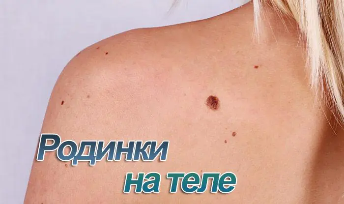
A mole (birthmark, nevus) is a benign neoplasm that almost every person has. Some have only a few of them on their body, while others have more than 100. Many moles on the body are a reason for self-examination and regular visits to the doctor, but not for panic.
Why did many moles appear on the body in a short time?
A pigmented mole is a collection of melanocytes (cells that produce the pigment melanin) in the epidermis or dermis. This process is regulated by melanocyte-stimulating hormones, which are produced in the intermediate lobe of the pituitary gland. The number of moles on a person’s body is determined mainly by heredity. Certain gene loci contain information about the number and location of all formations on the skin.
The appearance of new formations can be associated with the influence of many factors - hormones, ultraviolet radiation, infections, injuries.
What does this mean
An increase in the number of nevi may be associated with hormonal changes. For example, this is often observed during puberty, due to a sharp increase in the production of sex hormones. Many small moles can appear in a woman during pregnancy. This is a normal reaction of the body and does not pose a danger to human life.
Less commonly, endocrine pathology associated with disruption of the production of pituitary hormones can provoke the appearance of new formations.
The growth of melanocytes occurs under the influence of ultraviolet radiation; with increased insolation, moles can become larger. Thus, prolonged exposure to direct sunlight and frequent visits to the solarium can be dangerous to human health.
Skin injuries and infections
Sometimes many moles appear in response to injury or viral infection. Biologically active substances that are formed during inflammation can stimulate the growth of melanocytes.
Many large and small moles on the body: is it dangerous?
New moles can appear anywhere: face, scalp, stomach, back, arms, legs. Their color can also be different - brown, black, red, purple. The shape can be flat, in the form of a spot, or convex. These characteristics have little effect on the risk of melanoma. Only quantity matters, as well as the so-called. danger criteria - asymmetry, edge, color, size and dynamics.
What does this mean, normal indicators
Changes that cause concern
To assess symmetry, it is necessary to draw a conditional line along the center of the nevus. If the halves are approximately the same, this is a good sign.
Clear boundaries of the nevus are considered normal. The edge should normally be even and smooth.
Fuzzy and blurry edges, jagged edges, irregularities.
It is not the color of the nevus that is assessed, but the uniformity of color. It’s good if the color is uniform, without areas of lightening or darkening.
Uneven color - there are lighter and darker gaps and inclusions.
Small formations are considered prognostically more favorable.
The diameter of the neoplasm is more than 6 mm.
It is very important to monitor the dynamics of neoplasms - how quickly they grow, whether their color and surface change.
Increase in size in a short period of time. The appearance of unpleasant sensations (itching, pain, numbness of the skin).
A large number of birthmarks is associated with an increased risk of developing melanoma, a malignant neoplasm of the skin. If there are more than 100 moles on the body, the risk of malignancy increases 5 times.
To roughly determine the number of formations on the body, you need to count how many moles are located on 1 arm (hand, forearm, shoulder). If there are more than 7, most likely the total number of nevi is more than 100.
The number of large moles on the body is increasing: what to do
Question: what to do if large moles constantly appear? The first thing to do if many birthmarks appear on the body is to examine the skin and evaluate the symmetry, edge, color and size of the new growths.
If you identify at least one danger criterion, you should consult a doctor. The doctor will conduct an examination, and only after that a decision is made about the need to remove the nevus.
If moles appear in large numbers, a medical examination is necessary in any case.
At home
The main thing to do at home is to regularly inspect new formations. In most cases, this is enough to prevent complications. There is no need to try to get rid of birthmarks with the help of medications or folk remedies, this can have a bad effect on your health.
Some important skin care tips:
- Avoid direct sunlight on your skin (it is not recommended to sunbathe between 11 a.m. and 4 p.m.).
- Use sunscreen - it is better to choose a cream with a protection factor of at least 15 (SPF 15).
- Cover large birthmarks from the sun with a bandage.
- Do not scratch or injure the formations.
- Do not exfoliate areas of the skin where there are nevi.
When to see a doctor
In addition to the large number of formations on the body, risk factors include:
- male gender (men are more at risk of developing melanoma);
- age over 50 years (melanoma develops more often in adults);
- fair skin and hair, freckles;
- skin sensitive to sunlight (such people often burn in the sun);
- family history - one of your close relatives has been diagnosed with melanoma.
People who are at risk should visit a doctor at least once a year. You should also consult a doctor if any danger criteria appear (asymmetry, uneven edges, rapid growth, uneven color).
Is it worth removing nevi?
Not all skin lesions can be removed. Whether or not to remove a mole should be decided after consultation with a dermatologist, including dermatoscopy.
When a formation must be removed:
- there are signs of malignant degeneration;
- often injured;
- creates discomfort;
- localized on the face (cosmetic defect).
The remaining nevi are only observed; they do not need to be removed.
Several methods can be used for treatment: electrocoagulation, cryotherapy, surgical excision, laser therapy.
How it goes, what it involves
What formations can be removed
Advantages and disadvantages
The method is based on the action of electric current. A special instrument (coagulator) is used to cauterize the nevus. The procedure is performed under local anesthesia.
Only benign, small and medium
The advantages include the minimally invasive nature of the method and its accessibility. The main disadvantage is that melanoma cannot be removed. A scar may remain on the skin.
During cryodestruction, liquid nitrogen is used to remove a nevus. It freezes and is destroyed. A bubble forms at the site of exposure to liquid nitrogen, and after it is opened, a crust forms.
Small benign formations
Advantages: painless, minimally invasive. Disadvantage: several sessions are often required; large nevi cannot be removed.
Using a scalpel, the surgeon removes the formation, sutures the wound, and sends the material for histological examination.
Advantages: you can remove any formations, even malignant ones. Disadvantages: the method is traumatic, a scar remains after the operation.
The procedure is performed under local anesthesia, and the formation is removed using a laser.
Only benign, small and medium
The advantage is that there is no scar left after removal. Disadvantages: high cost, impossibility of removing malignant tumors.
Video
We offer you to watch a video on the topic of the article.

Education: Rostov State Medical University, specialty "General Medicine".
Found an error in the text? Select it and press Ctrl + Enter.
74-year-old Australian resident James Harrison has donated blood about 1,000 times. He has a rare blood type whose antibodies help newborns with severe anemia survive. Thus, the Australian saved about two million children.
People who eat breakfast regularly are much less likely to be obese.
Scientists from Oxford University conducted a series of studies in which they came to the conclusion that vegetarianism can be harmful to the human brain, as it leads to a decrease in its mass. Therefore, scientists recommend not completely excluding fish and meat from your diet.
It was previously believed that yawning enriches the body with oxygen. However, this opinion has been refuted. Scientists have proven that yawning cools the brain and improves its performance.
A person taking antidepressants will, in most cases, become depressed again. If a person has coped with depression on his own, he has every chance to forget about this condition forever.
There are very interesting medical syndromes, for example, compulsive swallowing of objects. One patient suffering from this mania had 2,500 foreign objects in her stomach.
If your liver stopped working, death would occur within 24 hours.
Tooth decay is the most common infectious disease in the world, which even the flu cannot compete with.
The rarest disease is Kuru disease. Only members of the For tribe in New Guinea suffer from it. The patient dies of laughter. The disease is believed to be caused by eating human brains.
Our kidneys are capable of purifying three liters of blood in one minute.
Each person has not only unique fingerprints, but also tongue prints.
Regular use of a solarium increases your chance of developing skin cancer by 60%.
Smiling just twice a day can lower your blood pressure and reduce the risk of heart attacks and strokes.
More than $500 million a year is spent on allergy medications in the United States alone. Do you still believe that a way to finally defeat allergies will be found?
The highest body temperature was recorded in Willie Jones (USA), who was admitted to the hospital with a temperature of 46.5°C.
The first wave of flowering is coming to an end, but the blossoming trees will be replaced by cereal grasses from the beginning of June, which will disturb allergy sufferers.
It's rare to see a person without small dark marks on their body. Is it worth paying attention to these points? Only a doctor will distinguish between dangerous and normal moles - malignant melanoma or harmless nevus - and give recommendations on what to do with them. Is it worth worrying about the appearance of new formations, when immediate contact with specialists is required, what are the signs of cancer development - the answers to these questions remain to be found out. No one is immune from disaster, and early diagnosis will protect you from severe consequences.
What is a mole
The first tiny spots may appear in children in infancy. A mole is a small formation on the skin - a nevus - that is considered benign and harmless. The basis for their appearance is melanocyte cells that accumulate the natural pigment melanin. Depending on its quantity, a difference in color is observed. Available colors:
The shape of the tumors depends on the location and concentration of melanin. They may have a stalk or be located under the skin, be flat and convex. The most common type is round, but there are exceptions. The development of neoplasms is provoked by ultraviolet radiation - natural from the sun, in a solarium. Hereditary factors cannot be excluded. A common cause of growth is hormonal imbalance, characteristic of periods:
- puberty;
- pregnancy;
- menopause.
What types of moles are there?
One person may discover very different tumors. Types of moles are classified according to several criteria. This helps in correct diagnosis in case of changes. They differ in:
- origin– congenital, newly acquired;
- structure– pigment, vascular;
- place of education – in depth, on the surface, in the boundary layer;
- raised above the skin – flat – even, protruding as a hemisphere, pedunculated, larger birthmarks;
- potential threats – dangerous, degenerating into melanoma, non-dangerous.

Safe moles
Those who have dark spots on their skin should be wary of their changes. In time, detected signs of degeneration into melanoma contribute to the timely removal of the formation and preservation of health. Safe moles are different:
- the presence of a stalk – it cannot be formed by malignant cells that grow randomly;
- long-term condition without changes.
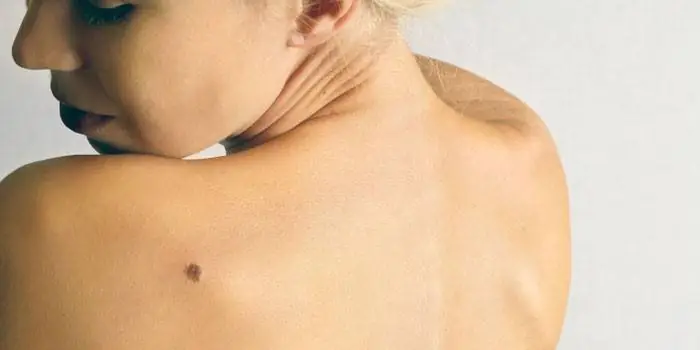
Spots that appear shortly after birth are not considered dangerous. It is important that they are small in size. Good – non-dangerous – signs of neoplasms include:
- flesh tone;
- unchanged pattern of the skin of the nevus and adjacent tissues;
- soft consistency;
- hair on the surface of the neoplasm - growing from the skin, indicates the absence of pathologies;
- diameter no more than 5 mm;
- symmetry;
- nevus in the form of a spot.
Which moles are dangerous?
Why do people with nevi on their bodies need to monitor their changes? There is always a threat of degeneration of non-dangerous tumors into a cancerous tumor. What moles are dangerous to health? Key signs you need to know:
- change in shades towards the dark side, the appearance of multi-color;
- rapid increase in size - exceeds two millimeters per year;
- occurrence of cracks;
- the formation of asymmetry due to uneven growth;
- lack of elasticity;
- the appearance of itching, burning;
- presence of discomfort.
The appearance of dangerous moles requires an immediate visit to a specialist to clarify the nature of the changes and the likelihood of developing skin cancer. Pathological transformations provoke:
- injury to the nevus due to negligence;
- self-removal;
- abuse of exposure to the sun, use of a solarium;
- location of the formation in places of frequent contact with clothing - on the neck, head, genitals, legs;
- placement in the hair, on the face, palms - where there is a high probability of injury;
- previously removed melanoma.
Why are moles dangerous?
Not a single person is protected from the sudden proliferation of cells of a harmless mole. Melanoma is an extremely serious disease. Changes not detected at the initial stage can result in death. The provoking factor is unsuccessful independent removal of tumors. Moles are dangerous because of their ability to:
- transform into an atypical – precancerous form;
- grow to large sizes;
- turn into cancerous;
- with minor external changes, metastases actively spread throughout the body through the circulatory and lymphatic channels.
How quickly does melanoma develop from a mole?
The transformation of a nevus into a cancerous formation can occur in different ways. The process depends on the stage of the disease and the type of tumor. Instant metastases are dangerous. Begins:
- growth of cancer (oncological) cells in the deep layers of the epidermis;
- their entry into the blood and lymph;
- penetration into the lungs, liver, kidneys;
- growth in these organs;
- complete damage to the body;
- death.
The growth phases of pigment cells are observed, along which melanoma develops from a mole. There are varieties:
- horizontal– damage to the upper layers of the skin occurs, lasting up to 10 years, but metastases do not appear;
- vertical– accompanied by the spread of cancer cells throughout the organs, can last two years, has an unfavorable prognosis;
- nodal – especially dangerous – characterized by deep spread within two months.
The first signs of melanoma
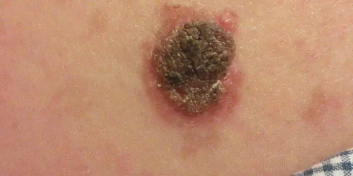
The patient can be assisted only when suspicious changes begin to be identified. The diagnosis, research, and referral for surgical treatment save a person’s life. The first signs of melanoma:
- increase in the height of the tumor;
- bleeding;
- the appearance of discharge;
- redness;
- burning, itching;
- swelling of tissues;
- softening of the nevus;
- the appearance of a crust;
- thickening;
- hair loss;
- expansion of pigmentation around the lesion.
With the further development of dangerous melanoma, the following are observed:
- significant change in size;
- the appearance of pain;
- enlarged lymph nodes;
- surface ulceration;
- formation of new foci;
- bleeding from places of pigmentation;
- liquid separation;
- skin thickening;
- the appearance of an earthy tint;
- signs of metastases are chronic cough, weight loss, cramps, headaches.
How to distinguish a mole from melanoma
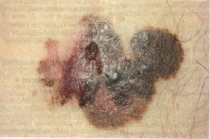
To recognize which moles are dangerous and which are not dangerous, you need to know what they look like. A person with nevi, in order to eliminate terrible consequences, must constantly monitor the appearance of new formations and changes that occur. You can distinguish a mole from melanoma by its signs. Non-dangerous neoplasm:
- symmetrical;
- with smooth edges;
- uniform in color;
- with dimensions not exceeding 6 millimeters.
Features of dangerous melanoma that require seeking help from dermatologists:
- growth in a short time;
- pronounced asymmetry of shape;
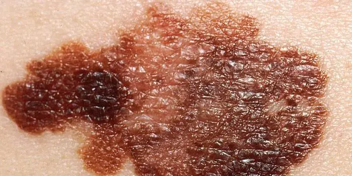
- heterogeneity in color - the presence of inclusions of several shades;
- lack of clear boundaries - the contour line is blurred, jagged, and looks like a coastline on a geographical map;
- increased diameter over six millimeters;
- variability of any parameters - color, size, shape.
What dangerous moles look like
What do nevi that are subject to pathological changes look like? Only a doctor can correctly distinguish between non-dangerous tumors. Dangerous formations look like this:
- blue– compactions under the skin with clear boundaries, with dimensions no more than 10 mm;
- nodal– round, flat in shape, color – brown, black;
- cutaneous– often pale, convex;
- halo nevus – pigment surrounded by a light or white rim;
- spitz- looks like a dome-shaped tumor of pink shades, with the possible presence of a hole through which blood and liquid leak;
- connecting- connect individual entities into a whole.
Mole with jagged edges
One of the signs of a non-hazardous formation turning into a dangerous one is a change in contours. It often has blurred edges and scalloped borders. There are non-dangerous types of nevi - dysplastic. Only a specialist can make a correct diagnosis. A mole with uneven edges can be dangerous if there are additional signs of melanoma:
- accelerated changes in size;
- the presence of clearly defined asymmetry;
- the appearance of highly indented boundaries.
Rough mole
Such a neoplasm is harmless if its diameter is no more than 5 mm and remains constant in size. Often its appearance signals a lack of vitamins and nutritional disorders. Doctors advise coming for a consultation if it is discovered that:
- the smooth nevus turned into a rough one;
- bothered by burning, itching, tingling;
- irregularities and compactions appeared in the middle;
- areas with different shades formed;
- diameter has increased significantly.
A dangerous rough mole requires immediate examination if:
- the appearance of bleeding;
- development of the inflammatory process;
- rapid change in size;
- formation of asymmetry;
- formation of purulent discharge;
- the occurrence of painful sensations when touched;
- the emergence of an irregular shape, blurred boundaries, along the edges of the neoplasm.
Large moles
Large formations on the skin are pigment spots. When they remain unchanged and do not cause inconvenience, this is a harmless phenomenon. It is important to constantly monitor their appearance, color, and size. To eliminate worries, you need to consult a dermatologist. During the visit, the specialist will conduct a diagnosis and give a forecast of the risk of developing a malignant neoplasm. Large moles become dangerous if they:
- injured;
- thickened;
- started to itch;
- were unsuccessfully removed independently;
- changed in size, shape;
- are bleeding.
What moles can be removed
Often nevi cause trouble for women when they are in a visible place - the face, neck. Even if they do not bother you, using removal will be the right decision - the appearance will improve significantly. After the procedure, the doctor must send the tissue for histological analysis to decide whether the mole is malignant or not. If the neoplasm is not dangerous, does not bother you, and does not change in size, surgery is not required. What moles cannot be removed? Experts believe:
- there are no contraindications;
- It is important to choose the right excision technique.
You should be careful about skin growths; it is unacceptable to remove them yourself. Only the doctor will determine whether a nevus is dangerous or not and decide what to do with it. You can delete it if:
- injured from clothing - on the neck, in the groin area, under the armpits;
- cause pain when touched;
- are located under the hair on the head and can be damaged when combing or cutting;
- change color, shape, outline;
- significantly increase in size;
- characterized by the presence of burning, itching;
- accompanied by inflammation and bleeding.
Skin without a single birthmark is a phenomenon. Brown, red, blue, big and small - everyone has them. They are not entirely correctly named, because babies are born with clear skin, and these nodules appear later. They consist of specialized cells of the dermis (melanocytes) that produce dark pigment, the cause of tanning. Sometimes they frighten you with their unpredictable behavior. They can develop into malignant melanoma.
p, blockquote 1,0,0,0,0 —>
p, blockquote 2,0,0,0,0 —>
About half of skin cancer cases occur due to malignancy of melanocytes in birthmarks. Let's consider their etiology, dangerous and non-hazardous types, photos, indications for removal, preventive measures.
p, blockquote 3,0,0,0,0 —>
Causes of moles
Small brown dots (nevi) most often occur in people with light or slightly dark skin tones. Their formation coincides in time with hormonal storms in the body that occur during certain periods of a person’s life:
p, blockquote 4,0,0,0,0 —>
- During adolescence.
- During pregnancy and menopause.
- After finishing taking oral contraceptives.
- After a solarium or a vacation at sea.
In an adult, the average number of such formations is from 12 to 20. Many moles on the body occur in people with a predisposition to this genetic trait. Experts perceive a large number of atypical nevi as a marker of melanoma if one of the close relatives has skin cancer.
p, blockquote 5,0,0,0,0 —>
Types of moles by color
The color of nevi is varied, but does not serve as a diagnostic symptom. There is a dependence on the phototype of the carrier’s skin and the characteristics of the cellular elements in the structure of the formation.
p, blockquote 6,0,0,0,0 —>
Red and pink
Strawberry-colored vascular moles are called hemangiomas. They are non-pigmented and develop when blood flow is disrupted. Upon careful examination, it is clear that they are plexuses of many capillaries. They turn pale when pressed. They are benign in nature and can disappear on their own. More often appear in children. They occupy the space between the skin itself and its epithelial layer. They can be convex or flat in shape. Able to grow, but only slightly. Appear for a number of reasons:
p, blockquote 7,0,0,0,0 —>
- Diseases of the heart and blood vessels.
- Vitamin deficiencies C, K.
- Failure in lipid metabolism.
- Injury to the skin, leading to the accumulation of capillaries.
- Problems with blood coagulation.
- Oncological and autoimmune diseases.
- Hepatitis, pancreatitis and other abnormalities in the digestive canal.
Red moles are associated with a hormonal factor. Dependence on ultraviolet radiation is impossible in principle, since they do not contain pigment and cannot appear from the sun. They form in pregnant women, disappear after the birth of a child, or remain unchanged. They are not dangerous, but for your own peace of mind it is better for the patient to visit a doctor. He will remove nevi if they create cosmetic problems, interfere with movement, bleed or itch. The excision process itself is painless, but it is better not to touch the tumor unless necessary.
p, blockquote 8,0,0,0,0 —>
Purple and blue
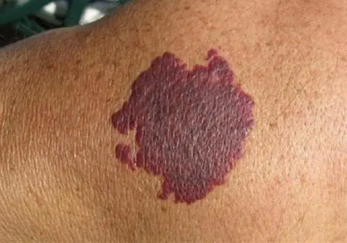
p, blockquote 9,0,0,0,0 —>
A group of melanoma-dangerous moles that appear more often in women. Localized on the legs, buttocks, face. They have a dense structure with a smooth, hairless surface. Rising above the skin. The maximum diameter is 2 cm. If the spot changes its color from blue to purple, and its outlines become blurred, then you should immediately visit a doctor. After the research, a decision will be made on further therapeutic actions.
p, blockquote 10,0,0,0,0 —>
Brown and black
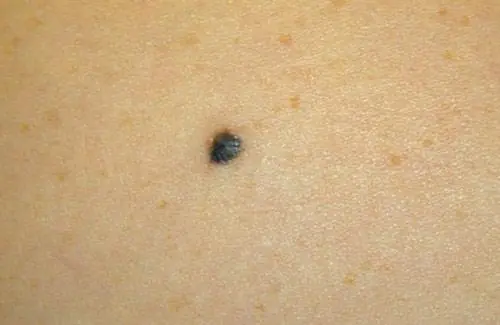
p, blockquote 11,0,1,0,0 —>
They arise as a result of the accumulation of many melanocytes. Their appearance is associated with solar radiation. Brown moles are usually benign, differing only in structure. They are localized throughout the body. If the nodules change shape, size, darken, or cause discomfort, you should consult a doctor to reduce the risk of malignancy. To prevent damage, stains located in inconvenient places, such as under the arms, must be removed.
p, blockquote 12,0,0,0,0 —>
Types of moles by shape
Nevi can be oval or round. In rare cases, they resemble animals, a star or a month. Shape, like color, is not a diagnostic sign by which one can judge the danger of a particular neoplasm.
p, blockquote 13,0,0,0,0 —>
Moles on the leg
Such nodules cause discomfort to a person due to constant contact with clothing and jewelry. They have a cone-shaped or round shape. After injury and excessive sun exposure they can become malignant.
p, blockquote 14,0,0,0,0 —>
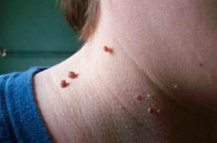
p, blockquote 15,0,0,0,0 —>
Their varieties include hanging moles that have a thin stalk. Location: skin of the face and neck. Occur due to infection with papillomavirus. If malignancy is suspected, pay attention to the following signs:
p, blockquote 16,0,0,0,0 —>
- The appearance of itching and pain.
- Bleeding from a nevus.
- Formation of purulent crusts.
- Change in structure and color.
- Fast growth.
If a mole on a stalk does not grow and does not cause any inconvenience, it is not necessary to visit a dermatologist, but you need to monitor its condition.
p, blockquote 17,0,0,0,0 —>
Flat moles

p, blockquote 18,0,0,0,0 —>
The following neoplasms of this type are distinguished:
p, blockquote 19,0,0,0,0 —>
- Simple. Formed in children and young people. Localized throughout the body.
- Sunny. Non-hazardous, they appear in areas exposed to strong insolation.
- Senile beige-brown shades, darkening over time.
These are the most common moles (lentigines). They lie in the superficial layers of the skin, so they are more often exposed to the ultraviolet part of the solar spectrum.
p, blockquote 20,0,0,0,0 —>
Convex
Dark structures with a smooth or rough surface with vellus or coarse hair protruding above the skin. There are 2 groups:
p, blockquote 21,0,0,0,0 —>
- Pigmented with a large number of melanocytes.
- Vascular or angiomas.
Such nodules require attention and careful handling to prevent their malignancy.
p, blockquote 22,1,0,0,0 —>
Benign moles
The first dots appear in babies older than 6 months. They grow up to 3 mm, are evenly pigmented, and have clear contours. They do not grow or increase only by 1-2 mm. They have a flat shape, no cracks or roughness. Varieties:
p, blockquote 23,0,0,0,0 —>
- Red moles or hemangiomas.
- Hanging formations or acrochordons.
- Yellow or brown patches called seborrheic keratomas.
Their change can be controlled if a person does not:
Malignant moles
Ultraviolet irradiation often triggers the malignancy of nevi. The time of insolation is individual. But prolonged exposure to the sun weakens the immune system and provokes changes in the structure and nature of tumors. Doctors note the important role of the genetic factor in this process.
p, blockquote 25,0,0,0,0 —>
Skin cancer (melanoma) is considered an insidious and aggressive pathology. It develops rapidly and has a high mortality rate. They recognize the disease by vertical growth, nodular formations, cracks, and red spots around the shiny surface. As malignant cells develop, they break away from the tumor and “scatter” throughout the body, affecting the heart, brain, eyes, lungs, and lymph nodes.
p, blockquote 26,0,0,0,0 —>
The number of melanoma patients is growing year by year. Among all types of oncology, this form of cancer accounts for 3-4% of cases. Treatment depends on the thickness of the tumor and the depth of its penetration. The prognosis is favorable for melanomas up to 1 cm in size. In other cases, it is alarming.
p, blockquote 27,0,0,0,0 —>
In women, the tumor process more often affects the skin of the lower leg (26.3%) and thighs (6.7%). Therefore, you cannot hide in the shadows and expose your feet to the sun. Light-colored trousers made of natural fiber block up to 72% of hard mutagenic radiation.
p, blockquote 28,0,0,0,0 —>
Melanoma can form due to injury to the nevus. If moles on the body are larger than 0.5 cm in size and have a varnished surface suddenly change color or shape, an urgent consultation with an oncologist is required. The main thing is to start treatment early.
There is no need to be afraid if there are smooth nodular formations or large spots with a lobed surface on the body. The cells of such nodules are located deep in the dermis and are little accessible to ultraviolet radiation. You can recognize a degenerated mole by the following signs:
p, blockquote 30,0,0,0,0 —>
- The pigmentation of the spot sharply increases or decreases, which becomes asymmetrical, with uneven outlines, and coal-black inclusions and stripes appear around them, forming a ring.
- The skin pattern disappears on the surface and peeling occurs.
- A red halo of inflammatory tissue appears around.
- The sizes are increasing.
- The nodule itches and tingles.
- Hair falls out from the surface of the neoplasm, ulcerations, cracks, and crusts appear.
At an early stage, melanoma has a favorable prognosis with 100% patient recovery. In case of metastasis to the lymph nodes, chemotherapy is performed along with surgical excision. Paying close attention to yourself and contacting specialists after the first alarming symptom is a guarantee of a good outcome.
p, blockquote 31,0,0,0,0 —>
Diagnostics
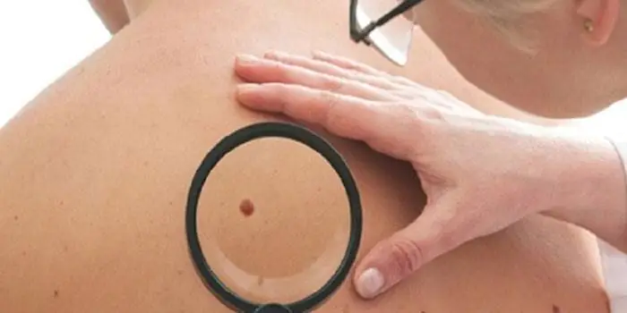
To detect melanoma, digital dermatoscopy and scanning methods for suspicious spots are used. If there are many nodules on the skin, a “photo session” is carried out, which is repeated six months later to record the changes that have occurred and draw up a map of the moles. As an addition, the ABCDE formula with feature analysis is used (in the Russian version AKORD):
p, blockquote 32,0,0,0,0 —>
- asymmetry;
- the edges;
- coloring;
- dimensions;
- dynamics.
Thermography can be used because the area of skin affected by melanoma is hotter than the rest of the skin. Wood's lamp helps determine the boundaries of the tumor.
p, blockquote 33,0,0,1,0 —>
If a mole bleeds or a substance is released from it, a cell culture method is used. But the final verdict on the nature of the nodules is given by histology. If the result is positive, the doctor prescribes an examination of the liver, lungs and other organs. If cancer has formed on the lips, diagnosticians study the condition of the oral cavity and nearby tissues. Ultrasound is used to determine the depth of the tumor. Metastases are detected using MRI.
p, blockquote 34,0,0,0,0 —>
p, blockquote 35,0,0,0,0 —>
Prevention
Measures to prevent the malignancy of moles have not been developed. There are recommendations that will help prevent the tumor process:
p, blockquote 36,0,0,0,0 —>
- Use sunscreen.
- Limit the time you spend sunbathing.
- Do not come into contact with chemicals.
- Do not visit the solarium.
- Wear loose-fitting cotton clothing that covers the body, wide-brimmed hats, and sunglasses.
- Do not take children under 3 years old to southern resorts. Burns received in early childhood increase the risk of developing skin cancer in adulthood.
- Do not try to remove the mole yourself. Entrust the procedure to a specialist who has the necessary equipment for this.
After swimming in the sea, take a shower and dry with a towel. This will get rid of water droplets and salt crystals, which, like lenses, enhance the harmful effects of ultraviolet radiation. It is dangerous to be in the sun between 10 a.m. and 3 p.m. At this time, even special creams do not help.
p, blockquote 37,0,0,0,0 —>
Elena Malysheva about moles in the program Live Healthy:
p, blockquote 38,0,0,0,0 —>
p, blockquote 39,0,0,0,0 —>
Mole removal
The following signs are indications for mandatory excision:
p, blockquote 40,0,0,0,0 —>
- Bleeding.
- Inflammation.
- Increase in size.
- Edge deformation.
- Darkening.
- Ulcerations.
- Itching and pain.
- Mole on the palm, sole of the feet, mucous membrane, anogenital area.
To prevent melanoma, complete (at a distance of 0.2-0.3 cm to the side of the spot) excision is performed. Postoperative scars are invisible if you use modern atraumatic suture materials and follow the rules of aesthetic surgery.
p, blockquote 41,0,0,0,0 —>
The removed nevi are subjected to histological examination. If tumor cells grow through the scar, a repeat operation is prescribed. Swelling at the surgical site goes away over time. A small bald spot may remain on your head. After a year, the remaining scars can be removed. In addition to surgical intervention, the following are used:
p, blockquote 42,0,0,0,0 —>
- Laser surgery – allows you to simultaneously seal vessels. Among its disadvantages: the impossibility of conducting histological examination.
- Electrocoagulation – burns the mole with low-frequency current. It is a traumatic manipulation with a high probability of scarring. It is used extremely rarely.
- Radio wave therapy is acceptable for small superficial nodules. It is carried out using a surgitron device. In this case, the tissues of the neoplasm itself are not damaged. They can be explored. A wound remains that heals quickly.
Treatment with traumatic methods or cauterizing compounds is unacceptable. Unchanged nevi should not be touched. If they are accidentally touched and injured, the wound is treated with an antiseptic to avoid secondary infection, and then visit a doctor.
p, blockquote 43,0,0,0,0 —>
Doctor's report
It's not the moles that are scary, it's our lack of awareness. A timely consultation with a dermatologist, an examination prescribed by him, and medical care based on best practices will help prevent a possible threat.
