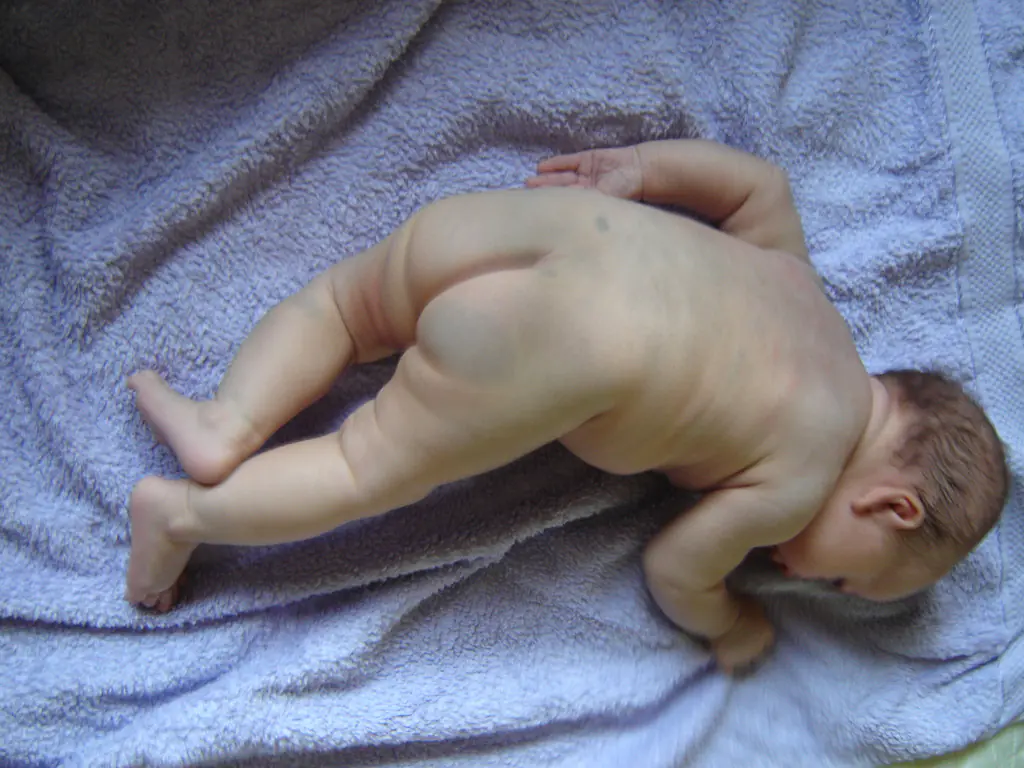
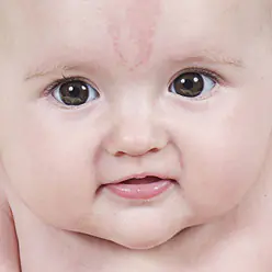
Almost every person has a mole, or non-tumor nevus.
This is an area of skin with a darker pigment color, of various shapes and sizes.
There are formations from light beige, almost invisible on human skin, to dark red and even black.
Most of them are completely safe for children (and adults).
But others are capable of causing fear in their rebirth, and bring trouble.
Moles most often appear between six months and four years. Then the appearance of nevi slows down and the next peak occurs in adolescence, which is due to hormonal changes in the human body. The baby is born, as a rule, with clear skin.
Exceptions also occur and are called birthmarks, which are usually more common in fair-skinned and fair-haired girls. Dark-skinned children are less likely to have moles from birth.
The formation of moles on human skin is a natural process. and is caused by the work of melanocytes, cells that determine skin color, provide tanning and give color to moles and other pigmented formations.
Reasons for appearance
- The most common and natural one is genetic. The number and location of nevi in a child directly depends on the number and location of them in his immediate family. Hence the name of education.
- Internal reasons. Hormonal. The peaks in the formation and disappearance of birthmarks directly depend on the peaks of hormonal changes in the human body, so the first occurs during the period of 2-3 years, the second during adolescence; often during pregnancy, a woman can also notice the appearance or disappearance of nevi. And this process is also natural.
- External reasons. First of all, the activity of melanocytes is influenced by solar activity and the effect of ultraviolet radiation in particular. Under the influence of this particular radiation, a mole can change its size, color, and even degenerate into a deadly disease - melanoma.
- A less common reason (and not yet fully proven) is traumatic. According to this hypothesis, the formation of foci of hyperpigmentation in children can form in the post-traumatic period, after suffering insect bites, skin injuries, as well as viral infections.
Doctor Komarovsky about moles in children
A mole, according to Komarovsky, is a formation in which cancerous (malignant) cells may appear. But it is not a reason to panic. Rather, it should serve as an impetus for obtaining more information about the peculiarities of the functioning of your body (and the child’s body).
A mole is in most cases completely safe; you can live with it (and even if there are many of them) quite comfortably all your life until a ripe old age. Moreover, Evgeniy Olegovich identifies two series of factors that determine the presence of moles in a person and monitoring their safety.
- Genetics or hereditary factor. We cannot influence her in any way. If the baby’s relatives have a scattering of areas of hyperpigmentation on their skin, it is not surprising that the child also has a lot of them.
- The second very important factor is solar Activity. Due to their direct effect on the activity of melanocytes, the sun and ultraviolet radiation in particular are directly related to the number of moles on the skin of both adults and children.
Moreover, ultraviolet light is an aggressive factor that contributes to the degeneration of nevus into a more dangerous form of hyperpigmentation - deadly melanoma.
The third factor that Dr. Komarovsky pays attention to is traumatic damage to moles. Moreover, permanent damage is dangerous. A one-time injury to a nevus, apart from more intense bleeding, cannot cause much concern.Which ones are dangerous?
For a qualitative assessment of the danger of a nevus Komarovsky recommends examining the child twice a year and make a map of the moles of him and his immediate family.
The doctor compiled an easy-to-memorize and quite informative “AKORD” system. What does this mean and how to use it?
Main characteristics of a mole, they also determine the degree of its safety for the body as follows:
- Asymmetry. If you virtually draw a straight line through the center of the mole, both parts should be mutually symmetrical. Otherwise, you should contact a specialist.
- The edges. Moles with even edges, smooth and rounded are safe and do not require contact with a specialist. You should consult a doctor if the edges are jagged or have unclear boundaries.
- Uniformity of color. The color of the nevus itself does not matter much, be it beige, deep brown, wine red or even black. The main thing is that it must be homogeneous.
- Size. A mole size up to 6 mm in diameter is considered safe (for comparison, this is the diameter of an eraser on a standard pencil).
- Growth dynamics. If the mole does not change significantly in size and there is no visible change within a month, it is safe. Anxiety can cause a noticeable increase within one month.
How to care?
Moles are smooth, do not protrude from the surface of the skin and are not subject to constant trauma and do not require special care. When visiting the beach they, like the rest of the skin, should be protected both by sunscreens and by not visiting the beach during peak solar activity (from 12 to 16 noon).
In the case where hair grows from a nevus, this is a cosmetic defect, but it is better to remove it simply by cutting it off, plucking it, or using creams or other methods of depilation.
What is a nevus map?
A mole map is needed to track when new moles appear and how old ones change. It is individual for everyone. It contains information about the location, size, color, symmetry, growth dynamics and edges of moles (that is, all information according to the AKORD system).
Thanks to this map it is possible to detect disturbances in areas of hyperpigmentation in a timely manner and contact a specialist, since although melanoma is often common, its cure with timely treatment reaches 95%.
Procedure for examining a child
- Examine the front surface of the body, right and left side with raised arms.
- Examination of the hands, the front and back of the shoulder, forearm and palm.
- Examination of the back of the body and buttocks.
- Examination of the legs, front and back of the thigh, lower leg and feet.
- Examination of the back of the head.
All enter the detected formations into the table indicating the location and characteristics of AKORD. Carry out an inspection twice a year.
How to prevent melanoma?
The best A way to prevent melanoma is the correct attitude towards the sun. And minimizing its negative impact on the child’s skin, including moles.
- Be sure to use a waterproof sunscreen with SPF 30+ or higher.
- Renew the protection every 2 hours (reapply the product to the skin of the body and face), as well as after each bathing.
- Do not specifically look for the sun, and also, if possible, do not sunbathe during the period of peak ultraviolet activity (from 12 to 16 pm).
- Stay in the shade if possible.
- Be especially careful where there is water, sand and snow. Because their reflective properties enhance the negative effects of sunlight on the skin.
- Avoid sunbathing. To give the appearance of tanned skin, use self-tanner. But under no circumstances should we refuse Sanskrins.
- Know your moles.
Thus, although a mole is a group of cells that can degenerate into a malignant form, if treated correctly, treated with care and regularly examined, they are not a cause for panic.
Watch a video on the topic:
In ancient times, people believed that birthmarks on a baby were signs of fate and predicted his future. Now scientists are considering more natural reasons for the appearance of such formations. Let's consider what factors influence the appearance of spots, and in what cases do they require removal? Why might a birthmark appear in a newborn?
Types of birthmarks
A child may have a wide variety of birthmarks on his body - smooth or covered with fluff, reddish or brown, convex or flat. The main types of birthmarks in newborns are nevi and angiomas.
What shade can nevi be?
Nevi are among the most common types of skin marks. They usually come in a variety of brownish shades, ranging from dark brown to pale. The basis of nevi are melantocytes. These epidermal cells contain melanin, a pigment that affects skin tone. It is necessary to protect the skin from ultraviolet radiation. Sometimes these cells are localized in one place, which leads to the appearance of a mole. Dark birthmarks indicate an abundance of melanin, while light ones indicate a lack of it.
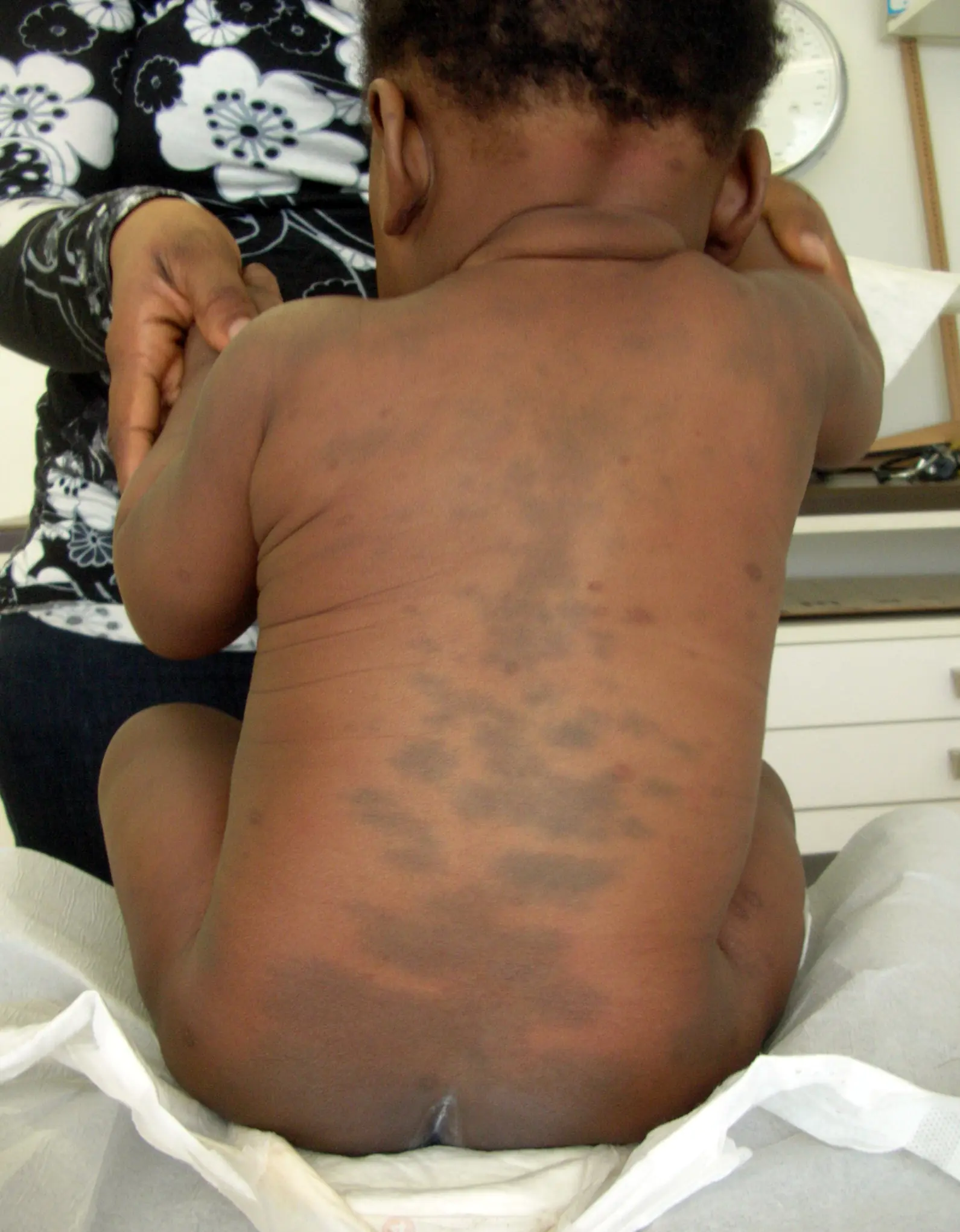
A Mongolian spot in a newborn should also not be a cause for concern for parents. It is also a place of concentration of melanin and is a spot, or several spots of different sizes from 1 to 10 cm in diameter, blue, green or even black. The most common location is the baby’s lower back, mainly the tailbone or butt. Mongolian spots are safe, they do not cause any discomfort to the child and go away on their own before adolescence. This type of nevus is named so because of their frequent detection in Mongolian children (90%), Mongolian spots are also often found in Asians, representatives of the Mongoloid and Negroid races.
There are also white formations. These include anemic nevi that arise due to underdeveloped blood vessels.
They need to be distinguished from millet grasses - milia. The latter look like convex dots filled with whitish content. They are a type of skin rash. Anemic nevi are a congenital phenomenon, and they are easy to identify: you need to rub the spot. The surrounding skin will turn red, but the formation will remain white.
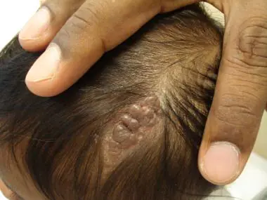
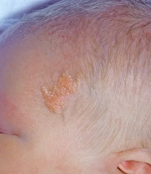
Light brown Jadassohn nevi indicate a congenital defect of the sebaceous glands. They are usually found on the baby's head, under the hairs. This occurs in 3 out of 1000 babies. It is recommended to remove it before adolescence, since in 10-15% of cases, they can subsequently develop into a cancerous tumor.
What if it’s a matter of blood vessels?
Another type of birthmarks is angiomas. They are of vascular nature. Congenital formations of small vessels on the skin are called hemangiomas. If such accumulations form in the lymphatic system, then they are classified as lymphangiomas. Even congenital, they appear externally only by the age of three.
In a newborn, only vascular hemangiomas can be detected. They are distinguished by a whole range of shades of red. Such formations are divided into several subtypes:
Strawberry (strawberry) hemangioma
These formations are convex, similar to red “berries”. They appear immediately after birth, usually on the face. The sizes can be different - from a millimeter to several in width. Strawberry hemangioma can increase in size, which is why it is dangerous, as it can affect the healthy tissues of the child.
Often this type of hemangioma stops growing, gradually brightens, shrinks and disappears completely by the age of 10.
Stellate (spider) angioma
It looks like a star with a bright base and “rays” extending from it. Most often it occurs on the child's neck. Disappears on its own in the first years of life.
Cavernous hemangioma
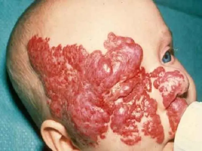
Loose, purple hemangioma, deeply embedded in the skin. It feels warmer to the touch than the surrounding epidermis. If you press, the baby will cry due to unpleasant sensations. This type of neoplasm requires treatment.
Flaming (fiery) nevus
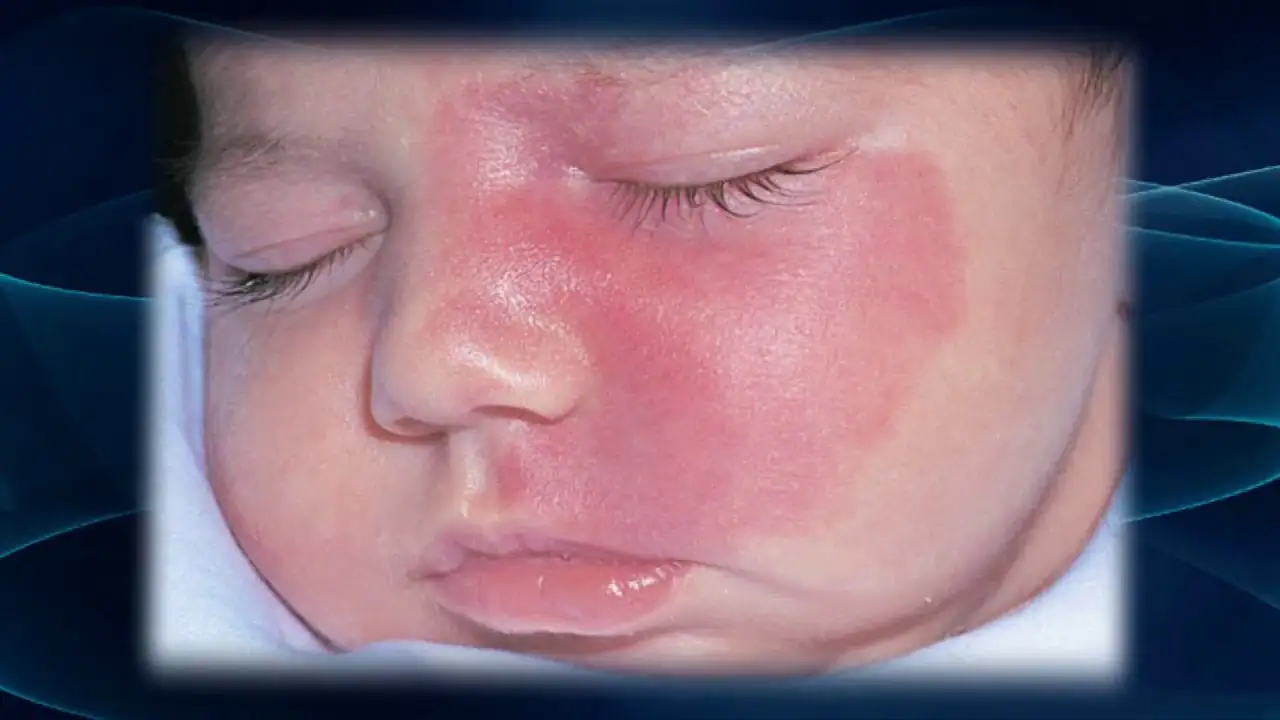
Looks like a red or purple stain from spilled wine. It can appear anywhere on the baby’s body. Such formations do not go away on their own. If they are not removed, they will remain for life. If the “wine stain” is in a visible place or continues to grow, it is better to take the trouble to correct the defect.
“Stork marks” (capillary hemangioma)
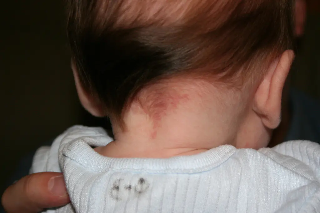
Such marks are also called “stork bites.” And if there is a mark on the baby’s forehead - “an angel’s kiss.” The formation is usually pink or red, but can also be orange, and resembles the mark of a bird's beak, which is how it gets its name. The formation is flat and does not rise above the skin. It is often found on the back of the baby’s head, in the neck area. When stressed, for example, when a baby cries, it acquires a brighter color. By the age of two, “stork marks” in most cases go away on their own.
In addition to the above, there are other types of birthmarks. But they are much less common.
If you notice that a child’s hemangioma is increasing in size, immediately contact a specialist (surgeon). He will be able to assess the danger of the condition and prescribe appropriate treatment or removal of the tumor.
Causes of skin formations

The reasons for a birthmark in a newborn, of course, are not that his mother loved to pet dogs and cats, as the ancients believed. However, scientists cannot say exactly why such marks may appear. Only risk factors for their occurrence have been identified.
Why do birthmarks appear in newborns? This is affected by:
- Hereditary factor;
- Hormonal surges in the expectant mother;
- Exposure to toxic substances on the body of a pregnant woman;
- Bad ecology;
- Climate change;
- Infections of the genitourinary system.
But it happens that a birthmark appears in a newborn even without exposure to risk factors.
Birthmark on a baby: what to do?
Is your baby's birthmark small, smooth, does not grow and does not cause concern to the baby? Everything is fine, nothing to worry about. But you need to take the new growth seriously. Observe the nevus and notice whether the mark grows or hurts. If changes occur, you should visit a pediatrician or pediatric dermatologist.
If a newborn has a birthmark on his body, several rules should be followed:
- Keep this area away from direct sunlight.
- Make sure that the baby does not scratch the area with the mark.
- Try to ensure that the nevus is never exposed to caustic substances, such as household chemicals.
In rare cases, marks on the skin can be fatal. Where can it appear? Under the influence of negative factors, a simple mole degenerates into a malignant formation - melanoma. Therefore, if the spot increases in size, you should urgently contact a specialist. If the formation is removed in time, there will be no health consequences.
Should moles be removed from babies?

It is recommended to eliminate formations in infants only if there is a danger to life. In babies, the immune system is not yet very developed, and any intervention can lead to serious consequences.
In what cases do doctors recommend surgery at an early age:
- The birthmark is very large;
- The formation rapidly increases in size;
- There are more than five marks, and they are concentrated in one place;
- The mole is located in a traumatic place (under the armpits, on the belt, on the skin of the eyelid, in the anus);
- Nevus interferes with the normal functioning of organs (on the hand, in the nose, in the eyes).
Particular importance should be given to those cases if a mole transforms - changes color or shape, grows, hairs fall out of it, it begins to bleed or itch.
How to get rid of formations?

The doctor may recommend one of the methods for removing nevi, depending on the size and condition of the formation, as well as the health of the baby:
Use of pharmaceuticals
Special medications are injected into the mole tissue to promote the death of overgrown cells. No anesthesia is required, but is not suitable in case of allergy to the active substances of the drug.
Using a laser
Excision of pathological tissues with a laser beam. It is quick and painless, but the procedure is not always possible for hard-to-reach areas.
Cryotherapy
Exposure of the mole to low temperatures. Suitable for eliminating small nevi.
Surgery
Removal of the formation using surgical instruments. It is used in cases where other methods cannot be used.
Carrying out the intervention under the supervision of a doctor, with preliminary examination of the tissues of the birthmark, reduces the likelihood of complications to zero. After removal of large formations, scars may remain. If they are located in a visible place, when the baby grows up, you can remove the scar using cosmetic procedures.
If you believe in fate, try telling your baby's destiny using moles. But pay attention only to happy signs:
- A mark on the baby’s cheek means love;
- A spot under the hair means high intelligence;
- Moles on the hands - to talents and good luck;
- Nevi on the back - to a life without worries;
- Mark on the leg - to hard work, calmness, confidence;
- A “sign” on the butt means success with the opposite sex.
As you can see, a mole is not a reason to panic at all. With the right approach, it will not be the cause of illness, but a happy sign that emphasizes the individuality of your son or daughter.
Remember that only a doctor can make a correct diagnosis; do not self-medicate without consultation and diagnosis by a qualified doctor.
Medical expert article
Many of us have heard that moles can be dangerous. However, as well as the fact that all birthmarks should be treated with extreme caution. That is why moles in children are not a groundless cause for concern on the part of parents. After all, all mothers and fathers want to see their babies healthy and beautiful. If the birthmark is small in size and located somewhere on the child’s arm, back or bottom, it causes emotion in the parents. Another thing is large, irregularly shaped spots located on the face and other places not hidden by clothing. Not only are they aesthetically unattractive, but they can also pose a hidden threat to the child’s life.
[1], [2]
ICD-10 code
Causes of moles in a child
Moles are unusual growths on human skin. The mystery of these pigment spots is that they can appear at any age, starting from the moment the child is born. True, the presence of moles (nevi) in newborns is a rather rare phenomenon that occurs in one baby out of a hundred. However, the fact remains that the baby may already be born with a mark called a birthmark. The birthmark may have a more or less deep brown or red color and be of various sizes.
Usually, moles in children begin to appear on the skin starting from the age of six months, but in most cases this process starts at 2-3 years. By the age of four, most children can see about 10 moles of various sizes on their skin. Then, for some time, the increase in the number of pigment spots does not occur or is inhibited. The next peak in the increase in the number of nevi occurs in adolescence, when the appearance of moles is associated with hormonal changes in the body.
In principle, the appearance of moles in humans is a natural process. This is due to the presence of special cells in human skin - melanocytes, which in some cases cause various changes in the pigmentation of the skin.
The reasons for the appearance of moles in a child can be either hereditary or the result of internal (changes in hormonal levels during puberty) and external (exposure to sunlight) influences. If the baby has had numerous birthmarks in his family, then, most likely, he will also have many moles. Moreover, they appear mainly in the same places as in relatives, which, by the way, determines the name of such neoplasms.
During adolescence, hormonal surges can cause an increase in the production of melanin, the substance responsible for skin pigmentation. At the age of puberty, nevi can either actively appear or disappear. Moreover, this behavior of moles does not at all indicate pathological processes in the body and directly on the skin. This is a normal, natural reaction.
There is also a theory that changes in skin pigmentation can cause a traumatic effect on the skin, for example, from an insect bite, or the influence of viral infections that trigger the process of melanocytes grouping and coming to the surface. There are moles that are almost invisible on the skin. A child may accidentally scratch it and it will change color to a darker color.
The influence of ultraviolet radiation on the skin can also provoke an increase in the number of nevi, as well as a change in their appearance (color, size, shape). Moreover, this happens at any age, both in childhood and adolescence, and even in adulthood. It is the effect of ultraviolet radiation that can subsequently trigger the pathological processes of modification and degeneration of moles.
Some studies show that newborns are more likely to develop a birthmark if the baby is premature or has very light skin. Light-skinned children often have more moles than those with dark skin. There is a dependence of the number of nevi on the gender of the child. As a rule, girls are more likely to develop birthmarks.
[3]
Symptoms of moles in a child
As mentioned above, moles can come in different shapes, sizes and colors. The color range of moles in children ranges from dark beige, almost invisible on the skin, to deep red and even black. Common safe moles in children have a regular round shape with smooth edges, brown color and small size up to 1.5 mm. They can be completely flat or stand out slightly above the surface of the baby's skin. Parents should not worry about such neoplasms.
Moles of medium (up to 10 mm) and large (more than 10 mm) sizes are more likely to be damaged and scratched, and accordingly, they are more likely to degenerate into a malignant neoplasm. A good indicator is the presence of hair on the mole itself, regardless of its size. Such moles are not prone to degeneration unless the hairs on them are pulled out.
In addition to this division, in medical practice there is a division of moles according to appearance and method of formation into ordinary and vascular nevi. Common moles are smooth, light pink or brown growths. Sometimes their color is darker, but this should not scare parents.
A black smooth mole in a child is more the norm than a deviation. A rich dark color in this case does not indicate its danger to the baby’s life. It’s another matter if the mole changes color to a more or less saturated shade, there are a lot of such moles, or if there is only one black mole, but it is large in size (more than 1.5 cm). This is already a reason to consult a dermatologist.
A red mole in a child indicates its vascular origin. Vascular moles are so named because they are composed of a large collection of blood vessels and are therefore red in color. They can have different shapes, and their color varies from light pink to deep red.
Vascular moles in children come in various types and forms:
- Hemangioma
- “Stork bite” - markings on newborns are a rich red-orange color.
- Port-wine stains are brownish-red or burgundy growths (flaming nevus)
Hemangioma is a benign formation on the skin, despite its unaesthetic appearance. Their appearance may not be noticed immediately. This can happen 2-3 weeks after the baby is born or even a year later. This mark can have different sizes and locations. Its peculiarity is the possibility of growth. Even if such a mole grows very quickly in a child, it does not pose a danger to life, except perhaps discomfort from an aesthetic point of view. Usually, by the age of one and a half years, hemangiomas become much lighter, and by the age of 10 they disappear completely.
There are 2 types of hemangioma: “strawberry” and “cavernous”. The “strawberry” mole is soft to the touch, has a convex structure and a color similar to the berry of the same name. Such moles most often appear on the child’s face, as well as on the head, back of the head and neck, but it is possible that they may appear in other places, including even internal organs.
“Cavernous” hemangioma looks a little different. It has a purple, rich burgundy or bluish-gray tint, a denser structure that goes deep into the layers of the skin. Often this is an irregularly shaped spot, consisting of one or several lesions close to each other. May appear on different parts of the body.
The greatest frustration for parents is caused by moles on the child’s face and head. But you just have to be patient, as such tumors disappear on their own. They are usually not treated. It is simply necessary to take all measures so that the baby does not damage such a mole or scratch it. After all, the main reason a mole turns into a life-threatening neoplasm is its injury. And the larger the mole and the more prominent it is above the surface of the skin, the higher the likelihood of damage.
Most often, on the face and back of the baby’s head you can find such a mark as a yellow or creamy-red birthmark in a child, jokingly called “stork bite” (or “angel kiss”). This can be a single large pink or cream spot or a cluster of several spots. Usually these marks disappear by the year, but there are cases that remain for a longer period.
The situation is more difficult with “port-wine stains” - flat, smooth neoplasms of a red-burgundy color. Such moles in children also tend to increase as the child grows, but do not disappear with age. They cannot be deleted. You can only try to make them less noticeable using home remedies to lighten spots on the skin or professional cosmetics. In some cases, a course of infrared or laser therapy may be recommended.
It is worth noting that some parents mistakenly believe that such a spot can be hidden behind a tan and allow their children to be in the sun for a long time. Such a careless attitude can only lead to a change in the color of the stain to a more saturated one, but will not hide the defect in any way. In addition, increased exposure to ultraviolet rays from the sun can lead to degeneration of the mole.
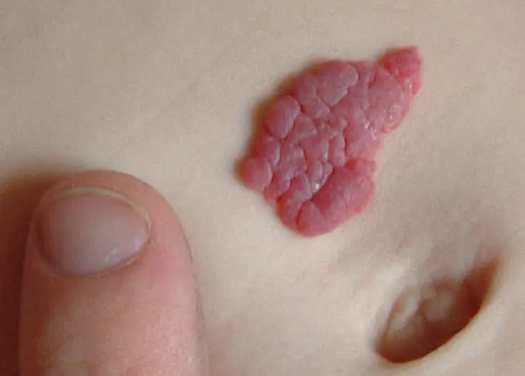
A hanging mole in a child occupies a special place among birthmarks. It can be located on the baby's neck or under the arms. It can appear at any age. This mole looks like a small piece of hanging skin of a natural or darker color. The whole danger of a hanging mole is that it cannot be torn off and injured, while it can become the object of close attention of your baby. It’s also not worth removing such a mole yourself if you care about your child’s health. The most correct decision would be to consult a dermatologist for examination and consultation, as well as close monitoring of the behavior of the hanging mole: changes in the color and size of the nevus.
Symptoms of mole degeneration
In general, if a mole is not injured during life and does not undergo changes noticeable to the eye, it exists on the body of its owner for a long time, without causing harm to his health. This is typical mainly for small moles up to 6 mm in diameter. A dangerous mole in a child is one that is larger than 6 mm. It is dangerous not in itself, but because the risk of injury in such neoplasms is higher than in small spots.
The same applies to a raised mole in a child. The baby, having felt an unusual tubercle on the body, will show special attention to it. He can constantly touch it, try to rip it off. The risk of injury to such moles is very high, so you need to carefully monitor not only the behavior of the mole, but also the baby’s actions in relation to it.
A large mole in a child, whenever it appears and no matter how it looks, is undoubtedly a reason to show the child to a dermatologist. The doctor will be able to assess the likelihood of a mole degenerating into a malignant one, and will definitely give advice on caring for nevi.
The pathogenesis of the transformation of harmless birthmarks and moles in children into dangerous malignant neoplasms has not yet been fully studied by doctors, however, the reasons causing these changes have been reliably determined. These include injury to the surface of the mole, unsuccessful attempts to remove birthmarks using dubious methods and means, as well as prolonged exposure to the sun without the necessary protection.
The consequences and complications of the influence of these causes can be the most tragic. Trauma to a mole can lead to ulcers and bleeding from the nevus, which are very difficult to stop. At the site of the lesion, in this case in the area of the mole, a malignant tumor (melanoma, or skin cancer) can develop, which develops very quickly with multiple metastases to all parts of the body. At the same time, early detection of the initial symptoms of melanoma guarantees a 95% chance of successful treatment. If the disease is started, this probability drops to 20%; the remaining 80% of cases lead to the death of the patient.
Any moles on the baby’s body require attention from the parents. Periodic examination of moles will allow you to notice the first signs of a mole changing and turning into a malignant tumor. These signs include:
- Asymmetricity of the neoplasm (asymmetry). Ideally, a mole is a circle or oval, the two halves of which are symmetrical (similar) to each other. If one side of the mole grows more than the other, this is a reason to examine it.
- Uneven borders of the nevus (border irregularity). A normal healthy mole always has smooth edges. If the boundaries of the nevus become blurred, with jagged edges, this is already one of the signs of the development of melanoma.
- Change in color (color). A uniform color of the pigment spot is considered normal. Particles of any color on the uniformly colored surface of the nevus become visible to the eye. Any strange mole in a child with an unusual color or shape should alert caring parents.
- Mole diameter (diameter). If the mole does not exceed 6 mm in diameter, then contacting a specialist is not required. Regular periodic monitoring of it is sufficient. It is better to immediately show moles with a large diameter to a dermatologist to assess their development and growth.
- Behavior variability (evolving). As a rule, a mole does not undergo any significant changes during a person’s life. If any of the above characteristics or several of them at once begins to change, it is better to immediately show the child to a dermatologist or oncologist in order to prevent sad consequences. The appearance of a large number of others similar to it around the nevus should also alert you.
This method of examining a mole for benignity and safety is usually called the ABCDE method.
