If the question arises, what is a pedunculated mole called, the answer is hemangioma or hanging nevus. It is a vascular formation that is located on the skin. The appearance resembles a collection of wart-like nodules. The appearance of formation is influenced by a congenital or acquired factor.
Why does it appear
Moles on the leg and the reasons for their appearance are as follows:
- The influence of ultraviolet rays. With prolonged and frequent exposure to open sunlight from 10 am to 4 pm, hanging formations may form.
- Heredity. If one of the parents has several hanging moles on the neck, a similar phenomenon can be inherited by the child.
- Hormonal imbalances. Pigment formations can form during pregnancy, puberty, a stressful situation, menopause, or termination of pregnancy.
- A pedunculated mole can occur as a result of an insect bite, as a result of which an infection has entered the body through an unhealed wound.
- When the protective crust is injured by an insect bite, the process of activation of melanocytes begins.
- Diseases of the endocrine system provoke the formation of hanging growths.
Is a mole on a leg dangerous?
Pedicled nevi are the most dangerous type of moles. The danger is based on the constant mechanical impact on the convex formation, as a result of which the risk of degeneration of a benign growth into a malignant one increases. Another danger is the similarity of the growth on the leg with papilloma. Papilloma is a viral disease caused by the human papilloma virus. Whether a pedunculated mole is dangerous or not can be determined when an examination is carried out, during which the presence or absence of pathology is confirmed.
Locations
Pedicled formations often appear in women on an open area of the body, resulting in aesthetic discomfort. Common places where hanging nevi are located:
- In the intimate area. The groin location of the growth is less dangerous because it is not exposed to ultraviolet rays and does not cause discomfort. It is necessary to observe the size and color of the nevus. Be careful when removing unwanted hair in the intimate area. If it begins to grow and become inflamed, you should consult a doctor.
- In the armpit. A mole on a leg under the arm causes inconvenience. Carefully remove hair in this area. If you accidentally cut off a mole, the risk of inflammation increases. Sweat glands are located under the armpit; sweat is intensely released, which causes the spread of bacteria. When a nevus is damaged, bacteria are able to penetrate the wound.
- On the neck. The danger comes from moles located in the collar area. A large formation on the neck is subject to constant contact with clothing or jewelry and is in direct sunlight. This can provoke a violation of their integrity, which will lead to the process of degeneration into a malignant convex formation.
Injury to the nevus will lead to heavy bleeding. If damaged, the area must be treated with a disinfectant and lubricated with brilliant green. To avoid re-injury, you should consult a surgeon for surgery to remove the growth.
When to see a doctor
Regardless of the reason for the appearance of a mole on a stalk, it is necessary to know the symptoms that may indicate that the process of transforming a nevus into a malignant formation has begun. These include:
- Rapid increase in the size of a raised mole.
- Discharge of blood or clear fluid from the formation.
- Change of shade. When degenerated, the nevus may turn red or black.
- Itching and burning in the area of the growth.
- The surface of the mole is covered with small nodules.
- The appearance of small spots around the mole.
- A dry, shiny crust appeared on the surface.
- The raised mole began to peel off.
- A white spot appeared around the formation.
The above symptoms may be a signal of an inflammatory process, caused by a hormonal imbalance, prolonged exposure to the sun in the summer, or thyroid disease. To determine the cause of the appearance of a mole on a leg, you need to visit a dermatologist or oncodermatologist. After the examination and the necessary tests, the doctor will determine treatment tactics or prescribe removal of the raised mole.
Removal methods
To get rid of a pathological or awkwardly located mole, you can choose one of the following methods:
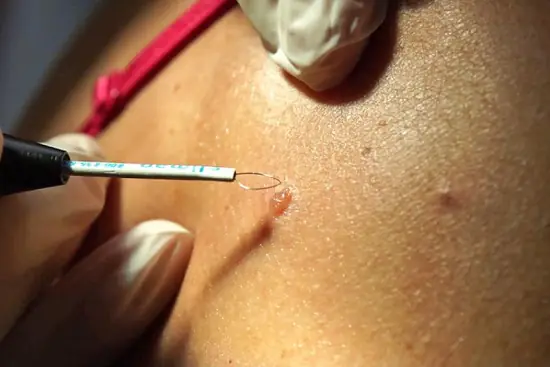
- Removal by radio wave. The thermal effect is able to remove the nevus without contacting it. The method is safe and painless, which can be taken on spots located on different parts of the body. Layer-by-layer removal of the nevus leads to its reduction and disappearance. After the operation, a protective crust will remain, which is not recommended to be peeled off. After two weeks it will dry out and fall off.
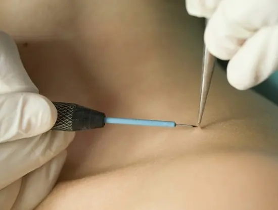
- Surgical removal. It is advisable to use surgical removal if the nevus is large or malignant. The doctor uses a scalpel to remove the nevus and its deep part. The sutures that are placed after removal are removed after a week. The disadvantage of this method is the presence of scars and scars, so you should not use this method to remove moles located on a visible area of the body.
- Laser removal. It is a popular but expensive method. The laser action is directed at the base of the convex nevus, during which layer-by-layer disappearance occurs. You can remove small or medium growths with a laser. The advantage of the method is the absence of contraindications and possible penetration of infection into the wound.
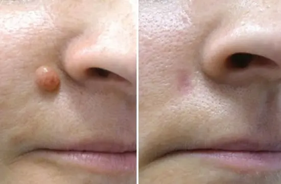
- Electrocoagulation method. The build-up is removed using the thermal effect of electric current. The operation is safe and painless. The patient is given a local anesthetic to prevent pain during surgery. The advantage of the method is cauterization of the wound, which prevents infection.
- Cryodestruction method. The removal process uses liquid nitrogen. The operation must be performed by an experienced doctor, since liquid nitrogen can cause an inflammatory process if it gets into a healthy area of the skin.
In addition to medical methods, it is possible to get rid of a nevus on the stem yourself. These methods include:
- Three times a day, cauterize the formation with iodine solution.
- In the morning and at night, lubricate the nevus with a swab dipped in celandine juice. When lubricating, do not violate the boundary of the formation. Celandine juice can be used in pure form or mixed with Vaseline in equal proportions.
- Bandage the stalk of the nevus using your own hair. The method is aimed at gradually stopping the flow of blood to the nevus, as a result of which it will begin to die. After the bandaged mole falls off, you should consult a dermatologist. The doctor will examine the area for any pathological tissue changes.
How to protect a pedunculated mole from degenerating into cancer
To avoid the conversion process, some rules must be followed. These include:
- Take careful care of the area of skin where the raised nevus is located to avoid drying out.
- Avoid prolonged exposure to the summer sun on the mole.
- Protect from mechanical impact.
- Do not allow household chemicals and cosmetics that contain aggressive components to come into contact with the growth.
- Visit a dermatologist every 6 months to examine and examine the hanging formation.
If you follow the above rules, you don’t have to worry about the growth degenerating into a malignant tumor.
What is a pedunculated mole, what are the reasons for its development and why is the appearance of such a neoplasm dangerous?
How to treat a pedunculated nevus at home and what methods can be used to remove it in the clinic.
Among all the nevi, it is the pedunculated mole that gives its owner the greatest discomfort.
- All information on the site is for informational purposes only and is NOT a guide to action!
- Can give you an ACCURATE DIAGNOSIS only DOCTOR!
- We kindly ask you NOT to self-medicate, but make an appointment with a specialist!
- Health to you and your loved ones!
Why are they dangerous? such neoplasms?
They most often come into contact with the edges of clothing, jewelry, and hygiene items.
The resulting damage, as well as prolonged exposure to ultraviolet rays, can lead to malignancy (malignancy) of the mole.
In order to avoid the development of complications, it is necessary to consult a doctor to decide on the removal of the tumor.
Knowing the reasons for the formation of tissue proliferation, the most common places of its development and situations when such a tumor poses a real threat will help in determining the moment of consultation.
What it is
Dermatologists consider two types of neoplasms to be pedunculated moles:
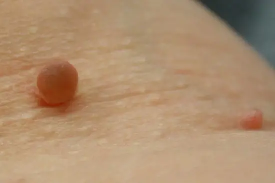
- hanging mole or papillomatous nevus. It is a formation on a thin stalk, has a different shade from pink to dark brown and can appear from birth or throughout life. In most cases, like all melanoma-dangerous nevi, it appears at birth but, due to its small size, is invisible until a certain age;
- acrochordons. Pathological growth of tissue caused by infection of the body with the human papillomavirus (papillomavirus or HPV). These are conical or rounded growths on a stalk, in most cases appearing on the skin in the facial area and on the neck.
It doesn’t matter what the pedunculated tumors are called, they are quite similar in appearance. They seem to hang from the body, have a thin stem and an uneven surface in the form of a cauliflower head.
Based on photographs of images of papillomas, warts and nevi on a leg, only a qualified specialist can distinguish them at first glance. However, the structure of these formations is different, so it can only be determined after a series of diagnostic manipulations.
Types of nevi
A pedunculated mole is significantly different in appearance from other types of nevi.
All moles are divided into:
- congenital - appear on the body and face even in the prenatal period and are least susceptible to malignancy;
- acquired - develop throughout human life; according to statistics, they become malignant much more often than congenital ones.
Regardless of the time of appearance of the mole, based on its histological structure, experts distinguish the following types of nevi.
Melanoma-free
They almost never become malignant and are removed mainly for aesthetic reasons.
These include:
- intradermal pigmented mole - most often develops in adolescents, predominantly localized on the skin in the neck, groin, armpits, and inframammary fold;
- papillomatous (a mole on a stalk, its photo differs sharply from all other types of nevi);
- halonevus or Seton's mole - a round, acquired formation in most cases, slightly protruding above the surface of the skin and having a pale rim around it, develops in people with severely reduced activity of the immune system;
- Mongolian spot is a genetically determined pigment pathology that appears in newborns, named after the predominant nation in which it appears (up to 90 Mongolian babies are born with this type of nevus);
- fibroepithelial mole is the most common type of nevus, appearing at any age, having various sizes and a round shape and color from pinkish to light red, consisting of connective tissue cells.
Melanoma-hazardous
Photo: melanoma-dangerous blue nevus
These are moles that have a tendency to become malignant.
Before removing such a formation, it is necessary to conduct a cytological examination or biopsy.
There are several types of melanoma-dangerous neoplasms:
- Jadassohn-Tiche nevus (blue or dark blue) is a single, acquired accumulation of melanocytes located deep in the skin, having a bluish color and a low tendency to malignancy; occasionally there are cases of the development of 3-4 such nevi;
- borderline pigmented nevus is a congenital (occasionally develops throughout life) formation that grows with the body, characterized by a high content of melanin, which provides the mole with a rich color (from purple to dark brown);
- giant pigmented - an exclusively congenital pathology, characterized by large sizes (from 3-4 cm to cover an entire area, for example, the cervical), strong elevation above the level of the skin and an uneven surface of the spot; cracks, furrows, outgrowths and hair growth can form on the surface, the color of the neoplasm is grayish or brown;
- nevus of Ota - a black-bluish spot located on the skin of the facial area;
- dysplastic - in half of the cases this is a congenital pathology, sometimes of a hereditary nature, has a different but heterogeneous color, uneven edges and a flat, smooth surface.
Video: “Dangerous moles! Is it worth removing and how to recognize melanoma in time?”
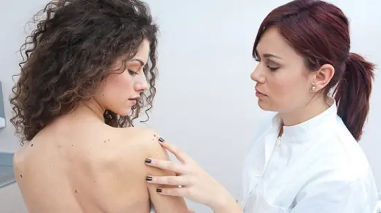
Should I see a doctor?
If throughout life the neoplasm does not cause discomfort, has the same size, or grows at an insignificant rate, and its color and surface practically do not change, it is not necessary to visit a doctor. It is necessary to constantly and very carefully monitor the condition of the tumor.
At the slightest suspicion of degeneration, you should immediately contact a specialist.
Which doctor should you contact - of course, a dermatologist; if necessary, he will give you a referral for a consultation with a dermato-oncologist.
If there is a hanging mole on the body or face, it is necessary to know exactly the signs that raise suspicion of malignancy of the neoplasm, these include:
- rapid increase in tumor size;
- change in the shade, structure (the mole becomes too loose or dense) and the appearance and color of the mole (it turns red, blackened, or, conversely, becomes too light);
- the appearance of discomfort - pain or severe itching in the mole area;
- bleeding from a nevus;
- the surface changes its pattern, bloody or purulent crusts appear on it.
If any of the above changes occur, especially if the mole becomes inflamed, becomes red or black and causes discomfort, do not think twice about who to turn to.
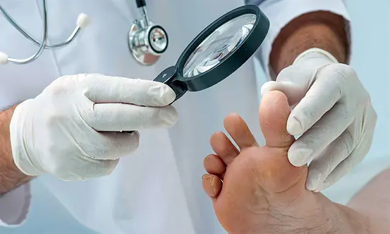
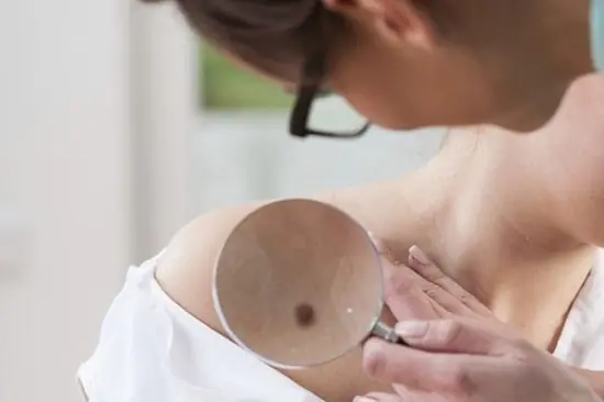
Photo: computer diagnostics
It is better to first consult with a specialist, and not self-medicate according to the recipes of traditional healers, because such treatment can lead to an acceleration of the transformation of a benign neoplasm into a malignant one.
How to remove a mole on a leg
Before getting rid of a boring tumor, you need to consult a doctor, even if it is preferable for a person to use traditional methods of treatment.
Only a specialist can distinguish a malignant neoplasm from a benign one and perform complete removal of the nevus.
On one's own
There are several proven traditional medicine recipes to get rid of a hanging mole:
- use of celandine - every morning and evening, the new growth is smeared with celandine, trying not to go beyond its boundaries (use plant juice, pure or mixed with petroleum jelly in a 1: 1 ratio);
- cauterization of the nevus with an alcohol solution of iodine;
- A rather ancient method is to bandage the base (leg) of the growth with your own hair as close to the skin as possible, this will disrupt the blood supply to the mole and cause its gradual death. If a mole falls off after using this method, you need to make sure that there are no changed tissues left, and for this you will still have to visit a doctor.
Why does a mole on my back itch? Find out here.
What does a mole on the stomach mean? Read on.
In a medical facility
It is better to remove a mole on a leg in a clinic, and not in a cosmetology office or beauty salon.
In specialized hospital departments, they can select a method for removing a tumor that corresponds to the individual characteristics of its development.
Ways to remove moles:
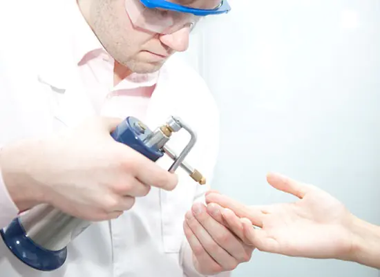
- electrocoagulation method. The skin around the nevus is exposed to high-frequency electric current;
- laser removal. Painless procedure, after which there are no scars;
- cryodestruction. Freezing formation using liquid nitrogen;
- exposure to a radio knife. A modern method of non-contact removal using radio waves;
- surgical intervention. Used for large or malignant tumors.
Photo: nevus removal using electrocoagulation
If the mole comes off on its own, it is necessary to stop the bleeding and visit a dermatologist for examination and consultation; removal of the remains may be necessary.
Reasons for appearance
Reason for appearance neoplasms on the skin - the growth of its surface layer caused by various factors, which include:
- local anomaly or developmental defect;
- genetic predisposition;
- prolonged, aggressive exposure to ultraviolet rays;
- chemical, thermal and mechanical damage to the skin;
- changes in hormonal status during various diseases, during adolescence, as well as during pregnancy and menopause;
- infection - viral (papillomavirus) or bacterial (the causative agent of syphilis can cause the development of a neoplasm on thick leg).
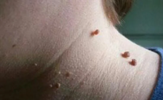
Dangerous locations
The most dangerous localization of such tumors is considered to be on the neck, under the armpit and in the groin area. Moles located in these places are more often injured during dressing and hygiene procedures.
Photo: birthmark under the armpit
Prevention of degeneration
In order to avoid malignancy of a mole, you must adhere to the following rules:
- do not expose it to excessive sunlight;
- protect from mechanical, chemical and thermal injuries;
- take care of the skin, preventing it from drying out;
- consult a doctor in a timely manner and, if necessary, treat skin diseases.
Questions and answers
Which doctor should I contact?
If there is a nevus on the stalk, you should consult a dermatologist or dermato-oncologist.
Appeared during pregnancy
The cause of the appearance of neoplasms on the skin during pregnancy is most often a change in hormonal levels.
But you need to warn your doctor about this event.
In all likelihood, he will prescribe examinations to rule out infection with papillomavirus.
Sore under the armpit
If a mole becomes inflamed under the arm, it is advisable to remove it to avoid further injury and the risk of degeneration.
What does a mole on the right cheek mean? Read here.
What do cancerous moles look like? See here.
What to do if it falls off
If a mole comes off, you still need to consult a doctor to find out the reason for its disappearance and possible removal of the remains.
To maintain your health you need to:
- observe the appearance of new moles on the body;
- changing old ones;
- follow the rules for preventing degeneration;
- If necessary, you should visit a dermatologist and have the disturbing nevus removed.
Video: “Laser removal of moles, warts, papillomas”
Literally every person knows what a pedunculated mole looks like - it is a small flesh-colored or dark brown formation attached to the surface of the skin with a small “leg”. People generally believe that touching or tearing off such moles is very dangerous. But is this really so? And why do doctors strongly advise against trying to remove them yourself? Let's figure this out together.
What is a mole on a leg?
In medicine, such a neoplasm is usually called a hemangioma, which can have several types:
- papillomatous nevus – is a neoplasm of light or dark color on a thin stalk. It may be present at birth or appear as you grow older. It is considered melanoma-free and can be present on a person’s skin throughout his life.
- acrochordon - outwardly practically no different from papilloma. However, it appears due to the entry of the human papillomavirus (HPV) into the body.
A pedunculated mole is characterized by a soft composition and an uneven surface, which can sometimes be rough or have a slight hair covering.
The most common locations for such moles are the face, neck, armpits and groin. All of them are quite traumatic and cause a person to constantly experience discomfort in the presence of large tumors. After all, every time you put on or take off clothes, they are somehow touched, which can lead to damage and a subsequent series of negative consequences.
Causes of nevi
Most often, hanging moles appear in adolescents during puberty and in women during pregnancy, which is associated with a sharp change in hormonal levels.
In addition, papillomas can appear due to:
- genetic predisposition
- diseases of the endocrine system
- frequent sun exposure
- excessive use of the solarium
- skin damage accompanied by bleeding and a sharp proliferation of melanocytes
- cancer and other pathological changes in the body
Which nevi are the most dangerous?
Everyone knows that moles are basically harmless. However, with the slightest disturbance in their structure or activation of transformation processes, there is always a risk of malignancy of nevi, that is, their malignancy. If we compare papillomas from this point of view, the most dangerous are those that appeared during life. Those that have been present on the human body since birth are practically harmless if they are not injured in any way.
When is it necessary to remove moles?
It’s good if the mole causes only aesthetic troubles. However, there are times when removing a mole is simply necessary in order to maintain health. These include the following situations:
- the mole begins to grow rapidly and increase in size (especially if its diameter has become more than 3 cm),
- it hurts and itches,
- the papilloma is located in a dangerous area (for example, in the armpits) and can easily be injured during shaving,
- a mole on a leg is located under a bra strap or right on the neck of a blouse, so it is constantly injured and painful,
- the nevus began to bleed or changed its structure (darkened, lightened).
In a word, if you notice sudden changes in the condition of a mole or it constantly interferes with you during some bodily movements, this is a good reason to visit a qualified dermatologist and consult with him about the possible removal of the tumor.
If you do not react to these signals in time, you risk becoming one of the cancer patients, because from a benign hemangioma to its malignancy is literally one step. And it is almost impossible to predict when this will happen and as a result of what.
Ways to remove moles
As we said above, today there are several ways to remove a mole:
- surgically,
- using cryodestruction,
- laser therapy,
- radio waves
- and electrocoagulation.
Surgical removal
It is the most common and cheapest method, the essence of which is to excise a mole from the surface of the skin using an ordinary scalpel. The patient is given general or local anesthesia, and then the tumor is excised along with a small area of skin around it.
Advantages of the method:
- allows you to remove fairly large moles along with the roots
- effective in the presence of malignancy processes
- eliminates relapses and complications
Flaws:
- a scar remains after the procedure
- the place where the mole was may take several days to heal and will hurt
- involves the use of anesthesia, which is not indicated for everyone
Cryodestruction
Its essence lies in the use of liquid nitrogen, with the help of which the mole is literally frozen, after which it simply falls off. This process takes from 1 to 2 weeks.
Advantages of the method:
- the procedure takes about 3 minutes
- no traces remain at the site of the mole
- it is performed absolutely painlessly
It has practically no disadvantages.
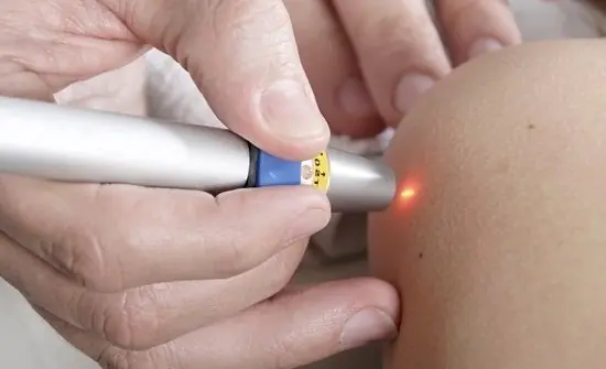
Laser removal
It is a process of evaporating benign tissue using a laser beam. The latter is configured on a strictly individual basis and does not pose any danger to humans.
Advantages of the method:
- complete painlessness
- speed of execution
- no scars or bleeding
Radio wave therapy
A relatively new method that is now just gaining popularity. Its essence lies in the effect of radio waves of a specially selected frequency on the surface of the skin.
Advantages of the method:
- non-contact influence
- absolute safety for healthy tissues
- leaves no scars
Flaws:
- Contraindicated in people with heart rate stimulants
- suitable for removing nevi with clearly defined boundaries
Electrocoagulation
Removal of a mole occurs by hooking the last one with a special loop connected to the electrodes. It allows you to carefully cut off a nevus of any diameter and size.
Advantages of the method:
- leaves no traces
- performed painlessly (under local anesthesia)
- suggests rapid recovery
Any of the above methods is contraindicated if:
- pregnancy,
- the presence of infectious diseases,
- inflammation of the skin in the area of the mole,
- and also during breastfeeding,
- presence of diabetes mellitus,
- blood clotting pathologies
- and individual intolerance to certain drugs used to remove nevi.
Only a doctor can choose the most appropriate method. It all depends on the individual characteristics of the body and the type of mole that needs to be removed.
It is very important to seek help from qualified clinics, where they perform all the necessary tests and know how to properly remove hemangiomas. The same cannot be said about simple beauty salons, the knowledge of whose employees may be insufficient to make a correct diagnosis and perform mole removal procedures properly.
