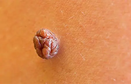
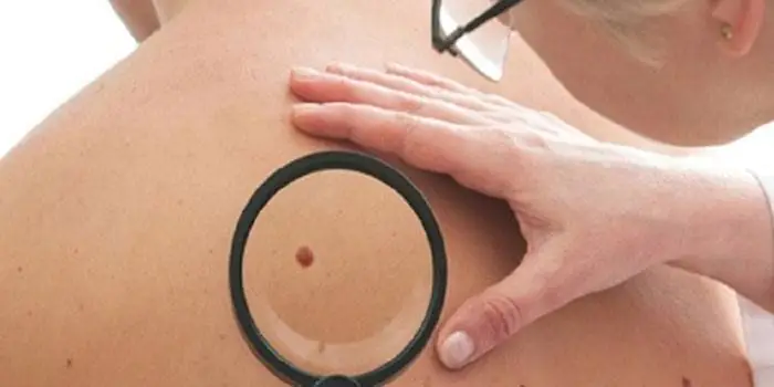
A pedunculated mole or dark spot (nevus) is a growth on the skin that hangs on a thin stalk. A mole on a thin stalk is most often dark and convex. What is the medical name for a pedunculated mole? Experts call it a hemangioma or hanging mole. This type of mole is the most insidious and dangerous. A pedunculated mole is dangerous because it is very fragile and can be easily torn off accidentally. Common places for hanging nevi to form are on the neck, face, armpits or arms, in places where it is very easy to rip or rub it.
Mole on a leg
Many patients confuse a hanging mole with a papilloma. It is really difficult to distinguish them; in order to make a correct diagnosis, the doctor must take tests and examine the tumor. The reason for the appearance of a neoplasm can be different. There are moles that you have from birth, and some that appear throughout your life. To establish the exact cause, it is necessary to undergo tests and find out the structure of the cells. You can see in more detail what hanging moles look like in a photo or video.
In what cases should you consult a doctor?
- hanging mole grows quickly
- the color of the nevus has changed
- hemangioma itches or hurts without external irritants
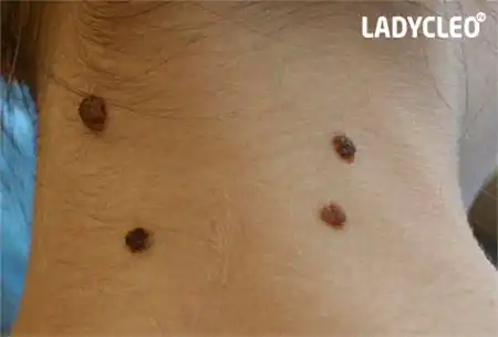
Common places where hanging moles appear
A pedunculated mole may appear on the neck. At this location, a mole can be dangerous, since the neck is most exposed to friction. Frequent friction on the body can cause inflammation. Sometimes it happens that a small mole comes off due to strong friction. If the nevus reaches a large size, then for women it seems unaesthetic. According to statistics, women are most often affected by this tumor.
It is necessary to remove a mole as soon as possible so that it does not further increase in size. You won’t be able to get rid of it at home; you need to consult a dermatologist. A hanging mole on a stalk can be removed by laser or surgical methods. Removal of a pedunculated mole takes place in specialized clinics and this procedure is not dangerous.
Armpits
In terms of the level of discomfort, the appearance of hemangioma in the armpit area is equivalent to that in the neck. There is always friction in the armpit area and the nevus is easy to catch. Removing such formations is impossible using traditional methods. You can only get rid of it using modern removal methods. The appearance of a nevus in the armpits limits you from shaving, hair removal and playing sports. Due to excessive sweating, many bacteria accumulate in the armpits. If moles are located there, they can become inflamed.
Groin area
The reasons for the appearance of a mole on a stalk in the groin area can only be determined through tests and examination by a dermatologist. In the groin area, hemangioma causes the least discomfort. This zone is considered the safest. Most often, benign skin formations appear in the groin area. The inconvenience of the location of the nevus causes discomfort mainly for women, since the appearance of moles is a direct prohibition on shaving and depilation in the bikini area.
Causes:
- changes in hormonal levels
- stress
- individual predisposition
- excessive exposure to sunlight
- various viruses
Is it necessary to remove a hanging mole?
Why are pedunculated moles dangerous and should they be removed? Many people who have nevi are not supporters of various removal methods and are afraid that by removing the tumor they will not harm themselves. Each patient has his own reason for the appearance of hanging formations, and together with the attending physician, you can decide whether it makes sense to remove them or not. Before any removal method, the nevus is checked whether it is benign or another formation.
After the results of the analysis are obtained, which also establishes the cause of the formation, the doctor decides whether to remove the mole or not. In any case, the final decision is yours and only you will decide whether to remove the nevus or not. The doctor gives you recommendations and advice regarding the course of treatment and tells you about the possible consequences. Before making a decision, weigh all the pros and cons; in some cases, leaving a nevus is even dangerous.
How to get rid of a hanging mole?
You can remove a mole on a leg using various and modern methods. Each patient, in consultation with the attending physician, chooses the most optimal method for himself. Each removal method has its own nuances. Every year, more and more people are wondering how to remove a mole on a leg quickly and effectively, so that there are no scars left in this place.
Radio wave method
Radio waves have been used relatively recently, but many customers have been satisfied with this method. The procedure lasts on average 15-20 minutes. The radio wave method can be used even if there are nevi throughout the body. Exposure of the stain to radio waves is considered a safe and effective method. Radio waves do not leave scars or scars on the body. Radio waves remove nevus without contact using a thermal effect.
With each layer, the hemangioma becomes smaller and disappears. Only a small crust remains at the site of exposure, which will disappear in 7-10 days. The radio wave method has several contraindications. First of all, removing a nevus using radio waves is not recommended for pregnant women, people suffering from diabetes, glaucoma or mental disorders. Radio waves have a negative effect on electronic sensors in the body.
Surgical method
The method of surgical intervention is considered the most radical. It is recommended to use a surgical method if the nevus has grown to a large size or the neoplasm is malignant. This method is carried out exclusively in a hospital or hospital in the presence of an experienced doctor. Compared to other methods, removing a nevus surgically is much cheaper. The essence of the method is that the surgeon removes the pedunculated mole and sutures the affected area.
The entire procedure lasts from 30 to 60 minutes, depending on the complexity and location of the nevus. The disadvantage of surgical removal is that the procedure leaves a scar or scar. When a nevus is removed, healthy tissues that are located nearby are also affected. During surgical removal, anesthesia is used, which can have various consequences for the body. This method has a number of contraindications. The surgical removal method should not be used by pregnant and lactating women, as well as people suffering from hemophilia, diabetes mellitus or infectious diseases.
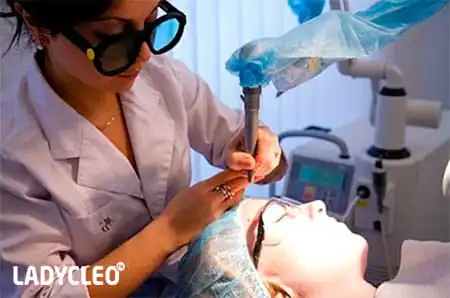
Laser removal
Removing nevi using a laser is currently becoming increasingly popular. The fact is that laser removal is considered the least traumatic and dangerous. The laser acts directly on the center of the hanging mole and removes it layer by layer. A laser is used to remove small or medium-sized nevi. A non-contact laser beam guarantees protection against infections and bacteria entering the wound. Compared to other methods, laser removal is considered expensive, but the most painless and effective. Laser removal is suitable for many patients, as it does not have many contraindications.
Please note that laser removal does not leave any scars or scars.
Choosing the right doctor and clinic
Whatever treatment method you choose, the most important thing is a qualified specialist. In order not to make a mistake with your choice, it is recommended to study all the reviews about this clinic and look at photos of patients before and after treatment. Trust your health only to experienced doctors in large clinics, where they are required to have a license and other necessary documents. Listen to the advice of a professional; if the doctor insists that a pedunculated mole should be removed, then perhaps he knows what is best for you.
Every person has their own set of moles on their body, some of them are hereditary, others were acquired after birth. Often moles look pretty and are a unique distinguishing feature. But not all of them can be safe, in particular, a pedunculated mole is one of the dangerous types of this neoplasm.
What is such a mole?
A mole (nevus) is a new growth of the skin, usually dark in color and slightly convex, but there may be variations. Moles can be harmless, but there are often cases when a malignant tumor forms from an inconspicuous spot on the skin.
A pedunculated nevus is also called a hanging mole or hemangioma. It is considered the most dangerous among all other species. Firstly, this is due to the likelihood of it rubbing with clothing or jewelry, which can lead to its degeneration into a malignant neoplasm. Secondly, such a mole can be very easily confused with a papilloma, and this is already a serious viral disease; they can be distinguished from each other only after testing; the difference cannot be seen with the naked eye.
When a new hemangioma forms, it is advisable to immediately consult a dermatologist to find out the nature of its occurrence and the degree of danger it poses. If, due to some circumstances, a visit to the doctor is impossible, the following factors may be alarm bells for which you must make time:
- the mole began to actively grow;
- the shade has changed;
- without external influences, she began to itch or hurt;
- noticeable bleeding;
- inflammation or suppuration has appeared;
- the mole has become denser.
In this case, you should pay attention only to large neoplasms; flat spots can be safely ignored. They can only interfere cosmetically if they are somewhere in a visible place.
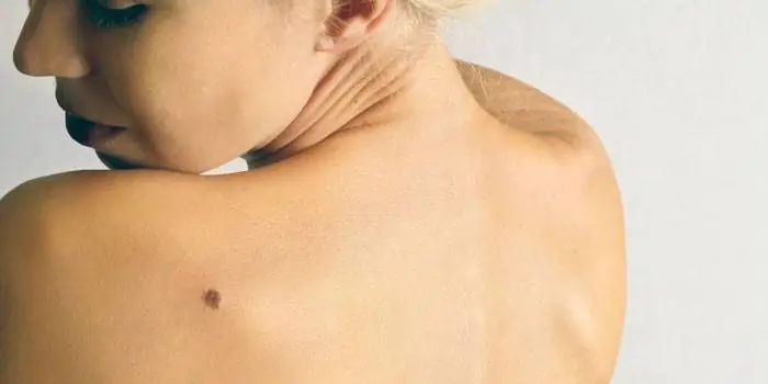
Location on the body
A pedunculated nevus can be located on any part of the body, but there are several common areas where it occurs.
Here, hanging moles pose the greatest discomfort and danger, since frequent friction is possible; in this area, such moles often degenerate into malignant tumors.
In addition, if the mole is large, it may look unaesthetic, forcing the girl to disguise it in every possible way. The female half of the population is more likely to have such a tumor than the male half.
Armpits
In terms of the degree of discomfort and danger, hanging moles in this area are almost as good as their counterparts on the neck. Their formation imposes restrictions on epilation of the area and active sports.
The armpits are the most favorable area for the formation of bacteria, so if a nevus appears on the leg, you must consult a specialist.
Groin area
Here, hemangiomas determine the least discomfort, since the danger is created only for girls who epilate the bikini area. But you can choose the procedure in such a way as not to injure the tumor in any way.
Removal in a cosmetology clinic
If a mole has become a problem that needs to be removed, it is better to seek professional help. Modern cosmetology does not stand still, so it can offer several different options for removing hemangiomas.
Laser removal
The effect of lasers has already firmly established itself in most areas of cosmetology, since this gentle effect effectively eliminates various problems without leaving any traces. This method is ideal for removing moles, since it is absolutely painless and safe, and the laser does not leave scars.
Depending on the individual characteristics of the patient, a specific laser beam is selected. It is directed to the mole, and it begins to gradually evaporate layer by layer. At the same time, the laser acts on the blood vessels, cauterizing them to eliminate the possibility of bleeding. After the procedure, only a thin crust remains at the site of the mole; within a week it peels off, and you can see clean skin without a trace of the mole.
Contraindications to the laser mole burning procedure:
- Excessively sensitive skin. In this case, redness, irritation, blisters and burns may occur after exposure to the laser beam. The same principle applies to sun exposure of the skin of people with allergies to ultraviolet rays.
- In people with a tendency to form pigment spots of various natures, they may form after the procedure.
- The presence of inflammation on the skin and unhealed damage in the area of laser exposure.
- Only a mole whose diameter does not exceed three centimeters can be removed with a laser.
- Pregnancy and lactation period.
- Diabetes.
- painlessness;
- lack of anesthesia, which eliminates the possibility of a negative reaction of the body to it;
- the likelihood of a new mole forming in the same place is minimal;
- weak rehabilitation period;
- absence of visible signs of a mole after removal;
- the likelihood of infection is very low.

Electrocoagulation removal
This method also does not involve damage to the skin, so it cannot help but gain popularity. It is based on the effect of high-frequency current on the mole; the procedure is painless, but slightly unpleasant sensations still occur. The procedure is as follows: the doctor administers local anesthesia, when it begins to take effect, he cuts off the mole with a special metal loop, which is under the influence of current. The loop not only cuts off the tumor smoothly, but also immediately affects the wound, preventing bleeding. As with the laser, only a crust remains at the site of the mole, which peels off within a week.
The following factors are contraindications to electrocoagulation:
- Pregnancy and lactation period.
- If the neoplasm is malignant.
- Inflammation and infectious dermatological diseases.
- Diseases affecting blood clotting.
- Diabetes.
- Intolerance to procedures based on the influence of current.
This method of removing hanging moles has some advantages:
- the procedure takes no more than 20 minutes;
- only one session is required to achieve the final result;
- there are no visible consequences of the procedure;
- absence of bleeding, which eliminates the possibility of postoperative shock, blood loss and infection;
- Even miniature nevi can be removed.
Cryodestruction method
In simple words, this procedure can be called freezing a mole. To do this, it is exposed to low-temperature liquid nitrogen, after which it is rejected by the body and peels off. Cryodestruction can be performed by two methods - hardware and manually. The choice of method is based on the degree of the problem; if the mole has not penetrated into the deeper layers of the skin, it is removed with a regular cotton swab soaked in nitrogen.
Contraindications to the procedure:
- Inflammation and unhealed damage in the affected area.
- Infectious diseases accompanied by fever.
- Malignant tumors.
- Individual intolerance to the method.
Among the advantages of cryodestruction the following can be noted:
- exposure time is no more than 3 minutes;
- painless, the patient feels only slight numbness;
- immediately after the neoplasm is rejected under the influence of cold, the wound heals, preventing bleeding and the possibility of infection.
Radio wave method
Radio waves began to be used in cosmetology relatively recently, but they have already acquired an impressive list of satisfied clients. This can be explained by their gentle effect and safety. This technique involves non-contact removal of tumors using the warm effect of high-frequency waves. Layer by layer, the mole evaporates, leaving only a barely noticeable crust at the site of the former nevus, which peels off within a week.
This method also has contraindications, including:
- Pregnancy and lactation period.
- Inflammation and infectious rashes in the affected area.
- Malignant tumors.
- Diabetes.
- Epilepsy.
- Glaucoma.
- The presence of heart rate sensors in the body.
The advantages of the radioknife method include the following factors:
- the procedure takes on average 20 minutes;
- absence of unpleasant and painful sensations;
- eliminates the possibility of bleeding;
- no visible consequences from removal;
- easy rehabilitation period;
- the ability to remove a mole on any part of the body.
Surgical method
The most radical way to solve any cosmetic problem has been and remains surgery. It can be carried out both in a state oncology clinic and in any private cosmetology clinic. The essence of the operation is as follows: healthy skin around the mole is grabbed, and it itself is excised, after which a suture is applied. The entire procedure takes from 30 to 60 minutes, the time depends on the characteristics of the individual patient.
Unlike hardware methods, surgical methods have several disadvantages. Firstly, damage to healthy tissue occurs, which does not always have a beneficial effect on the overall situation. Secondly, general anesthesia is used, and not all patients tolerate it easily, even if a preliminary test did not reveal any contraindications to the drug. Thirdly, noticeable scars may remain from the mole, which does not always look aesthetically pleasing.
Contraindications to this method are standard for many operations:
- Pregnancy and lactation period.
- Diabetes.
- Hemophilia.
- Infectious diseases.
- Inflammation in the affected area.
The advantages of the surgical method include the following factors:
- the ability to remove a mole of any size;
- malignant neoplasms can be removed;
- comparatively lower cost of the procedure.
Whatever decision you make, you should take care of choosing a clinic and a doctor who will perform the removal. It is better to rely on patient reviews and their photos from the specialist’s portfolio. Although a mole may look like an inconspicuous problem, improper removal can lead to serious consequences, so you should only trust experienced cosmetologists or surgeons.
In addition, you should not make the decision alone; the specialist you choose must conduct a preliminary consultation, collect your medical history and identify possible contraindications to a particular method. Only based on your individual characteristics and the results of the tests taken, the doctor can make an informed decision about prescribing a specific procedure.
It's rare to see a person without small dark marks on their body. Is it worth paying attention to these points? Only a doctor will distinguish between dangerous and normal moles - malignant melanoma or harmless nevus - and give recommendations on what to do with them. Is it worth worrying about the appearance of new formations, when immediate contact with specialists is required, what are the signs of cancer development - the answers to these questions remain to be found out. No one is immune from disaster, and early diagnosis will protect you from severe consequences.
What is a mole
The first tiny spots may appear in children in infancy. A mole is a small formation on the skin - a nevus - that is considered benign and harmless. The basis for their appearance is melanocyte cells that accumulate the natural pigment melanin. Depending on its quantity, a difference in color is observed. Available colors:
The shape of the tumors depends on the location and concentration of melanin. They may have a stalk or be located under the skin, be flat and convex. The most common type is round, but there are exceptions. The development of neoplasms is provoked by ultraviolet radiation - natural from the sun, in a solarium. Hereditary factors cannot be excluded. A common cause of growth is hormonal imbalance, characteristic of periods:
- puberty;
- pregnancy;
- menopause.
What types of moles are there?
One person may discover very different tumors. Types of moles are classified according to several criteria. This helps in correct diagnosis in case of changes. They differ in:
- origin– congenital, newly acquired;
- structure– pigment, vascular;
- place of education – in depth, on the surface, in the boundary layer;
- raised above the skin – flat – even, protruding as a hemisphere, pedunculated, larger birthmarks;
- potential threats – dangerous, degenerating into melanoma, non-dangerous.

Safe moles
Those who have dark spots on their skin should be wary of their changes. In time, detected signs of degeneration into melanoma contribute to the timely removal of the formation and preservation of health. Safe moles are different:
- the presence of a stalk – it cannot be formed by malignant cells that grow randomly;
- long-term condition without changes.
Spots that appear soon after birth are not considered dangerous. It is important that they are small in size. Good – non-dangerous – signs of neoplasms include:
- flesh tone;
- unchanged pattern of the skin of the nevus and adjacent tissues;
- soft consistency;
- hair on the surface of the neoplasm - growing from the skin, indicates the absence of pathologies;
- diameter no more than 5 mm;
- symmetry;
- nevus in the form of a spot.
Which moles are dangerous?
Why do people with nevi on their bodies need to monitor their changes? There is always a threat of degeneration of non-dangerous tumors into a cancerous tumor. What moles are dangerous to health? Key signs you need to know:
- change in shades towards the dark side, the appearance of multi-color;
- rapid increase in size - exceeds two millimeters per year;
- occurrence of cracks;
- the formation of asymmetry due to uneven growth;
- lack of elasticity;
- the appearance of itching, burning;
- presence of discomfort.
The appearance of dangerous moles requires an immediate visit to a specialist to clarify the nature of the changes and the likelihood of developing skin cancer. Pathological transformations provoke:
- injury to the nevus due to negligence;
- self-removal;
- abuse of exposure to the sun, use of a solarium;
- location of the formation in places of frequent contact with clothing - on the neck, head, genitals, legs;
- placement in the hair, on the face, palms - where there is a high probability of injury;
- previously removed melanoma.
Why are moles dangerous?
Not a single person is protected from the sudden proliferation of cells of a harmless mole. Melanoma is an extremely serious disease. Changes not detected at the initial stage can result in death. The provoking factor is unsuccessful independent removal of tumors. Moles are dangerous because of their ability to:
- transform into an atypical – precancerous form;
- grow to large sizes;
- turn into cancerous;
- with minor external changes, metastases actively spread throughout the body through the circulatory and lymphatic channels.
How quickly does melanoma develop from a mole?
The transformation of a nevus into a cancerous formation can occur in different ways. The process depends on the stage of the disease and the type of tumor. Instant metastases are dangerous. Begins:
- growth of cancer (oncological) cells in the deep layers of the epidermis;
- their entry into the blood and lymph;
- penetration into the lungs, liver, kidneys;
- growth in these organs;
- complete damage to the body;
- death.
The growth phases of pigment cells are observed, along which melanoma develops from a mole. There are varieties:
- horizontal– damage to the upper layers of the skin occurs, lasting up to 10 years, but metastases do not appear;
- vertical– accompanied by the spread of cancer cells throughout the organs, can last two years, has an unfavorable prognosis;
- nodal – especially dangerous – characterized by deep spread within two months.
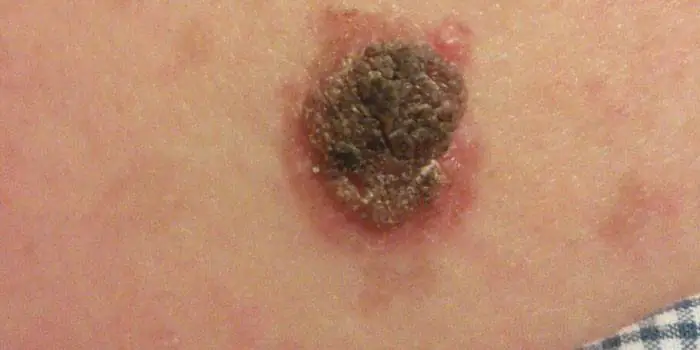
The first signs of melanoma
The patient can be assisted only when suspicious changes begin to be identified. The diagnosis, research, and referral for surgical treatment save a person’s life. The first signs of melanoma:
- increase in the height of the tumor;
- bleeding;
- the appearance of discharge;
- redness;
- burning, itching;
- swelling of tissues;
- softening of the nevus;
- the appearance of a crust;
- thickening;
- hair loss;
- expansion of pigmentation around the lesion.
With the further development of dangerous melanoma, the following are observed:
- significant change in size;
- the appearance of pain;
- enlarged lymph nodes;
- surface ulceration;
- formation of new foci;
- bleeding from places of pigmentation;
- liquid separation;
- skin thickening;
- the appearance of an earthy tint;
- signs of metastases are chronic cough, weight loss, cramps, headaches.
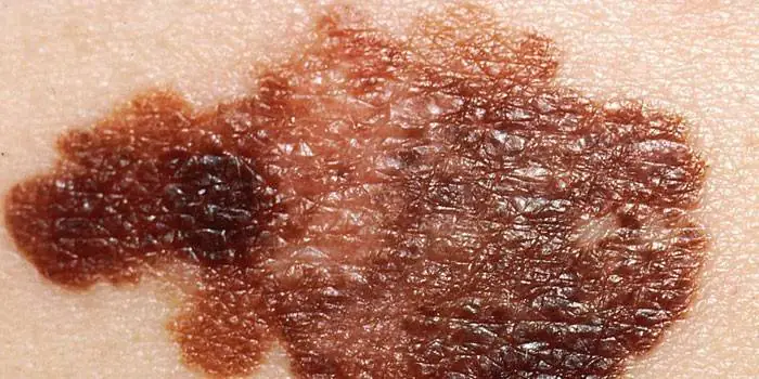
How to distinguish a mole from melanoma
To recognize which moles are dangerous and which are not dangerous, you need to know what they look like. A person with nevi, in order to eliminate terrible consequences, must constantly monitor the appearance of new formations and changes that occur. You can distinguish a mole from melanoma by its signs. Non-dangerous neoplasm:
- symmetrical;
- with smooth edges;
- uniform in color;
- with dimensions not exceeding 6 millimeters.
Features of dangerous melanoma that require seeking help from dermatologists:
- growth in a short time;
- pronounced asymmetry of shape;
- heterogeneity in color - the presence of inclusions of several shades;
- lack of clear boundaries - the contour line is blurred, jagged, and looks like a coastline on a geographical map;
- increased diameter over six millimeters;
- variability of any parameters - color, size, shape.
What dangerous moles look like
What do nevi that are subject to pathological changes look like? Only a doctor can correctly distinguish between non-dangerous tumors. Dangerous formations look like this:
- blue– compactions under the skin with clear boundaries, with dimensions no more than 10 mm;
- nodal– round, flat in shape, color – brown, black;
- cutaneous– often pale, convex;
- halo nevus – pigment surrounded by a light or white rim;
- spitz- looks like a dome-shaped tumor of pink shades, with the possible presence of a hole through which blood and liquid leak;
- connecting- connect individual entities into a whole.
Mole with jagged edges
One of the signs of a non-hazardous formation turning into a dangerous one is a change in contours. It often has blurred edges and scalloped borders. There are non-dangerous types of nevi - dysplastic. Only a specialist can make a correct diagnosis. A mole with uneven edges can be dangerous if there are additional signs of melanoma:
- accelerated changes in size;
- the presence of clearly defined asymmetry;
- the appearance of highly indented boundaries.
Rough mole
Such a neoplasm is harmless if its diameter is no more than 5 mm and remains constant in size. Often its appearance signals a lack of vitamins and nutritional disorders. Doctors advise coming for a consultation if it is discovered that:
- the smooth nevus turned into a rough one;
- bothered by burning, itching, tingling;
- irregularities and compactions appeared in the middle;
- areas with different shades formed;
- diameter has increased significantly.
A dangerous rough mole requires immediate examination if:
- the appearance of bleeding;
- development of the inflammatory process;
- rapid change in size;
- formation of asymmetry;
- formation of purulent discharge;
- the occurrence of painful sensations when touched;
- the emergence of an irregular shape, blurred boundaries, along the edges of the neoplasm.
Large moles
Large formations on the skin are pigment spots. When they remain unchanged and do not cause inconvenience, this is a harmless phenomenon. It is important to constantly monitor their appearance, color, and size. To eliminate worries, you need to consult a dermatologist. During the visit, the specialist will conduct a diagnosis and give a forecast of the risk of developing a malignant neoplasm. Large moles become dangerous if they:
- injured;
- thickened;
- started to itch;
- were unsuccessfully removed independently;
- changed in size, shape;
- are bleeding.
What moles can be removed
Often nevi cause trouble for women when they are in a visible place - the face, neck. Even if they do not bother you, using removal will be the right decision - the appearance will improve significantly. After the procedure, the doctor must send the tissue for histological analysis to decide whether the mole is malignant or not. If the neoplasm is not dangerous, does not bother you, and does not change in size, surgery is not required. What moles cannot be removed? Experts believe:
- there are no contraindications;
- It is important to choose the right excision technique.
You should be careful about skin growths; it is unacceptable to remove them yourself. Only the doctor will determine whether a nevus is dangerous or not and decide what to do with it. You can delete it if:
- injured from clothing - on the neck, in the groin area, under the armpits;
- cause pain when touched;
- are located under the hair on the head and can be damaged when combing or cutting;
- change color, shape, outline;
- significantly increase in size;
- characterized by the presence of burning, itching;
- accompanied by inflammation and bleeding.
