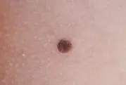
Moles: what are they?
Nevi are pigmented or vascular formations on the skin. They appear throughout life. A sharp increase in body size can be caused by various changes in the body, be it hormonal imbalances, pregnancy, certain diseases, long-term treatment with antibiotics, or constant exposure to the sun without protective cream. In most cases, emerging moles are not dangerous. And the desire to get rid of them is of an aesthetic nature, or a discomforting sensation.
Experts divide all moles into three types:
— Quiet ones are nevi that do not pose a danger. They are of normal size, up to 5 mm, with a smooth surface and uniform color. They do not grow, do not change, and do not cause physical discomfort. There are almost 99% of such moles on our body. The formation can be removed if the patient thinks that it looks unsightly.
— Borderline moles are moles that can potentially pose a threat to human health. However, only a doctor can determine exactly how dangerous they are. By external signs, degeneration can be determined:
- by rapid growth;
- color change;
- pain on palpation;
- itching and peeling.
If you notice such signs, you should consult an oncologist. After this, removal of the suspicious tumor will be scheduled.
— A dangerous mole is a formation that has already degenerated into skin cancer and requires immediate treatment. Such moles turn into ulcers, they bleed and hurt. For more information about what melanoma is and how to treat it, read our article “Skin cancer: melanoma.”
The doctor decides whether a particular mole needs to be removed. It all depends on how it behaves, where it is located and whether it is possible for it to degenerate into a malignant tumor.
Is it dangerous to remove moles?
Yes, it is dangerous if you decide to get rid of it at home using folk remedies without consulting a doctor. This can cause irreparable harm to your health. Because damaging a nevus and not completely removing it can provoke degeneration into melanoma. It is impossible to ensure 100% sterility at home and there is a risk of infection in the wound, which will lead to inflammation. It is also dangerous to remove tumors in unverified private beauty salons. In such a place, the doctor may not have special qualifications and, along with the procedure, may also cause harm to the body.
If you decide to get rid of a growth on your body, then this can only be done safely in a specialized clinic, with a certified surgeon or dermatologist. Read about what steps you need to take before removing a mole in our article “I want to remove a mole: where to start?”
In itself, excision of a nevus is nothing terrible. It is important for a doctor to take biomaterial and send it for histological examination to find out for sure whether there are cancer cells. If they are detected, the patient will be prescribed additional examination and treatment.
If the mole was removed only for aesthetic reasons, then there is no threat to health. Although doctors do not recommend this procedure if the tumors do not cause discomfort. Because, despite the low invasiveness, it is still an operation.
During pregnancy, many new formations may appear. Women are afraid of undergoing procedures at this time. Doctors also recommend waiting until after the baby is born. If the mole continues to interfere after childbirth, then you can part with it. You can learn more about this topic in our article “Moles during pregnancy.”
How to safely remove a mole?
Today, there are several safest methods for removing tumors. Among them:
— Laser therapy is one of the most effective procedures for getting rid of unwanted growths on the body. With its help, formations such as:
The laser gently acts on the skin without damaging healthy tissue. Only the neoplasm itself is destroyed. There is no risk of infection, since the beam coagulates the vessels. The advantages of this method include:
- Painless;
- Speed of implementation;
- Short rehabilitation period;
- There are no scars or scars left on the skin.
More information about how the procedure is carried out can be found in the article “Laser removal of benign skin tumors.”
— Cryodestruction - scientists have discovered that low temperatures of liquid nitrogen can be used to remove various formations, including moles. The process is simple and painless. A cotton swab soaked in nitrogen is applied to a small nevus, and for large growths a special apparatus with a needle is used. Liquid nitrogen acts instantly, freezing cells and preventing them from developing further. The removal site is covered with a crust, which subsequently separates itself. The disadvantage of the procedure is the hole that remains after the procedure. More details about cryodestruction can be found in our special article “Removing tumors. What is better: laser or liquid nitrogen?
— Electrocoagulation is the use of high-frequency alternating current, but you should not be afraid of it, since the procedure takes place under anesthetic. Only the cells of the neoplasm are damaged, water evaporates from them, so their further development is impossible. A crust appears at the removal site; it should not be torn off or scratched; it will fall off in a few days. However, a small white spot may remain on the skin. Find out more about electrocoagulation in the article “What is better to remove tumors: laser or electrocoagulation?”
- Radio wave method - an electrode emitting high-frequency radio waves is used here. With their help, you can excise a mole at its base. In this case, coagulation of the blood vessels occurs, which significantly reduces the risk of introducing bacteria into the wound. After the procedure, swelling and redness are possible, which will subside within 1-2 days. Read more about the radio wave method in the article “Laser or radio wave removal of tumors.”
All of these methods are carried out under the supervision of a doctor and using modern technologies. That's why they are safe. It is possible, and in some cases even necessary, to remove moles and other neoplasms, the main thing is to choose the right specialist and choose the method.
Probably, each of us has heard a story about someone who removed or injured a “mole” and died a month later. Let's figure out what is true in this story and what is from the realm of “horror stories”.
There really is such a tumor as melanoma. It develops from pigment cells of the skin; in rare cases, melanoma may develop in the cells of the membranes of the brain or eye. Old school oncologists call melanoma the “queen” of tumors. In stages 3 and 4, the patient's death occurs in 80-90% of cases. But we should also not forget that with stage 1 of the disease, with adequate treatment, the patient’s recovery can be guaranteed in 95 cases out of 100.
As already mentioned, in most cases, melanoma develops from pigmented nevi or what are popularly called “moles.” A pigmented nevus is a cluster of pigment cells - melanocytes, which on the skin look like formations from light brown to black-blue. It is also necessary to pay attention to the fact that there are a lot of other formations of different colors on the skin that have nothing to do with pigmented nevi and melanomas. In many cases, only an oncologist can determine their nature.
What are the predisposing factors that cause a harmless “mole” to turn into melanoma?
1. Constant trauma to the nevus, especially when it is located in areas of friction with clothing.
2. The presence of a large number of pigmented nevi on the human body (the risk of developing melanoma increases several times if there are more than 15 pigmented nevi on the human body).
3. Prolonged exposure to ultraviolet radiation (for example, in Australia, skin melanoma is an occupational disease among people working outdoors). Solar insolation is especially dangerous for children and adolescents.
Let's look at the first signs of malignancy of a pigmented nevus, or what should make you urgently contact an oncologist:
1. Itching and peeling in the area of the nevus.
2. Change in the size, shape, color of the nevus, and the color can change in both dark and light directions.
3. Bleeding and destruction of the nevus.
4. The appearance of a blue or purple rim around the “mole”.
5. The appearance of small brown or black formations around the main tumor.
Remember that timely consultation with a doctor will save your life.
If the patient does not consult a specialist when the first symptoms appear, the malignant transformation of the nevus will take its course, in accordance with the biological laws of tumor growth. Manifestations of the disease will sooner or later lead the patient to the doctor, but as already mentioned, at 3-4 tbsp. recovery occurs in only 5-20% of cases. There will be another reason to tell your friends a terrible story that after the removal of a “mole” a person died.
Measures to prevent malignant transformation of pigmented nevus:
1. Wearing loose clothing that does not compress the nevi.
2. Avoid exposure to ultraviolet radiation on nevi (when in the sun, cover the “moles” with an adhesive plaster or use sunscreen with a protection factor of at least 50 IU).
3. Preventive removal of nevi (it is especially recommended to remove nevi in areas of friction with clothing and in open areas of the body).
A radically removed nevus will never become a source of melanoma development.
Methods for removing nevi:
1. Surgical method: includes excision of the formation with a scalpel followed by suturing. The most reliable method for removing any skin formations. Allows you to radically remove the formation and obtain material for histological examination. The method is well suited for removing large formations, especially on the body. When used on the face, it can cause significant cosmetic defects.
2. Cryodestruction (removal with liquid nitrogen): a good method for small formations, as well as for the removal of vascular tumors in children. Not a bad cosmetic effect. Disadvantages - it is impossible to obtain material for histological examination.
3. Electrocogulation (removal using an electric loop). The method is quite easy to use, bloodless, there are no stitches, a short rehabilitation period, it is possible to take a biopsy. Disadvantages - poor cosmetic effect: white spots remain, high risk of keloid scars.
4. Laser destruction (removal using a high-energy laser). An excellent method for removing small skin formations. When removing large nevi, there is a high risk of scar formation. It is difficult to take material for a biopsy.
5. Radio wave excision (removal using devices generating high frequency radio waves). Allows you to remove both small and large formations. Very good cosmetic effect. The risk of scarring is minimal. Short rehabilitation period (10-15 days). Allows you to take a biopsy.
In any case, no matter what method you choose to remove a nevus, it is important that it be done by a specialist, preferably an oncologist or a dermatologist with oncological training. Remote lesions must be sent for histological examination. You also need to know that pigment formations are always present. are completely removed. and then sent for histological examination. It is unacceptable to remove the pigmented nevus in parts, because if there are areas of malignancy in the nevus, this will be enough for metastasis.
If a histological examination reveals areas of a malignant tumor in a remote formation, then within 1 month from the date of intervention it is necessary to perform surgical excision of the formation with sufficient deviation from the edges of the tumor. This will prevent recurrence of melanoma.
Do not forget to contact specialists in a timely manner, even if your worries are completely groundless.
A mole is an acquired or congenital formation on the body, accompanied by a change in skin color (pigmentation). Birthmarks can have different diameters and shapes. For many patients, the urgent question is: what is the danger of removing moles? After all, many have heard frightening stories about the development of skin cancer after removing or damaging a spot. Is this really true?
Causes of moles and indications for removal
A birthmark (mole, nevus) is a benign formation on the human dermis. The mechanism of development of moles is the degeneration of dermal cells into melanocytes (skin cells that synthesize the pigment melanin).
Causes of nevi:
- The influence of ultraviolet radiation. Prolonged exposure to the open sun not only contributes to the formation of new birthmarks, but also causes the risk of transformation of benign formations into malignant ones. According to statistics, people whose skin is regularly exposed to ultraviolet radiation are more likely to suffer from cancer.
- Hormonal imbalance in the body. It has been proven that hormonal surges often provoke the appearance of new nevi or their disappearance.
- Skin injuries lead to the formation of certain types of formations.
- Infectious diseases. In children and adolescents, spots often occur due to damage to the body by viral or bacterial infections.
- Hereditary predisposition. If parents have many birthmarks on their skin, the baby is also more likely to develop nevi.
- Other diseases. Pathologies of the thyroid, pancreas, liver, vitamin deficiency, radiation exposure, and hormonal disorders can provoke the development of moles.
The danger of moles lies in their ability to degenerate into malignant tumors. Experts advise paying close attention to birthmarks and regularly visiting an oncologist and dermatologist.


