I myself have a lot of moles and this is not a problem for me. there are those that appeared overnight and over several days. the most important thing is that they do not interfere.
and the idea that many moles are bad is complete nonsense. everything's OK. they should be proud.
I was putting the dishes away in the closet and suddenly felt a sharp pain in my palm. I still didn’t understand what caused this pain, since I didn’t hit myself. and the pain was strange, which I had never experienced before. After a few seconds it was all over. And the next day I discovered a mole on my palm. Before, I didn’t even know that they could be in the palm of your hand. 3 weeks have passed. Nothing bothers me. But I still didn’t understand why she appeared. Has anyone had anything like this??
Woman.ru experts
Find out the opinion of an expert on your topic
Juran Marina VladimirovnaPsychologist, Family child psychologist. Specialist from the site b17.ru
Korotina Svetlana YurievnaPsychotherapist. Specialist from the site b17.ru
Pukemova OlgaPsychologist. Specialist from the site b17.ru
Kostenich Lyudmila StanislavovnaPsychologist, Art therapist. Specialist from the site b17.ru
Marina Aleksandrovna BaydyukPsychologist, Analytical psychologist. Specialist from the site b17.ru
Valeria Bertnik-YuryevaPsychologist, Psychologist-guide. Specialist from the site b17.ru
Nevzorova Sofya IgorevnaPsychologist. Specialist from the site b17.ru
Alina SysoevaPsychologist, Coach and Trainer. Specialist from the site b17.ru
Ekaterina Alekseevna VasyukhinaPsychologist, Crisis counseling. Specialist from the site b17.ru
Spiridonova Nadezhda ViktorovnaPsychologist. Specialist from the site b17.ru
not a mole, but a subcutaneous bruise. Since the skin on the palms is thick, the bruise may not go away for years.
This is a small hematoma - you can carefully pick at the skin with a needle and pull out this lump of blood. Don’t forget to burn it with alcohol afterwards.
This is a small hematoma - you can carefully pick at the skin with a needle and pull out this lump of blood. Don’t forget to burn it with alcohol afterwards.
not a mole, but a subcutaneous bruise. Since the skin on the palms is thick, the bruise may not go away for years.
and if it appeared on the butt, you wouldn’t even notice and there wouldn’t be any “problems”
I read on the Internet that there are moles on the palms. It doesn’t look like a subcutaneous bruise... just like a mole(
Related topics
I read on the Internet that there are moles on the palms. It doesn’t look like a subcutaneous bruise... just like a mole(
Thanks for answers. Only a month has passed and nothing is happening.
This is how I got a mole on my palm, it’s been there for two years now.
appeared and appeared, I didn’t bother, but people are very surprised, they say they have never seen it, they see a secret sign)))
Here is the version of the occurrence - hematoma, very similar and probable.
Everyone here is smart, I didn’t even think of this version)))
Yes, my moles appeared unexpectedly, and on different parts of the body. And once on my finger. It just didn’t hurt when it appeared, but itched. I’m doing something, I feel it itching, I touch my hand - and suddenly there is redness, it immediately subsides and an ordinary mole remains, which was not there before. The usual thing. All those moles are still stable and appear normal. And no hematomas, that’s how they appeared suddenly.
This makes men's hair grow; women have moles. you need to have sex!
What are you doing? You can't touch this!
author, for reference, palm and washed. try holding Rosenthal's dictionary to your head, it helps
The palms and feet do not tan precisely because they cannot produce melanin. Any mole is its hyperconcentration, because they are from excessive tanning and spread all over the body. How can moles appear on the palms, can someone answer me?
As already written above, an ordinary hematoma,
They probably pinched it with something and didn’t notice, hence the sharp pain.
I pinched a piece of my finger like this in childhood, a very small spot appeared that looked like a mole, I thought it would stay forever, but then it all went away and the “mole” disappeared.
Thank you. I wish it were a hematoma
I don’t know, I have a mole on my little finger. Dark brown. As they already wrote here, it didn’t hurt before, but itched
You have developed stigmata. The second coming is just around the corner.
moles appear without pain
Maybe you got yourself a scoop (I don’t know what you call it)?
If not an organized blood clot, then variants of HPV, mycosis fungoides. A heavy dose of nicotinic acid and ascorutin will not hurt.
Well, if it really is a mole, then I constantly find new ones. Only today it wasn’t there, but tomorrow it’s there. But they don't hurt.
About 2 years ago, a mole appeared on my foot, it was also a very unusual place, I was worried that I would damage it, I went to TWO oncologists - in unison they said not to touch it, it’s subcutaneous, it’s okay, just observe.
I'm writing a sequel. Today the “mole” fell off. Many here were right - it would NOT be a mole))
Complaint
Moderator, please note that the text contains:
The complaint has been sent to the moderator
The page will close automatically
in 5 seconds
Forum: health
New for today
Popular today
The user of the Woman.ru website understands and accepts that he is fully responsible for all materials partially or fully published by him using the Woman.ru service.
The user of the Woman.ru website guarantees that the placement of materials submitted by him does not violate the rights of third parties (including, but not limited to copyrights), and does not damage their honor and dignity.
The user of the Woman.ru site, by sending materials, is thereby interested in their publication on the site and expresses his consent to their further use by the editors of the Woman.ru site.
Use and reprinting of printed materials from the woman.ru website is possible only with an active link to the resource.
The use of photographic materials is permitted only with the written consent of the site administration.
Placement of intellectual property objects (photos, videos, literary works, trademarks, etc.)
on the woman.ru website is permitted only to persons who have all the necessary rights for such placement.
Copyright (c) 2016-2019 Hirst Shkulev Publishing LLC
Online publication “WOMAN.RU” (Zhenshchina.RU)
Certificate of registration of mass media EL No. FS77-65950, issued by the Federal Service for Supervision of Communications,
information technologies and mass communications (Roskomnadzor) June 10, 2016. 16+
Founder: Limited Liability Company "Hurst Shkulev Publishing"
Many people are not interested in the meaning of moles on the body, and in vain, because these formations on the skin can be very dangerous for the body. There are many types of such formations, they differ in shape, size, color. Some people have a huge number of them, others only a few, but everyone has at least one. Read why these formations appear on the skin, how to understand which of them are dangerous, and learn about effective removal methods.
What are moles
Every person should know the nature of formations, which in scientific language are usually called nevi. Moles are a concentration of melanocyte cells. Melanin, the pigment that determines color, is concentrated in them. They come in different shades of brown, black, red, yellow and even purple. According to their shape, they are classified as flat, convex, hanging, lumpy, or with a stem.
Moles are of a similar nature to birthmarks. The difference is that the former can appear, change and even disappear throughout life (the most active period is from six months to 25 years), while the latter are given to a person from birth. They can be located on any part of the skin: both the face and the body. There are cases of their occurrence on mucous membranes.
Why moles appear
You have already read that pigmented formations can appear and disappear at any age, but what does this depend on? Factors that provoke the appearance of moles on the body:
- Heredity. Often in children, nevi appear in the same areas as in their parents, and sometimes in even greater numbers.
- Sun rays. Melanin under their influence is produced several times more intensely. Staying in the sun for a long time is dangerous not only because new ones can form, but also because old ones can transform into a tumor, even malignant.
- Viruses, injuries, radiation, x-ray exposure. Under the influence of each of these factors, melanocytes can group and come to the surface of the skin.
- Hormonal changes. Any surge in hormones (especially in women) can trigger the appearance or disappearance.
- Cluster of blood vessels. Nevi occur due to the accumulation of small processes of blood vessels.
- Dysfunctions of internal organs and systems. More often they lead to the appearance of vascular nevi. They can develop due to dermatological diseases, dysfunction of the large intestine, pancreatic dysfunction, and imbalance of lipid metabolism.
Reasons for the appearance of moles on the body in women
The occurrence of nevi is directly related to hormonal surges, of which there are plenty in the female body. The reasons for the appearance of moles on the body in women can be changes in the body associated with pregnancy (nevi often form on the skin of the abdomen, legs), menopause, and puberty. Sometimes, although rarely, they occur before or during menstruation.
How moles appear
Skin cells become more and more melanin pigment and transform into melanocytes. This occurs under the influence of one of the causes of nevi listed above (sun exposure, hormones, etc.). The accumulation of melanocytes is the reason why moles appear on the body. Whatever the nature of the nevus, the mechanism of its development always looks like this.
Types of moles
Formations can look very different, but according to a number of characteristics they are usually combined into several groups. The following types of moles are distinguished according to the nature of their occurrence:
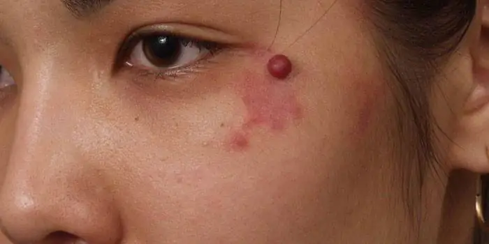
- Angiomas. Formed due to pathological transformation of blood vessels. They can be flat or convex, pink, red, purple. They will never become malignant.
- Hemangiomas. A type of angioma. They appear shortly after birth in a child, gradually turn red and slightly swell, and have clear boundaries. Most often localized on the neck and face.
- Vascular malformation. There are two types of defect. The first is called port-wine stains and affects the torso, face, and arms. At first they are pale pink, but then they become scarlet or crimson, and with any dilation of the blood vessels they acquire brightness. The second type of malformation is a stork bite. Deformation of blood vessels in a child due to excessive pressure from the mother’s pelvic bones. These are asymmetrical reddish spots that last up to a year.
- Lentigo. Flat, different shades of brown. Small, reminiscent of freckles, but slightly darker in color.
- Mongolian spots. Clusters of large bluish or brown nevi in the lumbar and sacrum areas, completely flat.
- Blue. Dense round small nodules. They can take on all shades of blue. Most often appear on the buttocks, face, and limbs.
- Coffee stains. Flat, light shade, different sizes.
- White. Appear due to the production of a reduced number of melanocytes.
- Sutton's nevi. Flat, the skin around which is not pigmented.
You read about the origins of moles. They also differ in the depth of the skin layer in which they are formed:
- Epidermal. In the top layer of skin, flat. They most often form in the groin, feet and palms. The shade can range from pale beige to coffee brown.
- Intradermal. Convex. They can be smooth or rough, often dark. If hair grows from the intradermal, then this is a sign that it is safe.
- Borderline. Flat, any shape, smooth. There is never any hair on them.
Classification by appearance:
- Flat. Dry and smooth, they do not pose a health hazard. The most common type for humans.
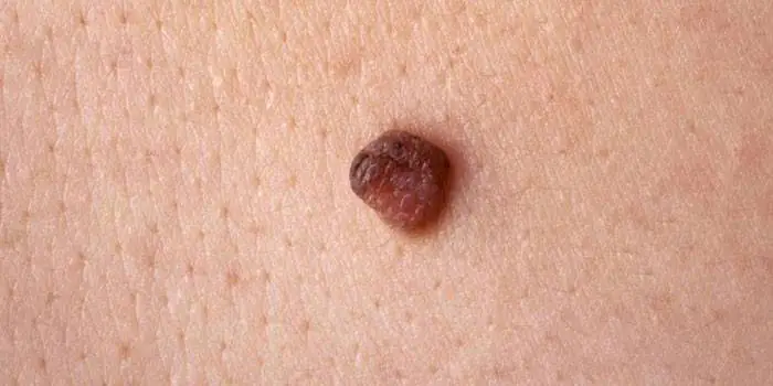
- Warty. Very dark and wart-like. Their condition must be constantly monitored.
- Convex. Dark, can be either smooth or rough, covered with coarse or vellus hair.
- small – up to 1.5 cm in diameter;
- medium – up to 10 cm;
- large – more than 10 cm;
- gigantic - cover significant areas of the body or face.
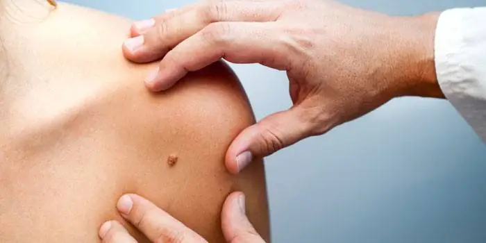
Dangerous moles
Some nevi, under the influence of one reason or another, can develop into cancerous tumors. As a rule, this occurs due to injury to the formation or prolonged exposure to ultraviolet rays. Dangerous moles are considered to be those that appear in adulthood, rapidly change their appearance and exceed a centimeter in diameter. A person should monitor such suspicious formations very carefully and regularly see a dermatologist.
People at increased risk of their occurrence include:
- fair-skinned, red-haired, with many freckles and age spots;
- have already removed malignant tumors;
- over 50 years of age;
- who have many dark ones;
- quickly “burn” in the sun;
- whose relatives had skin cancer.
What dangerous moles look like
Several types of formations are considered these:
- Nodal. A spot with a surface of uniform color, even black.
- Blue. A dense, smooth knot without hair, rising above the surface of the skin.
- Halo nevus. A colored formation on the skin surrounded by a colorless ring.
- Skin pigmentation. Slightly convex, pale, sometimes covered with hairs.
- Gigantic. Any formation of enormous size is dangerous.
- Nevus Ota. Dark brown or gray-blue formation, very large.
- Dubreuil's melanosis. Precancerous formation with uneven contours.
If dangerous moles look as described above, then there is a high risk of their degeneration into melanoma, a type of skin cancer. It is worth noting that such a tumor is highly treatable if detected in time. You should immediately contact a dermatologist if the following symptoms appear:
- deformations;
- discharge of blood or fluid;
- symmetry violations;
- burning;
- changes in surface texture;
- pain;
- hair loss;
- itching;
- the appearance of ulcers or cracks;
- seals;
- inflammation;
- color changes, appearance of inclusions.
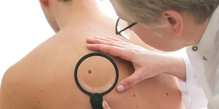
How to identify a malignant mole
You need to periodically examine your body in order to promptly consult a doctor if there are suspicious signals. To identify a malignant mole, use the basic ACORD rule:
- "A - asymmetry." If it is no longer the same shape, it may be reborn.
- "K - contour." Uneven, fuzzy, blurry edges are a warning sign.
- “O—shade.” Any changes in color, the appearance of dots, inclusions, stripes and specks may indicate a malignant formation.
- "R - size." If it suddenly begins to grow, consult a doctor immediately. The maximum acceptable diameter for the norm is 6 mm.
- "D - dynamics." If crusts, cracks appear on the formation, or blood or any substance begins to come out of it, then you need to visit a specialist. Suspect nevi that become too soft, become covered with nodules, become painful, or are surrounded by inflamed red skin. A sudden increase in altitude is dangerous.
How to remove moles on the body
Dangerous and suspicious formations are removed for medical reasons. A person can also remove moles on the body at his own request if they cause him aesthetic or practical discomfort (they cling to clothes, are constantly touched by nails). There are several ways to get rid of nevi: laser beam, surgery, radio waves, liquid nitrogen. Each of them needs to be discussed in more detail.
Laser removal
A very gentle and effective method of destroying formations with a directed beam. Laser mole removal can be performed using two techniques:
- Layer-by-layer evaporation. The beam gradually removes layers from the surface to the deep.
- Excision with a laser knife. The material after such an operation can be sent for histological examination.
- the method is absolutely safe;
- the risk of complications is minimized;
- there is no blood, because laser radiation immediately “seals the vessels;
- there are practically no contraindications;
- As a rule, one session is enough;
- painless (local anesthesia is performed);
- no recovery time after surgery is required;
- non-contact technology ensures complete sterility;
- the procedure is carried out very quickly.
- infection may occur;
- The wound takes a long time to heal, leaving a scar.
- diabetes;
- sun allergy;
- infections in the body;
- heat;
- oncological diseases;
- epilepsy;
- any skin inflammation;
- pregnancy.

Surgical method
A very affordable method, the only one suitable when there is no possibility of alternative procedures. The surgical method is reliable and is often used for formations with suspected malignancy. The formation and a small area of skin adjacent to it are excised with an ordinary scalpel under general or local anesthesia. The material can be immediately sent for histological examination.
- whatever the size of the formation, it will be removed in one go;
- low price;
- relapses almost never occur;
- complete absence of contraindications;
- the method is safe.
- A scar remains, although modern techniques for applying cosmetic sutures make it possible to make it as thin, even and invisible as possible. In addition, the use of modern anti-scar ointments will help reduce it to nothing.
- The wound takes a long time to heal. It needs to be processed regularly and carefully.
Cryodestruction
This is the name for the process of destroying formations with liquid nitrogen. To put it simply, the mole is frozen and its cells die from the cold. Cryodestruction is performed without anesthesia at all or with local anesthesia. This procedure will be most effective for flat formations on the body that do not go into the deeper layers of the skin. Nitrogen is applied by lubricating the surgical site with a cotton swab or using a special applicator.
- inflammatory, infectious processes;
- pregnancy;
- malignancy of formation;
- convulsions;
- epilepsy.
- removal is painless;
- the risk of complications is very small;
- cryodestruction is carried out quickly;
- the operation is inexpensive.
- the formation may not disappear completely, because nitrogen does not act on the deep layers of the skin;
- very high risk of scars;
- there is a possibility of damaging healthy tissue and causing a burn;
- for large sizes, several cryodestruction sessions may be required;
- recovery takes a very long time and during this period the use of cosmetics and exposure to the sun is prohibited.
