Red moles are presented in the form of a benign formation, which consists of many blood vessels. Statistically, they appear more often in childhood. In adults they occur several times less frequently. The color of the nevus is bright scarlet. This is explained by the fact that in the formation of overgrown vascular tissue. If we talk about ordinary moles, then these are skin growths, and the red spots on the body are blood vessels that have come to the surface of the skin.
Reasons for the appearance of red moles on the body and their locations
More often, red spots appear in the face area. Medical theory suggests that this phenomenon is associated with the presence of a large vascular network on the face. There are a number of reasons why red moles can form on the body. These include:
- Regular damage to capillaries, which indicates a violation of the vascular system.
- Hormonal imbalance in the body. This occurs during pregnancy, treatment with hormonal drugs, changes in climatic conditions, taking birth control pills, and diseases of the thyroid gland.
- Diseases of internal organs. In this case, nevi may not be located in the part of the body where the diseased organ is located.
- Lack of ascorbic acid affects vascular tissues.
- Exposure to ultraviolet rays.
- Viral infection.
- Penetration of chemical compounds into the body.
In addition to the surface of the skin, spots can be located in the tissues of organs such as the brain, liver, and kidneys. In different places of localization there can be either a single red spot or several pieces. One formation does not threaten a person in any way, but multiple inclusions signal a malfunction in the functioning of the entire organism.
Angiomas are found on the back, on the lip, on the legs and arms, on the face, and palms. There are cases when they can be found on the genitals. For a woman, a red mole on her chest gives them aesthetic discomfort. A woman should remember that a single incident of education does not threaten her health. But if there are several pieces together and they start to itch and hurt, this is a reason to visit a doctor immediately.
A mole on the head under the hair also causes inconvenience. Constant combing and cutting of hair can cause injury to it. Some women wear shapewear, which can cause irritation if the mole is located on the stomach.
Red spots on the body are divided into several types. This division depends on what part of the body the mole is located on, why it occurred in a person or child, and what type of vessel it is that served as the initial cause of its occurrence.
Considering the composition of the tissue from which it is made, moles are divided into:
- Knotty. The formation is small in size, presented in the form of a dot. The cause is a vessel that has reached the surface of the skin.
- Flat. This nevus looks like a lump on the skin.
- Cone-shaped. This growth has a convex shape. It protrudes strongly above the skin.
- Branched. Blood vessels weave from the mole, forming a mesh.
The spots are distinguished by the vessels that cause the growths: lymphangiomas and hemangiomas. Lymphangiomas occur when the lymphatic system fails. This phenomenon is quite rare. Lymphangiomas are presented in the form of small nodes that are easily compressed upon palpation.
They are found in the collar area, in the oral cavity, under the arms and in the groin.
Hemangiomas cause malfunction of blood vessels. Unlike lymphangiomas, they are large. Based on the volume and visible signs, hemangiomas are:
- Branched. A blood-filled growth consisting of several common nevi. It has a swollen appearance and a pulsating character.
- Capillary hemangioma. A spot that has a rich red or dark burgundy color. It can be localized in any part of the human body. The reason for the formation is dilated capillary vessels.
- In the form of a point. They are among the smallest hemangiomas. They look like dots.
- Cavernous. Such hemangiomas are located above the level of the skin. They are quite large in size. This hemangioma contains many blood vessels that merge into one formation. There are cases when such hemangiomas are located on the kidneys, liver, and pancreas.
Treatment
Many people who have spots on their body often wonder how to get rid of red moles without causing harm to their health and appearance. Especially if these red moles are located on the face. Medicine proves that most of these formations tend to disappear on their own. Therefore, no treatment is required.
However, if the red mole swells, increases in volume or becomes red, it is recommended to consult a specialist. Modern medicine provides many ways to treat red growths. The doctor determines which method to choose based on the results of some tests and studies.
The most effective methods, which have many positive reviews from patients, include:
Laser removal and cauterization method
The most popular procedures to remove a red mole are the laser method and the cauterization method. Before performing the procedure, the place where the mole is located is injected with a special anesthetic drug, or lubricated with an anesthetic gel. This method is performed on several layers of skin. The advantage is that you can get rid of formations of varying depths.
The process does not even take a minute. 24 hours after removing a mole, a person will observe a crust formed in the place where the formation was. The crust will disappear 20–30 days after the intervention. When the healing process is complete, light scars will be visible at the site where the mole was.
A safe method of removing a mole is cauterization. After the procedure, the person will have no trace of the place where the nevus was. Nitrogen or carbon dioxide is used in the cauterization process. Carbon dioxide is used if the mole is small and located above the skin. Nitrogen is necessary to remove large nevi. Before starting, the person is injected with an anesthetic into the place where the growth is located.
Surgical method of removal
This method of removing formations is carried out only within a medical institution. The surgery is performed by an experienced surgeon. Once the procedure and wound healing process are complete, the person will be left with a scar. Surgery is performed if the growth is small. It is not recommended to use this type of mole removal if it is located on the face. After the procedure is completed, the person is immediately sent home, with further recommendations.
Cryodestruction method and X-ray treatment
During this method, liquid nitrogen is used. With its help, a qualified doctor will freeze a benign formation. This method of removal is necessary if the growth is small in size and located above the surface of the skin. The freezing method is aimed at ensuring that during the freezing of blood vessels, their death occurs. This is due to the effect of extremely low temperatures on the nevus, as a result of which the vessels and capillaries become brittle.
When X-rays are used, the size of the mole decreases and its color changes to lighter. If you conduct several sessions, it is possible to get rid of growths. However, this method is not popular due to the harmful effects of X-ray radiation.
Treatment with folk remedies
To get rid of angiomas, you can use not only medical, but also traditional methods. Many people are interested in how to remove red spots at home. The main folk methods include:
- Wash the dandelion (root) thoroughly in advance. Dry it well. Grind the dried product by hand. Apply part of the crushed root to the mole and wrap it with a sterile bandage on top. Leave for 1.5–2 hours. Apply daily.
- Before going to bed, lubricate the angioma with castor oil. Next, stick a patch on the growth or wrap it with a clean bandage. Remove the bandage only in the morning.
- Finely grate the radish. Apply the resulting mushy product to the nevus. Wrap with a sterile bandage. Carry out the procedure twice a day, in the morning and at night.
- Finely chop a small onion. Squeeze the juice out of the chopped onion using gauze. Generously coat the mole with onion juice. Carry out the procedure every day for three weeks.
- It is recommended to spread liquid honey on the formation several times a day. After three weeks, the growth will decrease in size and may fall off spontaneously.
The appearance of angiomas
Angiomas can occur not only in adults, but also in children. The question often arises as to why red moles appear on the body if a woman is expecting a child. The answer is simple - hormonal changes occur in her body during this period.
The child has
An angioma can form when a child is born. They can be small, medium and large. The volume varies from half a centimeter to ten centimeters. Parents need to closely monitor such formations. If the growth is small and does not have swelling, then it does not pose a danger. There is no need to carry out treatment or resort to a removal method. Often, as the child grows, it disappears on its own.
However, if certain factors are present, it is recommended to consult a doctor. These factors include:
- if the mole turns red;
- the growth itches and there is a burning sensation;
- the nevus quickly increases in size;
- Painful sensations occur on palpation;
- other spots with a bright red color appear nearby;
- the nevus becomes crusty;
- if the mole is red and swollen.
These factors indicate that the child needs consultation with an experienced specialist who can tell you what to do if the mole turns red. He will also advise whether the child needs surgical intervention, or whether other treatment methods can be used that can relieve redness around the mole. Seeing a doctor is also necessary because he will prescribe appropriate tests, during which it will become clear that there are no malignant cells in the formation, or vice versa.
During pregnancy
When a woman becomes pregnant, hormonal changes occur in her body. This causes red moles to appear. They can be localized on any part of the body. Doctors say there is no need to worry about this. But it is recommended to keep an eye on her. If it changes its size to a larger one, or begins to darken, it is important to consult a doctor.
The growth may also appear during breastfeeding. This does not indicate the presence of cancer or disease of internal organs. In this case, you do not need to do anything. After a woman’s hormonal levels are restored, it will disappear.
If the mole grows and increases rapidly
If the size of the nevus changes, there is a risk of its degeneration into a malignant formation. An increase in growth may indicate that the hormonal balance in the human body is disturbed or there is a pathology of internal organs. It is not recommended to deal with this phenomenon on your own. It is better to seek help from a specialized doctor.
A dermatologist or oncologist will prescribe some tests to diagnose the state of a person’s hormonal levels and the formation that has begun to increase.
Complications
Angiomas can be located not only on the skin, but also on the tissues of some internal organs. In the latter case, the risk of disruption of the functioning of this organ increases. Of particular danger is the growth of a red spot inside the affected organ. If a mole is located on the surface of the skin and is subjected to mechanical impact, the consequences will be as follows:
- Prolonged and heavy bleeding from the injured growth. It is more dangerous when a completely red mole that is large in size is torn or torn off.
- An inflammatory process may begin on the nevus. There are cases when, after inflammation ends, the mole disappears.
- Getting into the torn formation of infection.
Consequences of moles
The main danger of red moles is their ability to degenerate into a malignant formation. Such transformations can occur due to trauma to the growth, regular exposure to ultraviolet radiation, or penetration of infection. Moles that are located in the nasal cavity or in the eye area in children can cause a violation of the sense of smell and impair vision.
Are red moles on the body dangerous for humans?
Red spots on different parts of the human body generally do not pose a danger to him. However, if the growth is regularly injured, bleeding may begin. And the lack of proper treatment of the wound in the future will provoke infection into it, which will cause the formation of pus and the transformation of the mole into a malignant formation.
The main places where damage can occur are the neck, scalp, arms and legs, armpits, and abdomen. In addition to the risk of infection, spots may indicate the presence of diseases of the internal organs. Therefore, there is no need to neglect a visit to a medical facility, so as not to trigger the disease and get serious complications for the body.
What happens if angiomas are not treated?
If the angioma does not cause inconvenience to a person, does not itch, does not hurt, does not grow, it is not recommended to apply any actions to it. Over time, it may disappear spontaneously. When a growth spoils a person’s appearance, he may decide to have it removed.
If you feel pain, the mole turns red and enlarges, you should immediately seek help from a medical facility. This will help avoid inflammation, suppuration, and damage to the organ on which it is located. When removing a mole, you should regularly visit a doctor who will conduct an examination and monitor the healing process.
Moles rarely cause discomfort. But sometimes, as a result of exposure to various factors, they become inflamed and cause pain. If the mole turns red, this may be the first sign of infection, which must be treated promptly.
Causes of redness of a mole
You need to constantly monitor moles on your body. Redness of the nevus or the appearance of a halo around it is an alarming symptom that often appears during inflammation or the initial stage of oncology. In both cases, mole cells respond to health problems by activating or decreasing the production of melanin (coloring pigment).
In the worst case, inflammation of the nevus indicates the degeneration of the formation into a malignant tumor. But most often, redness appears as a result of trauma to the tissue of the mole and the penetration of dirt and pathogenic microbes into the wound. In rare cases, such a reaction can be triggered by taking medications during the treatment of acute forms of chronic diseases.
Signs of an infected mole
Infection in the nevus tissue can occur as a result of injury. Even minor damage, a light scratch, is a potential danger for the development of inflammation. The following signs indicate that pathogenic microflora begins to multiply in the mole or adjacent tissues:
- redness;
- swelling of the skin around the mole;
- itching and pain;
- bleeding or watery discharge.
If signs of infection appear, you should consult a doctor as soon as possible to prescribe treatment.
What to do if a mole turns red and inflamed
You should immediately consult a doctor (oncologist or dermatologist) for advice!
Pre-medical care for an inflamed mole should consist of keeping the affected area clean and dry. If blood or fluid is released from the nevus, it is necessary to treat it with hydrogen peroxide and cover it with a dry bandage.
If the doctor diagnoses the presence of an infection in the inflamed tissues, he may prescribe a course of oral antibiotics: Analgin, Nimesulide, etc. For local treatment of the infection, antibacterial ointments, for example, tetracycline, are used. Very severe inflammation may require treatment with intravenous antibiotics.
If there is a suspicion that a mole has degenerated into a malignant formation, the doctor may suggest doing a tissue biopsy. A biopsy takes a small sample of the growth and sends it to a laboratory for testing. The procedure is usually done in the office of an oncologist or dermatologist.
Mole removal
If there is a high probability of developing oncology in the nevus area, then it must be removed. It is also recommended to perform surgery if the mole becomes inflamed again or if it is located in a place where it is often injured.
Your doctor may remove the growth with a minor surgical procedure. But before it is carried out, it is necessary to undergo an examination, including a number of tests:
- general blood analysis;
- research to identify tumor markers;
- analysis of blood clotting indicators.
This examination is carried out to assess the general condition of the body and determine the cause of inflammation. Additionally, tests for drug reactions and allergy tests may be prescribed.
There are several ways to remove a mole:
- Laser surgery is considered one of the safest methods for removing nevi. Therefore, it is suitable even for sensitive skin. Allows you to eliminate a mole in one procedure. The ablation laser used for the operation is the most gentle tool, allowing you to gradually remove the stain layer by layer. The beam is directed to pigmented tissues, leaving healthy epidermis untouched. Performed under local anesthesia.
- Cryodestruction is a method of exposing tissue to low temperatures using liquid nitrogen. The cells of the mole die as a result of freezing, and a crust appears at the site of formation, under which the process of skin regeneration occurs. This is a rather extreme method, after which a long recovery period is required. Cryodestruction is usually not performed to remove moles on the face.
- The radio wave method is based on the use of a special tool in the form of a loop, which cuts off the build-up. And exposure to radio waves cauterizes the wound and prevents bleeding. This method is only suitable for small growths protruding above the surface of the skin.
- The surgical method is a proven and proven method for removing large moles. If there is a suspicion that the nevus is malignant, doctors recommend surgery.
Contraindications to mole removal
It is impossible to remove a mole using radical methods if you have the following diseases:
- acute dermatological diseases;
- HIV;
- autoimmune diseases;
- oncology;
- diabetes.
In these cases, the doctor prescribes therapeutic treatment for inflammation of the nevus.
Prevention of inflammation
Typically, adults have no more than 40 moles on their body. People with lighter skin tend to have more spots. These can change as a person ages. Some become darker or lighter, and may increase slightly in size.
Most moles are harmless, but people should check them for changes and follow simple tips to prevent breakouts:
- Protect skin from direct sunlight;
- Avoid excessive tanning and sunburn;
- Be extremely careful when visiting a solarium;
- Use sunscreen;
- Wear clothes that do not rub moles;
- In case of trauma to nevus tissue, carry out timely disinfection.
Taking steps to protect your moles will help reduce your risk of developing inflammation and skin cancer.
Most often, the moles that we have on our skin hardly change throughout our lives. But what to do if suddenly, without external influence on the mole, you discover characteristic symptoms of inflammation in its area? Redness? Soreness? Increase in size? Does this indicate that her malignant degeneration has occurred? The answers are in this article.
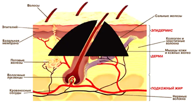
Structure of a mole
To understand the processes that occur when a mole becomes inflamed, you need to delve a little deeper into the structure of the skin. Why does hair grow from a mole? As we have already found out in this article, hair can grow from a mole (i.e., a pigmented nevus). This is due to the fact that, as a rule, the mole itself is located slightly above the level at which the hair follicles lie. It turns out that it is not the hair that grows on the mole, but rather the hair that grows through it and appears on the surface. She just happened to be in their way.
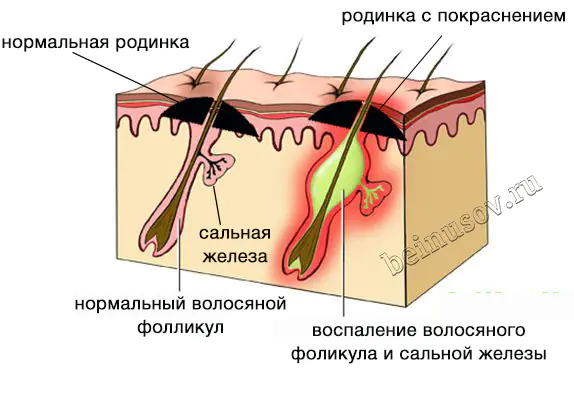
Why does a mole turn red, enlarge and become painful?
The fact is that inflammation can begin in the area of hair follicles and sebaceous glands. As a rule, the sebaceous gland becomes inflamed, the excretory duct of which opens into the hair follicle.
In medicine, inflammation of the sebaceous gland and hair follicle is called a boil, popularly - "pimple".
This happens for a variety of reasons, from pollution and microtrauma of the skin to metabolic disorders. As you can see in the picture below, all inflammation phenomena appear in the area of the mole:
Does inflammation of a mole indicate its malignancy?
Can talk. One of the signs of melanoma (No. 10) is “inflammation in the area of the nevus and in the tissues surrounding it.” However, do not panic if you suddenly discover this symptom. The fact is that this may well be inflammation of the sebaceous gland and hair follicle, and not at all malignant degeneration. How to distinguish one from the other?
What to do if your mole becomes inflamed and red?
The first thing to do is to stop further independently studying the Internet on this topic. The fact is that on the Internet you can almost always find confirmation of your worst suspicions about your health. This is not what we need now.
In addition, it must be remembered that this symptom alone is extremely rarely a sign of melanoma. In the vast majority of cases, there should be other symptoms - uneven (geographic, scalloped) edge, asymmetrical shape, bleeding, etc.
Then just follow these recommendations:
- Wet a regular gauze pad (not sterile) with a solution in the ratio of 1 part chlorhexidine, 1 part dimexide, 2 parts water
- Apply the resulting compress to the area of the inflamed mole for 30 minutes, 3 times a day, for 5 days.
In most cases, redness, pain, and enlargement of the mole disappear under the influence of these compresses within 5 days.
If the inflammation does not subside within 5 days, you should urgently see an oncologist. In this situation, the likelihood that these changes were caused by transformation into melanoma is very high.
Briefly about the main thing:
If your mole becomes inflamed, red, or painful without any external influence, do not panic. Apply compresses according to the above scheme and inflammation should subside within 3-5 days. If this does not happen, see an oncologist immediately.
If you don’t have the mental strength to wait 5 days, you can get my consultation online right now. There is also the opportunity to make an in-person appointment with me at the clinic in St. Petersburg (Asafieva 7/1).
