Moles do not always decorate the appearance; they often need to be removed. For this, the most proven method is often used - surgery. It will also help in cases where other methods are contraindicated.
Read in this article
Indications for surgical removal of moles
It is necessary to get rid of a nevus surgically under the following circumstances:
- it grows or has reached a size of more than 5 mm;
- the formation is deeply rooted in the skin;
- it consists of several parts;
- is located on an area of the body that is in close contact with clothing or shoes, and is therefore often injured;
- the mole bleeds, itches, changes color, its edges become uneven;
- she spoils the appearance.
Advantages of the method
The traditional method of excision of nevi with a scalpel has advantages that more modern methods do not have:
- safety, since the manipulation is performed by a surgeon and not in a beauty salon;
- painlessness, because the mole is removed under local anesthesia;
- the ability to examine the material using the histological method;
- a chance to get rid of a mole in a single operation;
- complete removal of formations that cryodestruction or laser cannot handle;
- affordable price;
- no absolute contraindications;
- almost zero risk of relapse.
Contraindications to surgical removal of a mole
There are no absolute prohibitions on intervention to get rid of a nevus with a scalpel. But there are relative contraindications:
- infection;
- inflammatory process in the body;
- acute period of chronic disease;
- pregnancy and lactation;
- herpetic rashes on the skin or mucous membranes.
Before removing a mole, you need to get rid of these problems.
Preparing for removal surgery
No special measures are required to prepare for surgical manipulation. A preliminary examination by a surgeon is necessary, who will explain the essence of the operation and give recommendations on how to behave before it.
The patient should not take blood thinning medications for several days, drink alcohol, and it is better to quit smoking. If a mole is being removed from the face, there should be no makeup on it.
How is surgical removal performed and how long does the operation last?
The intervention is carried out using one of two possible methods:
- Seamless. After disinfecting the surgical space and injecting the anesthetic, the formation is cut off with a scalpel just below the surface of the skin.
The wound is treated using special equipment to stop bleeding, and an antibiotic is applied to it. A sterile bandage is applied on top. This method is suitable for small, shallow moles.
- With sutures. The nevus and the skin around it are treated with an antiseptic and an anesthetic injection is given. Then the mole and adjacent tissues are excised. The wound turns out to be quite extensive and deep, which dictates the need for sutures. They are usually biodegradable, but if the moles are large, they can also use non-absorbable ones.
After suturing the deep and outer layers of tissue, the surface is treated with an antiseptic and a bandage is applied.
The operation, regardless of the method, will not take more than 30 - 60 minutes.
To learn how micro-operation to remove a mole takes place, watch this video:
Recovery after surgery and suture care
The end of the intervention begins the rehabilitation period. A wound or suture remains at the site of the excised formation, which should be taken care of:
- do not allow water to enter this area;
- treat it with antiseptics 2 times a day;
- do not tear off the formed crust, avoid contact of hands and clothing with it;
- after 7 - 10 days, go to the doctor to remove the stitches if they are non-absorbable;
- protect the area from the sun.
The latter is especially important when the crust falls off and the young skin is exposed. This will happen in about 2 weeks. But ultraviolet radiation is contraindicated for at least another 2-3 months. The area of the healed wound should be hidden under clothing or a protective cream should be applied.
During the rehabilitation period, it is worth giving up alcohol and smoking so that healing is not disturbed and proceeds faster.
Will there be a scar after?
Even an operation with suturing does not necessarily require the presence of pronounced marks in this place. With proper care, the scar after the intervention remains almost invisible. If adjacent tissues were removed, the mole “sat” deep, it is more likely that the mark from the operation will be pronounced. To smooth out the scar, absorbable creams are applied to it or a special patch is used. And if their effect is not enough, the scar can be removed surgically.
Other possible complications after surgery
Excision of a mole can lead to new problems:
- Wound infection. Possibly during surgery, but more often it happens due to poor care of the wound and suture.
- Keloid scars. The complication occurs when healing is slow or due to a predisposition to its occurrence.
- The appearance of education in the same place. This is possible if the mole was not completely removed, or it was melanoma.
Cost of surgical removal of moles
The price for the operation depends on the size of the nevus. If it is less than 5 mm, it will be 1500 - 3000 rubles. For larger moles you will have to pay more. Preliminary examination and consultation are indicated separately in the price lists of clinics.
Alternative options to surgical removal of a mole
If the formation is small, you can get rid of it using:
- Radio wave method of mole removal
laser;
cryodestruction (low-temperature nitrogen); electrocoagulation; radio wave method.These methods are not always allowed to be used, unlike traditional surgery.
Don't be afraid to remove a mole with a scalpel. If the doctor insists on this method, it means that in a particular case it is optimal. And recovery after surgery is no more difficult than after using more modern methods.
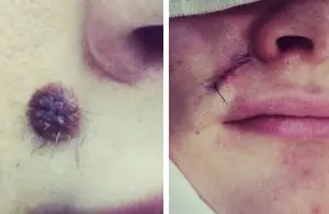
Moles on the face and body do not always fall into the category of harmless “decorations”. Often, nevi are located in places that are constantly injured - on the neck, in the armpits, in the groin, under the hair and on the bra line. There are often cases when, after intense tanning in a solarium or hormonal imbalance, a mole begins to degenerate - it increases in size, becomes inflamed, begins to hurt or itch, and rashes appear around it. In these situations, it is necessary to urgently make an appointment with an oncodermatologist in order not to miss the appearance of skin cancer - melanoma.
One of the most common methods of removing a mole is excision of the neoplasm with a scalpel. Typically the procedure can take up to 1 hour. To relieve pain, the doctor makes several injections into the extraction area. During the operation, in addition to the affected tissue, part of the healthy skin is captured, because otherwise suturing is impossible. When surgically removing a mole, the patient must be tested for syphilis, HIV, hepatitis B and C and other infections that are transmitted through the bloodstream.
In what cases is it really worth removing a mole with a scalpel?
- At the slightest suspicion of a malignant tumor;
- To remove large diameter nevi (1.5-2 centimeters in diameter);
- If it is necessary to remove moles that have broken up into several segments;
After removal, the material must be sent for histological examination, and sutures are placed on the wound.
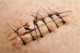
On the second day, dressing may be necessary; the stitches are removed after 6-10 days. During the postoperative period of several weeks, the skin area should be protected from sunlight and you should not try to remove the crust at the site of the mole yourself.
Disadvantages of the surgical method of nevus removal:
- If the doctor is poorly qualified or does not follow the rules of asepsis, complications are possible - bleeding, suppuration of the wound;
- A long recovery period, the need to apply and remove stitches, see a doctor, do dressings, etc.
- Highly traumatic, high likelihood of scar formation;
Radio wave surgery has become a modern alternative to the outdated surgical method in removing small moles on the face and body. With this method, high-frequency radio waves affect the tissue around the nevus, causing the thinnest layer of skin to evaporate. Removing one mole takes on average 3-5 minutes and does not require hospitalization.
Advantages of radio wave surgery for mole removal:
- The procedure is quick and virtually painless;
- There is no need to take preliminary tests;
- Minimal likelihood of complications;
- Rapid tissue healing;
- The material is sent for histological examination;
- Impeccable cosmetic effect;
- No stitches required;
- The only restriction after the procedure is that you should not take a bath or shower for 1-2 days.
Are you looking for a clinic where you can quickly and without complications remove a mole? Make an appointment with a professional oncodermatologist. If there are indications, we will remove the nevus using radio wave surgery - without complications and unnecessary hassle!
Summary:
Mole removal with a scalpel - a good, reliable, but outdated method. Nowadays it is used quite rarely - when a mole is suspected of being malignant and when the formation is large. In other cases, I recommend a more modern approach - radio wave surgery. It is much more convenient for the patient and has fewer complications.
All iLive content is reviewed by medical experts to ensure it is as accurate and factual as possible.
We have strict sourcing guidelines and only link to reputable sites, academic research institutions and, where possible, proven medical research. Please note that the numbers in parentheses ([1], [2], etc.) are clickable links to such studies.
If you believe that any of our content is inaccurate, out of date, or otherwise questionable, please select it and press Ctrl + Enter.
There are many methods for removing a mole, the most common being surgical. Let's consider the features of the procedure, indications for its implementation and possible consequences.
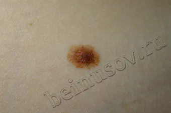
A mole is a skin growth. Its color (from light brown to black) depends on the number of melanocytes. Nevi are hereditary and can appear throughout life. If pigment cells are located in the deep layers of the skin, then convex moles are formed, if melanocytes are on the surface, then flat ones.
Such skin growths must be monitored. If they are large or have an unusual shape, then you should consult a dermatologist. Nevi up to 5 mm with smooth contours and uniform color are not dangerous and, as a rule, do not require removal. But their main danger is that they can degenerate into malignant formations - melanoma. Such formations must be urgently removed - excision.
Surgical removal of a mole is the most common and relatively cheap treatment method. It is suitable for destroying large and deep skin formations, but has a number of features.
- High efficiency - allows you to remove a mole in one procedure.
- The minimum of contraindications and the low cost of the operation make it accessible to many patients.
- Low risk of relapse - since the nevus is completely removed, a repeat procedure is not required.
- Safety – the procedure is carried out in a hospital setting under the strict supervision of a doctor.
- Long-term wound healing and recovery - after surgery you will have to give up sunbathing and solarium for a long time.
- Scars - the larger the wound surface, the higher the risk of scarring. In particularly severe cases, keloid scars may develop.
Surgery may be necessary if the mole is larger than 5 mm, has uneven color, uneven edges and irregular shape. The scalpel includes nevi that are constantly injured and cause discomfort or cosmetic inconvenience. If they are present, you need to contact a dermatologist who will determine the type of skin growth and the method of its removal.
[1], [2], [3]
Indications
Any operation, regardless of complexity, has its own indications. Excision is most often performed when malignant degeneration is suspected. Surgical removal of a mole is necessary if it:
- Increased in size.
- Has deep penetration into the skin.
- Broke into several pieces.
- Started to bleed.
- It causes pain and is often injured.
- Causes aesthetic inconvenience.
Despite the reasonable cost, excision is quite traumatic and can leave scars. But this method also has its advantages over more modern technologies: laser removal and cryodestruction. After surgery, the obtained materials can be examined for histology.
Excision prevents recurrence of skin growths, since areas of healthy tissue are captured. The procedure must be carried out only in specialized medical institutions, and not in beauty salons.
Preparation
Regardless of medical indications, preparations are made before the operation. First of all, the doctor explains the essence of the procedure, its possible complications and the nuances of the recovery period. After this, the patient takes a place on the couch. The neoplasm and the skin around it are treated with a disinfecting solution.
Anesthesia is mandatory to ensure that there is no discomfort during excision. Most often these are drugs with lidocaine (Anestakon, Xylocaine, Bactin, Zalactin-L). This does not take much time, but it reduces blood flow to the operated area. If there is a risk of bleeding, then Epinifrine is added to the local anesthetic.
After this, the mole and part of the healthy tissue are excised using a scalpel. The resulting wound is treated with a special solution and stitches are applied. The tissue obtained during the operation is sent for histological examination. At the end of everything, the doctor gives recommendations on skin care.
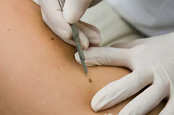
Surgical mole removal technique
Getting rid of nevi on the face and body is not only a cosmetic procedure, but also an effective method of preventing cancer. Surgical removal is used when dealing with deep and extensive growths or moles that have several segments. This is the only effective way to eliminate large tumors.
The technique for surgically removing a mole depends on its location and size. The procedure is carried out under local anesthetic and takes 40-60 minutes.
- Cutting method without sutures - using a scalpel, the growth is cut off slightly below skin level. If bleeding occurs, the wound is cauterized and a local antibiotic is applied to stop it. A bandage is applied and recommendations for further skin care are given.
- Excision with sutures – used to treat flat or dark moles. The doctor cleans the skin of the tumor and numbs the surgical surface. A scalpel is used to excise not only the mole, but also the surrounding tissue. Based on the depth of the operation, sutures are placed on the upper or deep layers of the skin. For this purpose, self-absorbable materials are used that do not require removal.
The healing speed usually takes 1-2 weeks. During this period, it is necessary to protect the operated area from any external influence.
Contraindications
Surgical removal of a mole is a cosmetic procedure, the advantage of which is the absence of absolute contraindications. This is explained by the fact that the operation is performed on a separate area of the body. But there are a number of relative contraindications, let’s consider them:
- Presence of infectious diseases.
- Acute inflammatory processes.
- Exacerbation of chronic diseases.
- Pregnancy and lactation period.
- Exacerbation of herpes.
In the presence of chronic diseases and after their relief, additional tests are required. This avoids complications and speeds up the recovery process.
[4], [5], [6], [7]
Consequences
Any operation can cause serious consequences. When using a scalpel on a nevus and applying sutures, the following pathologies are possible:
- Scarring - during the operation, not only the nevus is removed, but healthy tissue is also affected. Based on this, the size of the scar depends on the size of the skin growth and the professionalism of the surgeon. Over time, scars lighten, making them less noticeable.
- Keloid scars - most often occur in patients who have a tendency to form them. Appear after suturing large wounds.
- Recurrence - a repeated skin defect is possible if the doctor did not completely remove the mole. Degeneration is not excluded in melanoma-hazardous formations.
The likelihood of the above-described consequences is significantly reduced when treating moles with safer methods.
[8], [9], [10]
Complications
Surgical methods for removing moles, unlike alternative ones, have a high risk of complications. As a rule, these are painful sensations at the operation site, the appearance of scars and cicatrices. The procedure is not recommended for the face and other sensitive areas of the skin.
Most often, scars remain after surgery. This complication is inevitable, since cosmetic stitches are applied to the skin. Surgery is used if the mole is located on an inconspicuous area of the body and the scar can be hidden.
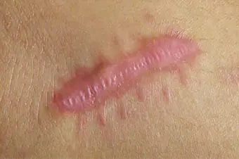
Another complication is subcutaneous hemorrhage. Slight redness around the removed tissues disappears after 7-10 days. Since after excision the wound remains open, there is a possibility of infection. All patients are warned about this, and it is this factor that causes the choice of less traumatic methods.
[11], [12], [13], [14]
Rehabilitation period
After removal of a mole, as after any surgical operation, the patient faces a rehabilitation period. This implies special care of the wound surface. A wound forms at the site of the nevus; its size depends on the diameter of the removed material. Gradually it becomes covered with a crust, which falls off after 1-2 weeks. Young pink skin appears at the wound site and needs proper care.
Skin care features:
- Do not wet the wound for 4-5 days, but maintain hygiene around the operated area.
- Do not touch or tear off the crust that covers the wound, as the healing process is taking place underneath it, and if it is disrupted, it can cause a large scar.
- Once the crust falls off and pink skin appears underneath, cover it from the sun.
After about a month, at the site where the mole was, the skin regains normal pigmentation. Pain may still occur for 1-2 months. Final healing depends on the individual characteristics of the body, but on average it takes 2-6 months.
Scar care
Healing of the skin after getting rid of a mole takes 2-4 weeks. During this period, it is necessary to treat the wound, and in the future, scar care will be required. After the operation, the patient may be prescribed wound-healing medications with antibiotics and mandatory skin treatment with brilliant green or potassium permanganate. If the operation was successful and medical recommendations are followed in full, then there are no undesirable consequences, that is, complications in the form of infection and bruising.
Within 5-10 days, a crust appears on the skin, which is replaced by young pink skin. Such areas must be protected from exposure to negative factors, especially from the sun. If it is impossible to hide the wound, then use sunscreen with a high level of protection before going outside. If you leave your skin unprotected, pigment spots may appear on it.
Surgical removal of a mole often leaves behind scars and scarring. In most cases, they resolve on their own, but if this does not happen, then the skin needs help. For this purpose, natural cocoa butter or a silicone patch is suitable (used only for medical prescription). If this does not give the expected result, then you should contact a cosmetic surgeon. After treatment, carefully examine your moles, especially if the removal was due to permanent injury to the nevus. Make sure that no changes occur.
[15]
Sick leave after surgical removal of a mole
Getting rid of nevi involves surgery and a long recovery period. Sick leave after surgical removal of a mole depends on its location and size, and the amount of work done. As a rule, patients are given leave from work for 1-2 weeks.
If the mole was large and sutures were placed, then the patient needs to go to have the wound dressed, and after it has healed, to have the sutures removed. Tissues obtained during surgery are sent for histology. Therefore, during the period of sick leave, the doctor familiarizes you with the results of this study. If there is evidence of degeneration of a skin lesion, the patient is registered to monitor the dynamics of moles and their treatment.
