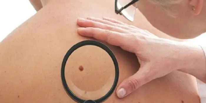
It's rare to see a person without small dark marks on their body. Is it worth paying attention to these points? Only a doctor will distinguish between dangerous and normal moles - malignant melanoma or harmless nevus - and give recommendations on what to do with them. Is it worth worrying about the appearance of new formations, when immediate contact with specialists is required, what are the signs of cancer development - the answers to these questions remain to be found out. No one is immune from disaster, and early diagnosis will protect you from severe consequences.
What is a mole
The first tiny spots may appear in children in infancy. A mole is a small formation on the skin - a nevus - that is considered benign and harmless. The basis for their appearance is melanocyte cells that accumulate the natural pigment melanin. Depending on its quantity, a difference in color is observed. Available colors:
The shape of the tumors depends on the location and concentration of melanin. They may have a stalk or be located under the skin, be flat and convex. The most common type is round, but there are exceptions. The development of neoplasms is provoked by ultraviolet radiation - natural from the sun, in a solarium. Hereditary factors cannot be excluded. A common cause of growth is hormonal imbalance, characteristic of periods:
- puberty;
- pregnancy;
- menopause.
What types of moles are there?
One person may discover very different tumors. Types of moles are classified according to several criteria. This helps in correct diagnosis in case of changes. They differ in:
- origin– congenital, newly acquired;
- structure– pigment, vascular;
- place of education – in depth, on the surface, in the boundary layer;
- raised above the skin – flat – even, protruding as a hemisphere, pedunculated, larger birthmarks;
- potential threats – dangerous, degenerating into melanoma, non-dangerous.

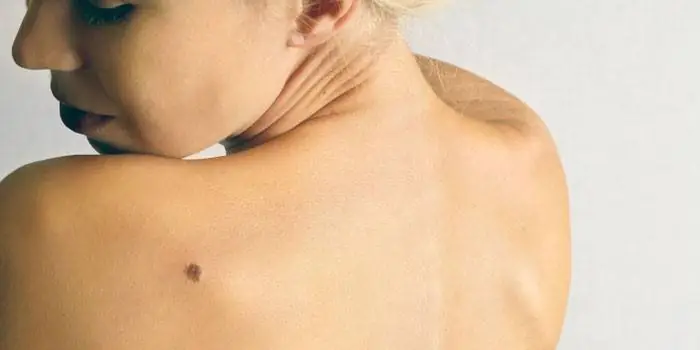
Safe moles
Those who have dark spots on their skin should be wary of their changes. In time, detected signs of degeneration into melanoma contribute to the timely removal of the formation and preservation of health. Safe moles are different:
- the presence of a stalk – it cannot be formed by malignant cells that grow randomly;
- long-term condition without changes.
Spots that appear soon after birth are not considered dangerous. It is important that they are small in size. Good – non-dangerous – signs of neoplasms include:
- flesh tone;
- unchanged pattern of the skin of the nevus and adjacent tissues;
- soft consistency;
- hair on the surface of the neoplasm - growing from the skin, indicates the absence of pathologies;
- diameter no more than 5 mm;
- symmetry;
- nevus in the form of a spot.
Which moles are dangerous?
Why do people with nevi on their bodies need to monitor their changes? There is always a threat of degeneration of non-dangerous tumors into a cancerous tumor. What moles are dangerous to health? Key signs you need to know:
- change in shades towards the dark side, the appearance of multi-color;
- rapid increase in size - exceeds two millimeters per year;
- occurrence of cracks;
- the formation of asymmetry due to uneven growth;
- lack of elasticity;
- the appearance of itching, burning;
- presence of discomfort.
The appearance of dangerous moles requires an immediate visit to a specialist to clarify the nature of the changes and the likelihood of developing skin cancer. Pathological transformations provoke:
- injury to the nevus due to negligence;
- self-removal;
- abuse of exposure to the sun, use of a solarium;
- location of the formation in places of frequent contact with clothing - on the neck, head, genitals, legs;
- placement in the hair, on the face, palms - where there is a high probability of injury;
- previously removed melanoma.
Why are moles dangerous?
Not a single person is protected from the sudden proliferation of cells of a harmless mole. Melanoma is an extremely serious disease. Changes not detected at the initial stage can result in death. The provoking factor is unsuccessful independent removal of tumors. Moles are dangerous because of their ability to:
- transform into an atypical – precancerous form;
- grow to large sizes;
- turn into cancerous;
- with minor external changes, metastases actively spread throughout the body through the circulatory and lymphatic channels.
How quickly does melanoma develop from a mole?
The transformation of a nevus into a cancerous formation can occur in different ways. The process depends on the stage of the disease and the type of tumor. Instant metastases are dangerous. Begins:
- growth of cancer (oncological) cells in the deep layers of the epidermis;
- their entry into the blood and lymph;
- penetration into the lungs, liver, kidneys;
- growth in these organs;
- complete damage to the body;
- death.
The growth phases of pigment cells are observed, along which melanoma develops from a mole. There are varieties:
- horizontal– damage to the upper layers of the skin occurs, lasting up to 10 years, but metastases do not appear;
- vertical– accompanied by the spread of cancer cells throughout the organs, can last two years, has an unfavorable prognosis;
- nodal – especially dangerous – characterized by deep spread within two months.
The first signs of melanoma
The patient can be assisted only when suspicious changes begin to be identified. The diagnosis, research, and referral for surgical treatment save a person’s life. The first signs of melanoma:
- increase in the height of the tumor;
- bleeding;
- the appearance of discharge;
- redness;
- burning, itching;
- swelling of tissues;
- softening of the nevus;
- the appearance of a crust;
- thickening;
- hair loss;
- expansion of pigmentation around the lesion.
With the further development of dangerous melanoma, the following are observed:
- significant change in size;
- the appearance of pain;
- enlarged lymph nodes;
- surface ulceration;
- formation of new foci;
- bleeding from places of pigmentation;
- liquid separation;
- skin thickening;
- the appearance of an earthy tint;
- signs of metastases are chronic cough, weight loss, cramps, headaches.
How to distinguish a mole from melanoma
To recognize which moles are dangerous and which are not dangerous, you need to know what they look like. A person with nevi, in order to avoid dire consequences, must constantly monitor the appearance of new formations and changes that occur. You can distinguish a mole from melanoma by its signs. Non-dangerous neoplasm:
- symmetrical;
- with smooth edges;
- uniform in color;
- with dimensions not exceeding 6 millimeters.
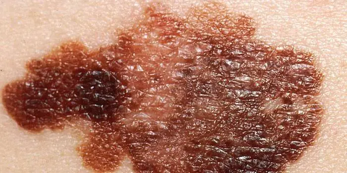
Features of dangerous melanoma that require seeking help from dermatologists:
- growth in a short time;
- pronounced asymmetry of shape;
- heterogeneity in color - the presence of inclusions of several shades;
- lack of clear boundaries - the contour line is blurred, jagged, and looks like a coastline on a geographical map;
- increased diameter over six millimeters;
- variability of any parameters - color, size, shape.
What dangerous moles look like
What do nevi that are subject to pathological changes look like? Only a doctor can correctly distinguish between non-dangerous tumors. Dangerous formations look like this:
- blue– compactions under the skin with clear boundaries, with dimensions no more than 10 mm;
- nodal– round, flat in shape, color – brown, black;
- cutaneous– often pale, convex;
- halo nevus – pigment surrounded by a light or white rim;
- spitz- looks like a dome-shaped tumor of pink shades, with the possible presence of a hole through which blood and liquid leak;
- connecting- connect individual entities into a whole.
Mole with jagged edges
One of the signs of a non-hazardous formation turning into a dangerous one is a change in contours. It often has blurred edges and scalloped borders. There are non-dangerous types of nevi - dysplastic. Only a specialist can make a correct diagnosis. A mole with uneven edges can be dangerous if there are additional signs of melanoma:
- accelerated changes in size;
- the presence of clearly defined asymmetry;
- the appearance of highly indented boundaries.
Rough mole
Such a neoplasm is harmless if its diameter is no more than 5 mm and remains constant in size. Often its appearance signals a lack of vitamins and nutritional disorders. Doctors advise coming for a consultation if it is discovered that:
- the smooth nevus turned into a rough one;
- bothered by burning, itching, tingling;
- irregularities and compactions appeared in the middle;
- areas with different shades formed;
- diameter has increased significantly.
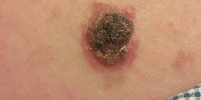
A dangerous rough mole requires immediate examination if:
- the appearance of bleeding;
- development of the inflammatory process;
- rapid change in size;
- formation of asymmetry;
- formation of purulent discharge;
- the occurrence of painful sensations when touched;
- the emergence of an irregular shape, blurred boundaries, along the edges of the neoplasm.
Large moles
Large formations on the skin are pigment spots. When they remain unchanged and do not cause inconvenience, this is not a dangerous phenomenon. It is important to constantly monitor their appearance, color, and size. To eliminate worries, you need to consult a dermatologist. During the visit, the specialist will conduct a diagnosis and give a forecast of the risk of developing a malignant neoplasm. Large moles become dangerous if they:
- injured;
- thickened;
- started to itch;
- were unsuccessfully removed independently;
- changed in size, shape;
- are bleeding.
What moles can be removed
Often nevi cause trouble for women when they are in a visible place - the face, neck. Even if they do not bother you, using removal will be the right decision - the appearance will improve significantly. After the procedure, the doctor must necessarily send the tissue for histological analysis to decide whether the mole is malignant or not. If the neoplasm is not dangerous, does not bother you, and does not change in size, surgery is not required. What moles cannot be removed? Experts believe:
- there are no contraindications;
- It is important to choose the right excision technique.
You should be careful about skin growths; it is unacceptable to remove them yourself. Only the doctor will determine whether a nevus is dangerous or not and decide what to do with it. You can delete it if:
- injured from clothing - on the neck, in the groin area, under the armpits;
- cause pain when touched;
- are located under the hair on the head and can be damaged when combing or cutting;
- change color, shape, outline;
- significantly increase in size;
- characterized by the presence of burning, itching;
- accompanied by inflammation and bleeding.
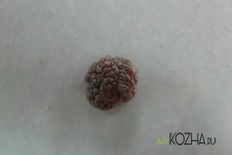
If you look at the photo, you will notice that a papillomatous nevus (especially if it has skin color) looks like a papilloma - a formation on a stalk that looks like a cauliflower inflorescence. This is where nevus gets its most common name. In medical terminology, this nevus is also called verrucous, hyperkeratotic, linear, hard and unilateral.
general characteristics
This mole is a lumpy nodule consisting of papillary warty growths, which is attached to the epithelium with a wide base (pedicle). The favorite location is the scalp, neck and face, but sometimes it can be found on other parts of the body. This form of nevus, like most others, is congenital, although a papillomatous birthmark can remain invisible for a long time. But unlike many moles, this one grows slowly and increases with age, reaching its final size at 20–30 years.
This type of nevus birthmark is classified as a benign, non-dangerous neoplasm. However, there are rare cases where papillomatous nevus develops into melanoma.
A raised mole causes inconvenience. Since it is most often located on the head, the risk of damaging it when combing, dressing, or washing the hair increases significantly. As you know, any injury to a mole can trigger the process of its transformation into a malignant formation - melanoma, and open the way for infection to enter the body, causing inflammation.
Symptoms of papillomatous nevus
The following signs are characteristic of a papillomatous neoplasm:
- The photo shows that the mole is convex, similar to a tubercle - it protrudes above the skin and is attached to the skin with a wide stalk;
- The integumentary epithelium is thickened - the birthmark is dense to the touch;
- Most often, the surface of the spot is lumpy, granular, as it consists of elongated epidermal processes - this is clearly visible during microscopic examination or in an enlarged photo;
- The boundaries of the neoplasm are clear, the outlines are arbitrary;
- Various photos show that papillomatous nevus can be of several colors and shades - from flesh to brown and even brown. Sometimes you can find a warty mole that is black in color - it all depends on the amount of melanin contained in the cells of which it is composed;
- Birthmarks of this type can be single or multiple;
- The size of the mole is large - from 1 cm in diameter.

Types of papillomatous nevus
A papillomatous birthmark can be combined with other neoplasms on the body - nevus of the sebaceous glands, cysts, pigmented moles. In medicine, it is customary to distinguish between two forms of warty nevus: organic and disseminated.
- Organic is the most common form, which is characterized by single formations, brown color, the shades of which vary from light gray to brown. The mole does not rise much above the skin and is located on a wide base. The photo shows that it consists of small papillary outgrowths that are covered with a thickened stratum corneum. To the touch, an organic warty mole is dense due to keratinized cells.
- The disseminated form consists of multiple formations in the form of plaques that are scattered throughout the body. There is a certain pattern in the location of disseminated nevus of papillomatous form - moles are located in the Zakharyin-Ged zone - along large vessels and nerve trunks (along the nerve). Moles of this type can spontaneously fall off throughout life and increase in size due to the thickening of the crusts that cover the surface of the mole. Very often, the appearance of multiple papillomatous moles can be a symptom of central nervous system disease and epilepsy. Therefore, treatment of moles of this type is carried out under the strict supervision of a neurologist who keeps a history of the underlying disease.
Treatment
It is recommended to get rid of a papillomatous mole only in the case of a serious cosmetic defect. To remove tumors of this type, all known methods are used:
- Laser and radio wave surgery - if the nevus has settled on the face and neck, since after removing moles using these methods there are almost no scars left;
- Evaporation with liquid nitrogen, surgical excision - if the nevus is in the scalp;
- You cannot remove a mole using the traditional method - tying it with thread at the base. In this case, there is a danger of not getting rid of it completely, but only damaging the integrity, thereby provoking its degeneration into a malignant formation. Therefore, it is better to contact specialized clinics for help.
It is advisable to send the removed nevus for histological examination. The study is especially necessary in cases where melanoma could not be completely excluded before surgery.
Papillomatous nevus is a benign formation that very rarely poses a health hazard. However, some scientists emphasize that cases of degeneration of a warty nevus into spinocellular cancer are not excluded. Therefore, you need to treat it carefully and at least sometimes see a dermatologist.
Every adult has moles on their body. They all look different and sometimes it can be difficult to figure out which ones are dangerous to health and which ones are not. In this article I will try to talk in simple human language about the main types of moles. Read this text to the end and you will be able to make a preliminary diagnosis yourself with a high probability.
It is important to understand that the word "mole" is not a diagnosis in the medical sense of the word. This is usually used to refer to everything that protrudes above the skin. Below are the most common benign lesions.
Pigmented nevus (mole, birthmark, age spot)
It is a collection of pigment-producing cells (melanocytes or nevus cells)

- Form: may take the form of a spot, nodule or plaque on a broad base, protruding above the level of the skin.
- Surface: smooth or cauliflower-like. Sometimes the skin pattern remains.
- Consistency: most often - elastic or soft.
- Color: Mostly brown. If the formation contains even the slightest shades of brown, it is most likely a pigmented nevus.
- Health Hazards: Trauma and sunburn can lead to the transformation of a nevus into melanoma or skin cancer.
- Possible changes that do NOT indicate malignant degeneration: may increase by 1-2 mm per year, may become convex.
- How to distinguish from other moles: the most characteristic signs for a nevus are brown color (from light brown to brown-black) and a wide base adjacent to the skin

Papilloma (hanging mole)
A benign skin tumor caused by the human papillomavirus.
Appearance: Frequent locations of papillomas are the eyelids, neck, armpits, area under the mammary glands, and perineum. In other places, papillomas are very rare.


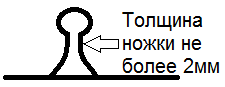
- Form: A distinctive feature of papillomas is the presence of a thin base (“leg” 1-2 mm thick), on which a larger nodule is located. Also, the shape of the papilloma can be cylindrical or cone-shaped, but in any case, the height of the formation will prevail over the width.
- Surface: smooth or cauliflower-like
- Consistency: most often soft, in rare cases - dense
- Color: flesh or brown
- Health Hazards: has no idea. Cases of tumors developing from papillomas have not been described.
- Possible changes that do NOT indicate malignant degeneration: can significantly increase in size while maintaining the stalk, the number of papillomas can also increase significantly.
- How to distinguish from other moles: The most characteristic sign of papillomas is a thin “leg”
A benign skin tumor caused by the human papillomavirus.


