Hemangiomas, better known as red moles, are benign growths that originate from blood vessels.
Red moles appear mainly in children of both sexes, less often in adults. It is impossible to prevent or predict their occurrence.
There is still debate as to which pathology a hemangioma is classified as - a vascular tumor or a congenital malformation. Recent data confirming the occurrence of tumors due to proliferation of vascular endothelium allow the neoplasms to be classified as vascular tumors.
What is a red mole?
Why are moles and dots red? Because it is actually vascular tissue filled with blood. If ordinary moles are skin growths, then red moles are several small (or single) overgrown blood vessels. In a pronounced process, the accumulation of blood vessels merges into a blue or burgundy spot.
Prevalence and localization
In most cases, vascular tumors are detected immediately after birth (87%), and 70% of the total mass are girls, who, accordingly, fall into the highest risk group. This pathology accounts for about 48% of all soft tissue and skin tumors in childhood. 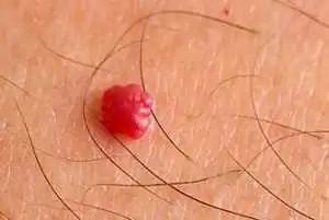
On the body, a red mole can be localized in any part; about 80% of tumors occur in the upper part of the body. Very rarely found in internal organs - liver, brain, lungs, bones.
- about 95% of all diagnosed vascular tumors are simple formations
- about 3% are cavernous
- and another 2% are mixed and combined variants of the disease.
Causes
No doctor can still give an exact answer as to why these formations appear. Why there are many red moles in the facial area is also difficult to explain. This is probably due to the abundant vascular network of facial tissues.
In children
It is generally accepted that a congenital tumor develops as a result of an intrauterine disorder in the formation of vascular tissue, which occurs against the background of incompetence and the processes of development and growth.
How does this happen? During the formation of organs and systems, vascular tissue penetrates all parts of the body without exception along a certain chain of pericytic cells. These cells, being a kind of conductors of information, react to the slightest lack of oxygen: if the fetal tissues experience hypoxia, the synthesis of special proteins that attract pericyte cells is immediately launched. These cells begin to pave new blood supply routes, thus eliminating hypoxia. In some cases, even after the cessation of hypoxia, the synthesis of specific proteins does not stop; the vascular system continues to develop, turning into voluminous tumor-like formations.
The second name for red moles is vascular hyperplasia. This means that the tumor arises as a result of disruption of the growth processes of vascular tissue, which lead to an increase in its quantity. How and in what way this process occurs is difficult to answer with 100% accuracy, since this requires monitoring the characteristics of intrauterine tissue development. The data presented are based on autopsy results of aborted and stillborn fetuses.
In adults
- Acquired pathology is associated with hormonal disorders, explaining the appearance of hemangiomas in adults (pregnancy, menopause, diseases of the endocrine system, as well as hormonal therapy or oral contraception).
- There are suggestions about the negative effects of ultraviolet and radiation exposure, viruses and chemicals that provoke tumor growth in adults.
- Microtraumas and skin cracks with permanent damage to the capillary network lead to such neoplasms.
- Long-term and uncompensated hypovitaminosis C, leading to thinning and fragility of capillaries, is also relevant among the causes.
- Red moles accompany the course of other diseases (for example, diseases of the liver, pancreas, cancer of internal organs). It is not uncommon for a cluster of red moles in a certain area of the body to indicate a predisposition to cancer in this area, a nearby organ.
Red moles in newborns
This is a common occurrence in babies, and if such a mole is noticeable in a newborn, then most often by the age of 3-5 the red mole may disappear. Since this is a benign tumor, it is not dangerous if:
- Does not bother the baby (skin itching, irritation, pain)
- Does not increase in size (in a month, for example, it doubled)
- Located in a non-hazardous place (if it is located under the eye, on the nose, genitals, on the face, then its removal is indicated)
Red moles are characterized by rapid peripheral growth, especially intense in the first months of a child’s life. Therefore, 10-12% of hemangiomas in children are removed for medical reasons. During the growth process, the tumor destroys tissue and leads to a cosmetic and sometimes functional defect, especially when located near or on vital organs (eyes, ears, brain). Impaired function of organs and tissues occurs due to compression of them by the tumor.
Features in adults
Primary hemangiomas do not occur in adults, i.e. They arise from existing, undiagnosed tumors. As a rule, visible formations are treated even before school age, so in adulthood either untreated superficial moles or tumors on internal organs are discovered.
Of particular danger is a vascular tumor on the spine, which is localized in the vertebral body and weakens its structure, sometimes leading to fractures.
Classification
According to morphology
Capillary. The histological structure of the neoplasm is compact layers or concentric groups of capillary vessels, closely adjacent one to one. The wall of each vessel consists of a basement membrane and 1 or several layers of epithelial-like cells. The lumens of fused capillaries are filled with formed elements of blood. In some cases, groups of vessels form lobules separated by stroma.
Cavernous. It consists of multiple cavities of various shapes and sizes, which are lined with 1 layer of endothelial cells, similar in structure to the endothelium of blood vessels. In some cases, rupture of the septa occurs with the formation of papillae in the lumen of the caverns.
According to location, vascular hyperplasias are divided into:
- Simple, with a subcutaneous location throughout the body;
- Cavernous, localized under the skin;
- Combined, having a supra- and subcutaneous part;
- Mixed, including other tumors, for example, lymphangioma, originating from lymphoid tissue.
By origin:
- Congenital, appearing immediately after birth or in the first months of life;
- Acquired, occurring in adults. Acquired red moles can only be of a subcutaneous location, i.e. simple. Complex forms of the disease, discovered due to complications or by chance, are congenital and not diagnosed in childhood.
With the flow:
Simple, not presenting a risk of complications or dysfunction of organs;
Difficult:
- near large vessels or vascular nodes;
- on or near vital organs and structures (eye, brain, ear);
- in places that are difficult to access (vertebrae).
Features of red moles
Vascular tumors have a number of characteristic properties that differ from other neoplasms:
- Rapid tumor growth during the first three months after birth.
- Accelerated (2-3 times compared to full-term) growth of education in premature babies.
- The likelihood of spontaneous regression of simple tumors (mostly small) during the first years of life. This explains the cessation of hemangioma growth when exposed to a number of factors, such as heat, cold, and certain chemicals.
- The impossibility of spontaneous resolution of cavernous, combined and mixed variants of pathology.
- Unpredictability of further development even after growth and involution have stopped.
Clinical picture
Simple angioma
This is a spot of varying sizes, predominantly red, rising above the skin. With simultaneous finger pressure on the edge of the tumor and healthy tissue, the angioma becomes pale and shrinks, and after the compression stops, it returns to its previous shape and color. In babies up to 3-4 months, peripheral growth of the vascular tumor is clearly visible. This can be verified by making an initial paper stencil of the tumor and applying it to the hemangioma after 15-20 days.
Cavernous angioma
This is a formation in the subcutaneous tissue with unchanged skin above it. It can be diffuse without clear boundaries or encapsulated. A bluish-colored formation is detected under the skin; in some cases, feeding vessels are visually visible. When pressing on the skin above the tumor, the formation decreases, and when the compression stops, it returns to its previous size.
The skin over the tumor may be warmer than the rest of the skin. No pulsation is detected above the formation. In some cases, upon palpation, the lobulation of the formation is noticeable. Cavernous hemangiomas located on the head, neck and near the ears are characterized by rapid growth with active germination into surrounding structures.
Combined angioma
This is a formation with a cutaneous and subcutaneous part; the subcutaneous part, as a rule, is larger.
Mixed tumors
These are various variations of the combination of a vascular tumor with lipoma, lymphangioma, keratoma and other neoplasms.
Spontaneous resolution
True regression of simple or superficial hemangiomas is observed in 10-15% of cases, especially when tumors are located in closed areas of the body. The brightness of the formation decreases, whitish areas appear, and peripheral growth completely stops. After 6-8 months. the hemangioma transforms into a smooth whitish-pink spot that does not rise above the surface of the skin. The skin over the spot undergoes atrophy, leaving only a small depigmented area by the age of 3-4 years.
Complications
Red dots are dangerous due to rapid growth and subsequent compression of nearby structures with disruption of their function, which is especially important when hemangiomas are localized in the brain, in the liver, or near the eye.
- Ulceration and inflammation during growth. Some types of red moles undergo reverse development after such complications.
- Bleeding due to injury, especially dangerous with extensive cavernous and combined hemangiomas, as well as tumors located on internal organs, since such bleeding is very difficult to stop.
- Infection (bleeding, ulcerated moles), i.e. the addition of a bacterial skin infection.
Diagnostics
With superficial hemangioma, the diagnosis is made on the basis of clinical and histological data. For extensive and deep processes, angiography is performed to determine the connection of the tumor with the vascular network, as well as radiography, which provides accurate data on the size and depth of the vascular tumor.
Treatment of red moles
Is it possible not to treat red moles? If the tumor does not interfere with organ function, is not dangerous for bleeding and does not grow, these marks of intrauterine life can be left without treatment, especially since these tumors do not carry the risk of malignancy. Moreover, it is not recommended to remove moles if they do not bother you, do not increase in size, or are located on closed parts of the body (they are not a cosmetic defect).
For extensive and deep processes, the doctor selects treatment - surgical or conservative; methods can be combined to increase efficiency. Therapy depends on the type of tumor, its location and size, growth rate, the presence of complications, and the age of the child.
Simple hemangiomas
Low-temperature destruction or cryodestruction is considered an effective method for treating small red moles. It can be performed in several ways: direct application of crystalline carbon dioxide to the surface of the tumor for 15-20 s or instrumental cryodestruction using liquid nitrogen. The effectiveness of treatment is up to 96%.
For simple angiomas of large size, hormonal treatment with prednisolone is advisable at the rate of 4-6 mg per 1 kg of weight, taking 1/3 of the dose at 6 a.m. and the remaining portion at 9 a.m. The duration of treatment is 28 days with the drug taken every other day. Gradual withdrawal of the drug is not required. During treatment, blood sugar and potassium are monitored.
Laser removal allows targeted action strictly on the tumor with minimal cosmetic defect. Modern laser systems with various types of pulses can coagulate both superficial and deep subcutaneous tumors without destruction of healthy tissue and complications.
Cavernous
When the process is located in a cosmetically unfavorable part of the face (cheek, nose, forehead, bridge of the nose), sclerosing therapy is used: special substances are introduced into the angioma, leading to aseptic necrosis and subsequent scarring of the tumor under the skin without scar formation and tissue deformation. Hydrocortisone, quinine-urethane, sodium chloride solution 10%, ethyl alcohol 70% are used as sclerosing agents. For complete sclerosis of the tumor, 10-15 injections are performed with breaks between each injection of 14-30 days, i.e. the process is quite lengthy.
When cavernous hemangioma is located on the thigh, shoulder, back and other closed parts of the body, surgical removal of the tumor is performed.
Combined
When the tumor is localized on closed parts of the body, radical surgical excision is advisable. Removing red moles rarely leads to any complications; the tumor is removed entirely with minimal cosmetic defect.
When localized on open parts of the body and face, microwave cryodestruction is recommended: irradiation of the hemangioma with an ultra-high-frequency electromagnetic field, followed by cryodestruction. This combination can significantly enhance the destructive effect of freezing, while maintaining the ability of epithelial cells to regenerate.
Hormonal, sclerosing and radiation therapy with Buki rays, which have a middle range between X-ray and ultraviolet irradiation, is also used.
Deep and extensive hemangiomas with dangerous localization
Such tumors are located on the neck, near the ears, on the head and are characterized by constant peripheral growth. The tendency to bleeding and ulceration of these types of angiomas does not allow the use of the treatment methods described above.
In case of such pathology, angiography is mandatory to determine the nature of the blood supply to the hemangioma and its anatomical relationship with nearby tissues and structures. One of the effective treatment methods is embolization of the tumor with a hydrogel, which reduces the blood supply to the tumor and its size.
Then cryodestruction is carried out without removing the tumor itself: after the necrobiotic process, the tumor partially resolves, leaving behind areas of atrophic skin, i.e. a cosmetic defect that can be eliminated by skin grafting if the patient wishes.
Red moles (angiomas) in children are a fairly common occurrence. They can even appear in newborns, and often the child is already born with such “decoration”. Red moles can be of different sizes and shapes - from a barely noticeable dot to a significant size convex spot.
Of course, having noticed such a mole on a child, parents panic: therefore, in this article we will talk about the features of such moles, find out what types they are, whether we should be afraid of these neoplasms, how to treat them, what preventive measures will help prevent their appearance.
Types of red moles in children
A red mole that appears on a child’s body is a formation of a benign nature. In essence, these are fused capillaries. Typically, red moles rise above the surface of the skin and have a convex shape. There are several varieties of red moles. Let's tell you more about them.
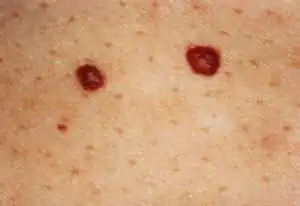
- Knotty. This neoplasm looks like a small point without additional capillary branches. In essence, it is simply a visible exit of a blood vessel to the surface of the skin.
- Pineal. Such a red mole can have different sizes, but in any case it will protrude significantly above the surface of the child’s skin.
- Branched. This angioma has various shapes - sometimes very bizarre (star, spider, flower, etc.). This unusual form of neoplasm is explained by the fact that blood vessels extend from the center of the mole in different directions.
- Flat. This red mole practically does not protrude above the surface of the skin and has the appearance of a flat plaque.
Hanging angiomas
This type of red mole is a new growth on a small stalk, due to which the mole seems to hang down. This angioma forms very quickly in children - it takes about a couple of months from its inception to full development. The size of such a neoplasm reaches about a centimeter in diameter.
It is impossible not to notice a hanging red mole - it protrudes significantly above the skin. If a child accidentally injures the angioma, it begins to bleed. Note that the surface of such a formation is uneven: it is lumpy, has lobules and papillae.
In children, such a mole usually appears as a result of trauma to the skin. Since the neoplasm begins to bleed quickly with any injury (even minor scratching), it is better to remove the hanging angioma.
Typically, the removal procedure is performed either surgically or using a laser. If the mole is still small, the removal procedure is carried out in more gentle ways: using liquid nitrogen or a solution of silver nitrate.
Vascular nevus
This red mole is the most common case. Vascular nevus can have different sizes: from a point to a spot of decent size. Sometimes a nevus can grow to such an extent that it literally covers the child’s entire limb.
Note that this formation is quite harmless, and if it is small in size, it often resolves safely on its own. A vascular nevus is formed in children as a result of injury to a blood vessel. However, sometimes the cause of its formation can be a hormonal imbalance or too aggressive sun tanning.
Especially often, such neoplasms appear on the child’s face, less often on the body. Sometimes a child is already born with such moles, and the reason for this is usually pathologies of pregnancy, injuries to the mother’s abdomen, or her addiction to alcoholic beverages during the period of gestation.
The method of removal is selected by the doctor individually. But most often it is removed with a laser, which is capable of selectively eliminating only affected tissues without affecting healthy ones.
Is there a danger to the child?
If a red mole is small in size and is not located in a visible place, then its appearance often remains unnoticed for a long time. However, if a neoplasm goes undetected for a long time, there is a danger of it developing into a tumor form.
If you notice a red mole on your child or notice that the mole has turned red, this problem should definitely be addressed. At a minimum, show the tumor to a dermatologist. Although angioma rarely develops into a malignant form, however, it is necessary to exclude all risks.
The main danger of red moles is their easy injury. Even from simple accidental scratching or scratching, the tumor can bleed profusely. Those angiomas that are subject to mechanical friction from clothing are often injured: in the area of the shoulders, chest, back, and abdomen.
Moles on the head also have an increased risk of injury, since the child can scratch his head, and in addition, these neoplasms have a risk of injury when the child is cut.
If several red moles appear at once or one large one, this may be a signal that there are hormonal imbalances in the child’s body, or gastrointestinal pathologies have arisen. In addition, red moles sometimes appear in adolescents as a result of hormonal changes.
If a child has an angioma located on the eyelids or close to the eyes, there is a possibility of decreased vision. The sense of smell worsens if the tumor partially blocks the nasal passages.
Is it worth removing the new growth that has appeared?
Usually, having noticed a red mole on a child’s body, parents immediately begin to think about how to remove this growth. But we note that if the angioma is small and located in a non-traumatic place, you don’t have to touch it - most likely, over time the neoplasm will resolve on its own.
However, a red mole should be removed if it:
- hurts;
- bleeding;
- located in a traumatic area;
- has significant dimensions;
- located on the face (spoils appearance);
- there is a risk of its degeneration into a malignant formation.
Rapidly growing angiomas must be removed. If the tumor has changed color, size or shape, this is also an alarming sign, and after examination, such a mole is also usually removed.
If the tumor is itchy, resulting in constant scratching, removal is usually also prescribed. Dermatologists also strongly advise removing red moles that appear on the scalp.
The fact is that this arrangement leads to frequent and almost inevitable injury to neoplasms. In this case, removal is usually done using a laser, sometimes with nitrogen.
After removal of the angioma, it is very important not to expose the child to ultraviolet rays for some time. The wound should be covered with a bandage so as not to further injure the vulnerable area and protect it from germs.
Removal methods
If the doctor has decided to remove a red mole from a child, there may be several options for this removal. Let's look at the features of each of them.
- X-ray. In this case, the angioma is irradiated with X-rays, after which it disappears. In order for the tumor to disappear, a course of several procedures is required. Experts do not recommend using this method to treat children, since the radiation in this case is quite strong.
- Surgery. In this case, the mole is destroyed mechanically: by direct excision. The method is not suitable for angiomas located on the face, as it leaves a scar.
- Carbon dioxide. Cauterization with carbon dioxide is used if the angioma is located superficially and does not have deep “roots”.
- Sclerosis. In this case, a substance is injected into the mole, which cuts off the neoplasm from the blood vessel that feeds it. As a result, after some time the formation dries out.
- Cryodestruction. This method involves the use of liquid nitrogen. The method is suitable for superficial formations and is capable of completely destroying the capillaries that form the angioma.
- Coagulation. This method is safe and effective and is often used in the treatment of children. Coagulation can be of different types:
- radio wave;
- infrared;
- light;
- electric.
The procedure requires preliminary anesthesia of the affected area.
Laser. This method is also often used to remove angioma in children. The laser leaves no scars, and the procedure itself lasts only a few minutes. In addition, after laser removal, the wound heals very quickly.Prevention
What measures will help prevent the appearance of angiomas in children? Let’s consider this issue in more detail.
- The most important preventive measure in this case will be protecting the child from unnecessary injuries, creating more comfortable and safe conditions for him. Of course, you cannot protect your baby from all falls, bruises and abrasions, however, if possible, it is necessary to prevent injury.
- Since sometimes red moles in children are the result of malfunctions in the gastrointestinal tract, an important preventive measure will be normalizing the child’s diet, including more healthy foods, vitamins, and fiber in the menu.
As well as strict adherence to the diet. Among other things, it is necessary to give the child more water to drink to cleanse the digestive tract.
If the weather is hot, Do not allow your child to spend a lot of time in the sun without protection. Aggressive rays of the sun can have a negative effect on delicate skin, contributing, among other things, to the appearance of red moles.So, we looked at the features of the appearance of red moles in children: we learned why they appear, what the danger is, and when it is necessary to remove these neoplasms. As you can see, there is no need to panic, but you should take your child to a dermatologist to have the mole examined.
In most cases, the entire problem is solved by self-resorption or a simple removal procedure.
Video on the topic
Doctor about nevi in general, and angiomas in particular.
By nature, birthmarks in children are not capable of harming a small organism. The appearance of pigment formation occurs after birth or after 10–15 weeks. Moles in newborns do not indicate the presence of a disease or pathology of the body. Nevus in infants is most often located on the back of the head, on the forehead, on the stomach.
Causes of birthmarks in newborns
The reasons for the appearance of birthmarks in a newborn should be considered:
- Hereditary factor. If a man or woman has a birthmark on the face or foot, there is a high probability that the child will be born with a similar location of the pigmented formation.
- Stress during childbearing. With nervous excitement, a drop in blood pressure occurs, as a result of which the blood vessels narrow and placental blood exchange is disrupted. Bursted blood vessels transform into a red birthmark in a newborn.
- Prolonged exposure to the sun. In babies born in the summer, the number of moles may increase or the existing formations will become darker in color.
- Changes in the hormonal background of a child are one of the reasons for the appearance of a mole in a newborn.
Medical statistics indicate that girls, premature and fair-skinned children are susceptible to the appearance of brown nevi after birth.
Types of nevi
A birthmark appears in a newborn at birth or within two months after birth. There are the following types of moles:
- Vascular formation - a large number of vessels, which are presented in the form of a convex or flat nevus. The color of the vascular spot is bright red or light pink. It is recommended to remove benign formations of this type, because after a while they increase in size and cause discomfort. The child is embarrassed about his appearance if the vascular nevus is located on the face.
- A simple mole has a smooth surface and is light brown or black in color. Appear after birth or in the first twelve months of life. The appearance of a simple nevus should not worry the parents of the baby, provided that the mole does not change shape, structure and color. If the nevus is located in a place that is subject to constant trauma, doctors recommend removing it.
Vascular formations are divided into the following types:
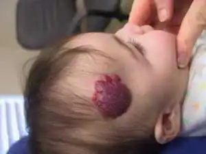
- A flaming mole or port-wine stain appears on the baby's scalp or face. The shape of such a spot is flat, the color is red. As the child grows, the size of the nevus also increases. It is not recommended to resort to removal of the formation, as it can be treated, which is based on the effect of a laser beam or infrared radiation.
- Hemangioma. It is not immediately detected on the baby’s body. After three to four months it will begin to manifest itself. Hemangiomas appear in various parts of the body and increase sharply. Medical statistics say that upon reaching the age of ten, the hemangioma will disappear, so there is no need to remove it when detected.
- The stork's bite is located on the back of the head, eyelid, and bridge of the nose. This type of nevus is pink in color. There are situations when many spots accumulate in one place.
Among the simple formations there are:
- A red nevus appears at birth or within three years and can be located in any area. Red nevi do not need treatment or removal unless there is a rapid increase in size.
- The hanging formation is benign. Because a hanging nevus involves epithelial cells, the appearance is similar to a growth. The color varies from flesh to dark brown. There are hanging moles in the armpits and groin. The presence of growths is dangerous for a child, so it is necessary to regularly visit a dermatologist for examination and examination, during which the absence or presence of cancer cells will be determined.
- The anemic spot is small in size and forms on the back or face. Vascular underdevelopment can provoke the appearance of an anemic spot. It is necessary to remove the formation using a surgical method.
- The flat blue nevus is large in size. Divided into simple and cellular. The color of the simple varies from blue to dark blue, the diameter does not exceed one centimeter, and has a smooth surface. Cellular nevus is malignant, exceeds three centimeters in diameter, and the surface is covered with nodules. Localization – buttocks, feet, hands. Removing blue spots is dangerous to health.
- The brown spot has a flat structure and can disappear on its own after five to six years. They do not pose a danger to the child's body.
- The Mongolian spot, which is located on the buttocks and thighs, is characteristic of a child with Asian roots. It is not dangerous and disappears two years after birth.
- Red spots appear on the baby's head. After the child turns one year old, the red spot will disappear without surgery or therapy.
- Strawberry hemangioma. Occurrence is rare. It has a soft structure and bright color. If the formation appeared at birth, after two years it will disappear on its own or change color to light.
When moles are dangerous
When moles appear in a newborn, you need to pay attention and regularly visit a dermatologist. The doctor will use a dermatoscope to examine the formation, determine its nature and the presence of cancer cells. When bathing or changing your baby, you should carefully examine new spots or changes in existing ones. Birthmarks in a baby are not dangerous if there are no alarming symptoms. These signs include: intense increase in size and change in color, localized on the face, clear boundaries have disappeared, blood or clear liquid is released. It is necessary to visit the office of a pediatric dermatologist if the nevus in the area that is swollen and irritated is covered with hair. A cause for concern is the appearance of a bumpy surface or compaction that begins to peel off.
After the examination, the dermatologist will determine the period during which parents need to monitor the spot. If the situation worsens, the doctor will prescribe surgery to remove the nevus to avoid degeneration into a malignant formation. It is allowed to remove the growth after two years if there is no need to get rid of the stain.
When is treatment required?
If a brown nevus appears in a newborn, there is no need to worry and conduct a comprehensive medical examination. It is necessary to monitor the size, shape, and surface of the birthmark. In the absence of intense enlargement, redness or darkening, or bleeding, it is not advisable to remove the spot. If there have been cases of melanoma along the family line, parents must observe:
- Avoid mechanical damage to the nevus.
- Regularly visit a pediatrician, who will examine the formation independently or refer you to an appointment with a pediatric dermatologist.
- Minimize the baby's exposure to direct sunlight. When visiting the beach, apply protective creams to the child’s skin and put a hat on the head.
If a newborn's birthmark rubs, rubs, or the child accidentally tears it off with his hand, the formation must be removed. Without surgical intervention, the risk of the spot transforming into a malignant tumor increases. Hanging moles, which become inflamed more often than others, must be removed. When scheduling removal, parents follow the dermatologist's instructions.
Medicine offers several safe ways to remove growths on a child’s skin:
- Laser removal. The procedure is performed under general or local anesthesia. The choice of anesthesia depends on the size of the spot.
- The cryodestruction method is indicated for older children, as the procedure is painful.
- Removal with a scalpel. A surgical method for excision of large formations, hemangiomas. The operation requires general anesthesia.
The development of melanoma in infants is a rare occurrence, but visiting a doctor should not be neglected. This helps control the development of nevus and detect malignant cells.
