Almost every person has one or more moles on their body. As a rule, they do not cause discomfort and do not affect health in any way. But lately, more and more often, many people have begun to develop cancerous moles, which are harbingers of a terrible disease - skin cancer. Unfortunately, few people can distinguish an ordinary mole from a malignant one, which leads to the development of the disease. In this article we will look in detail at what cancerous moles look like, what their features are and how to get rid of them.
What is a malignant mole?

A malignant mole is a cancer called melanoma. It can form anywhere on the body, but most often appears in exposed areas, as they are exposed to ultraviolet radiation.
Melanoma is the most dangerous form of cancer. It is very important to monitor all moles on the body, especially if there are a lot of them. If a malignant mole is detected early, the development of melanoma can be prevented.
Characteristic
To prevent the development of skin cancer, it is very important to know how to identify a cancerous mole. For comparison, consider the characteristics of ordinary moles and cancerous ones.
Common, harmless moles have a uniform color (brown or black) with a clear border that separates them from the rest of the body. Moles are round or oval in shape, their size is approximately 6 mm.
There can usually be from 10 to 45 moles on the human body. New ones can appear up to 40 years, and some, on the contrary, disappear with age.
Now let's talk about malignant moles. As a rule, there are a lot of them, and in appearance they are very different from ordinary ones in color, size, outline (more on this below). It happens that an ordinary mole can develop into a malignant one. In order not to miss this moment and start treatment on time, you need to undergo examination every six months or year.
Signs of malignant moles

Malignant moles (cancer cells) have some telltale signs that can help differentiate them from a typical mole. The initial stage of the disease - melanocytic dysplasia - is still treatable. Therefore, if a cancerous mole is identified and removed in time, the development of skin cancer can be avoided.
In 1985, dermatologists developed the abbreviation ABCDE, each letter of which represents one sign of a cancerous mole. Over time, this abbreviation was adapted into Russian, and it began to sound like AKORD (asymmetry, edges, color, size, dynamics). It is by these signs that a malignant growth can be identified. Let's take a closer look at each sign.
- Asymmetry. As mentioned above, ordinary moles are symmetrical. If you notice even the slightest asymmetry, you should urgently consult a doctor.
- The edges. Cancerous moles have jagged, blurry, and even jagged edges.
- Coloring. Common moles are usually one color (black or brown). Cancerous moles on the body can be of different shades, including red.
- Size. Ordinary moles do not exceed 6 mm in volume. If the mole is larger than 6 mm, then most likely it is malignant. In addition, cancerous moles quickly increase in size.
- Dynamics. If the mole is benign, then it does not change its color or size over the years. If you begin to notice changes, you need to consult a doctor for examination.
So we've looked at the characteristics and symptoms of a cancerous mole. If you notice at least one of these points, immediately run to the doctor to prevent the possible development of melanoma.
Risk factors
A person can live with moles all his life, and they will not bother him in any way. But there is always a risk that a standard neoplasm will begin to develop into a malignant one. Let's look at the most likely risk factors for a mole to become cancerous:

- Having severe sunburn or prolonged exposure to sunlight on common moles.
- People with white skin, light hair and eyes, and freckles are more likely than others to develop cancerous moles on their bodies.
- If there are a lot of ordinary moles on the body, then there is a very high risk that sooner or later they will begin to develop into malignant ones.
- Large sizes of standard moles. If an ordinary mole itself is large in size, then the risk of developing melanoma increases significantly.
- Hereditary factor. If your relatives have skin cancer, then you are also at risk.
To avoid the development of melanoma, it is important to take into account all these factors and, at the slightest suspicion that a mole is becoming malignant, go to the doctor.
How is the examination carried out?
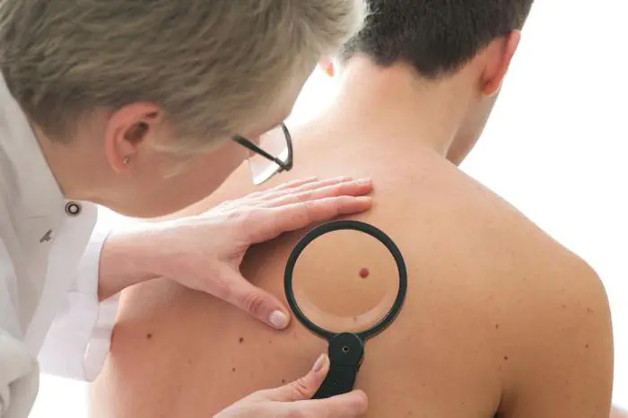
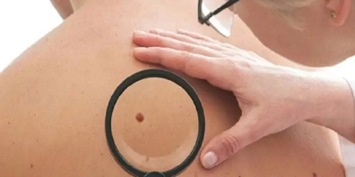
To diagnose cancerous moles, a dermatoscopy must first be performed. Using a magnifying glass and a dermatoscope, you can examine the signs of melanoma on the surface of the growth. This involves studying and evaluating the pigment of the skin and blood vessels by taking a sample of the growing mole.
The diagnosis is confirmed after a biopsy (histological analysis). Using local anesthesia, part of the mole is removed in order to carefully study its structure in the laboratory. This method is one of the most accurate.
It is possible to diagnose cancer at an early stage using a computer microdermoscopy system, but this method is not yet very widespread.
Most importantly, if you yourself notice even the slightest change in the appearance or size of your moles, you need to consult a doctor. The doctor will choose the necessary diagnostic method, and with timely examination, the risk of developing skin cancer is reduced.

Some facts you need to know about cancerous moles
If a person has more than 50 moles on his body, then he needs to monitor their condition very carefully and contact an oncologist at the slightest change.
In addition to the above signs, there are several factors that you should pay attention to:
- Darkening. A common mole may be black in color. But if it was originally brown and suddenly began to darken, then this is a cause for concern. Many people do not pay attention to the darkening of moles, since black is considered the norm.
- Inflammation. If the skin around the most common mole is inflamed or redness has formed, then you should urgently go to the doctor for examination. And under no circumstances should you treat inflamed areas of the skin with alcohol, this can only worsen the situation.
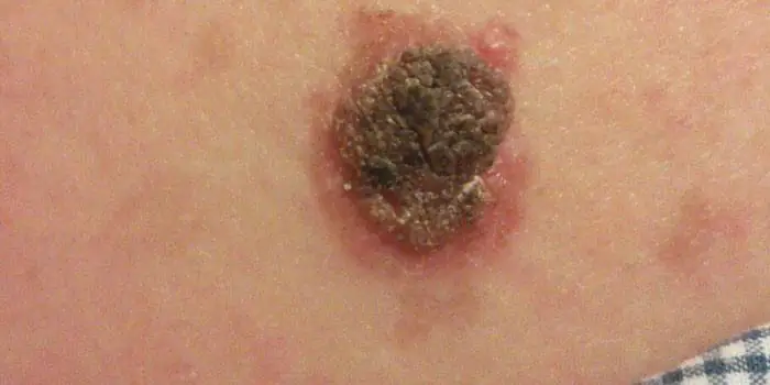
- Surface. The boundaries of the mole have already been discussed. But you should also pay attention to its surface. It should be smooth on top, without obvious roughness. If there are any, then this is a sign of the development of melanoma.
- If darkened areas of skin appear around an ordinary mole, then this is a big cause for concern. It is urgent to check with an oncologist.
As you can see, there are a lot of signs of melanoma development. It's very difficult to remember them all. Remember that any change in a standard mole may indicate that it is becoming malignant.

Treatment
Currently, the only possible treatment option for melanoma is the removal of cancerous moles. The complexity of the operation depends on the severity of the situation and the size of the formation. For small growths, half an hour will be enough.
When removing a cancerous mole, the surgeon cuts out a small area of skin (1 cm) around the mole to prevent new ones from appearing in the same place. The larger the malignant mole in volume and size, the more skin around it needs to be removed.
After the mole is excised, a sample is sent to the laboratory. There they study its prevalence level, that is, the likelihood that new such growths will appear on the body.
What forecasts do doctors give?
Tumor thickness is the main criterion by which oncologists make predictions. If the mole was small, then the risk of its reoccurrence is small, and the chance of life without melanoma increases.

The rehabilitation period after removal of the growth is short. A scar forms at the site of the removed mole, which heals quite quickly. The size of the scar depends on the method of removal.
Laser removal is the safest method, which leaves almost no marks or scars. But this method cannot be used in advanced cases.
It should be noted that if the operation is performed in a timely manner, then the risk of melanoma developing in the future is very small. In the future, you just need to be regularly monitored by an oncologist to avoid relapse.
Conclusion
In the article, we examined in detail what cancerous moles are, what are the methods of treating them, as well as signs that will help identify their development at an early stage. Take care of your body and be healthy!
It's rare to see a person without small dark marks on their body. Is it worth paying attention to these points? Only a doctor will distinguish between dangerous and normal moles - malignant melanoma or harmless nevus - and give recommendations on what to do with them. Is it worth worrying about the appearance of new formations, when immediate contact with specialists is required, what are the signs of cancer development - the answers to these questions remain to be found out. No one is immune from disaster, and early diagnosis will protect you from severe consequences.
What is a mole
The first tiny spots may appear in children in infancy. A mole is a small formation on the skin - a nevus - that is considered benign and harmless. The basis for their appearance is melanocyte cells that accumulate the natural pigment melanin. Depending on its quantity, a difference in color is observed. Available colors:
The shape of the tumors depends on the location and concentration of melanin. They may have a stalk or be located under the skin, be flat and convex. The most common type is round, but there are exceptions. The development of neoplasms is provoked by ultraviolet radiation - natural from the sun, in a solarium. Hereditary factors cannot be excluded. A common cause of growth is hormonal imbalance, characteristic of periods:
- puberty;
- pregnancy;
- menopause.
What types of moles are there?
One person may discover very different tumors. Types of moles are classified according to several criteria. This helps in correct diagnosis in case of changes. They differ in:
- origin– congenital, newly acquired;
- structure– pigment, vascular;
- place of education – in depth, on the surface, in the boundary layer;
- raised above the skin – flat – even, protruding as a hemisphere, pedunculated, larger birthmarks;
- potential threats – dangerous, degenerating into melanoma, non-dangerous.

Safe moles
Those who have dark spots on their skin should be wary of their changes. In time, detected signs of degeneration into melanoma contribute to the timely removal of the formation and preservation of health. Safe moles are different:
- the presence of a stalk – it cannot be formed by malignant cells that grow randomly;
- long-term condition without changes.
Spots that appear soon after birth are not considered dangerous. It is important that they are small in size. Good – non-dangerous – signs of neoplasms include:
- flesh tone;
- unchanged pattern of the skin of the nevus and adjacent tissues;
- soft consistency;
- hair on the surface of the neoplasm - growing from the skin, indicates the absence of pathologies;
- diameter no more than 5 mm;
- symmetry;
- nevus in the form of a spot.
Which moles are dangerous?
Why do people with nevi on their bodies need to monitor their changes? There is always a threat of degeneration of non-dangerous tumors into a cancerous tumor. What moles are dangerous to health? Key signs you need to know:
- change in shades towards the dark side, the appearance of multi-color;
- rapid increase in size - exceeds two millimeters per year;
- occurrence of cracks;
- the formation of asymmetry due to uneven growth;
- lack of elasticity;
- the appearance of itching, burning;
- presence of discomfort.
The appearance of dangerous moles requires an immediate visit to a specialist to clarify the nature of the changes and the likelihood of developing skin cancer. Pathological transformations provoke:
- injury to the nevus due to negligence;
- self-removal;
- abuse of exposure to the sun, use of a solarium;
- location of the formation in places of frequent contact with clothing - on the neck, head, genitals, legs;
- placement in the hair, on the face, palms - where there is a high probability of injury;
- previously removed melanoma.
Why are moles dangerous?
Not a single person is protected from the sudden proliferation of cells of a harmless mole. Melanoma is an extremely serious disease. Changes not detected at the initial stage can result in death. The provoking factor is unsuccessful independent removal of tumors. Moles are dangerous because of their ability to:
- transform into an atypical – precancerous form;
- grow to large sizes;
- turn into cancerous;
- with minor external changes, metastases actively spread throughout the body through the circulatory and lymphatic channels.
How quickly does melanoma develop from a mole?
The transformation of a nevus into a cancerous formation can occur in different ways. The process depends on the stage of the disease and the type of tumor. Instant metastases are dangerous. Begins:
- growth of cancer (oncological) cells in the deep layers of the epidermis;
- their entry into the blood and lymph;
- penetration into the lungs, liver, kidneys;
- growth in these organs;
- complete damage to the body;
- death.
The growth phases of pigment cells are observed, along which melanoma develops from a mole. There are varieties:
- horizontal– damage to the upper layers of the skin occurs, lasting up to 10 years, but metastases do not appear;
- vertical– accompanied by the spread of cancer cells throughout the organs, can last two years, has an unfavorable prognosis;
- nodal – especially dangerous – characterized by deep spread within two months.

The first signs of melanoma
The patient can be assisted only when suspicious changes begin to be identified. The diagnosis, research, and referral for surgical treatment save a person’s life. The first signs of melanoma:
- increase in the height of the tumor;
- bleeding;
- the appearance of discharge;
- redness;
- burning, itching;
- swelling of tissues;
- softening of the nevus;
- the appearance of a crust;
- thickening;
- hair loss;
- expansion of pigmentation around the lesion.
With the further development of dangerous melanoma, the following are observed:
- significant change in size;
- the appearance of pain;
- enlarged lymph nodes;
- surface ulceration;
- formation of new foci;
- bleeding from places of pigmentation;
- liquid separation;
- skin thickening;
- the appearance of an earthy tint;
- signs of metastases are chronic cough, weight loss, cramps, headaches.
How to distinguish a mole from melanoma
To recognize which moles are dangerous and which are not dangerous, you need to know what they look like. A person with nevi, in order to avoid dire consequences, must constantly monitor the appearance of new formations and changes that occur. You can distinguish a mole from melanoma by its signs. Non-dangerous neoplasm:
- symmetrical;
- with smooth edges;
- uniform in color;
- with dimensions not exceeding 6 millimeters.
Features of dangerous melanoma that require seeking help from dermatologists:
- growth in a short time;
- pronounced asymmetry of shape;
- heterogeneity in color - the presence of inclusions of several shades;
- lack of clear boundaries - the contour line is blurred, jagged, and looks like a coastline on a geographical map;
- increased diameter over six millimeters;
- variability of any parameters - color, size, shape.

What dangerous moles look like
What do nevi that are subject to pathological changes look like? Only a doctor can correctly distinguish between non-dangerous tumors. Dangerous formations look like this:
- blue– compactions under the skin with clear boundaries, with dimensions no more than 10 mm;
- nodal– round, flat in shape, color – brown, black;
- cutaneous– often pale, convex;
- halo nevus – pigment surrounded by a light or white rim;
- spitz- looks like a dome-shaped tumor of pink shades, with the possible presence of a hole through which blood and liquid leak;
- connecting- connect individual entities into a whole.
Mole with jagged edges
One of the signs of a non-hazardous formation turning into a dangerous one is a change in contours. It often has blurred edges and scalloped borders. There are non-dangerous types of nevi - dysplastic. Only a specialist can make a correct diagnosis. A mole with uneven edges can be dangerous if there are additional signs of melanoma:
- accelerated changes in size;
- the presence of clearly defined asymmetry;
- the appearance of highly indented boundaries.
Rough mole
Such a neoplasm is harmless if its diameter is no more than 5 mm and remains constant in size. Often its appearance signals a lack of vitamins and nutritional disorders. Doctors advise coming for a consultation if it is discovered that:
- the smooth nevus turned into a rough one;
- bothered by burning, itching, tingling;
- irregularities and compactions appeared in the middle;
- areas with different shades formed;
- diameter has increased significantly.
A dangerous rough mole requires immediate examination if:
- the appearance of bleeding;
- development of the inflammatory process;
- rapid change in size;
- formation of asymmetry;
- formation of purulent discharge;
- the occurrence of painful sensations when touched;
- the emergence of an irregular shape, blurred boundaries, along the edges of the neoplasm.
Large moles
Large formations on the skin are pigment spots. When they remain unchanged and do not cause inconvenience, this is not a dangerous phenomenon. It is important to constantly monitor their appearance, color, and size. To eliminate worries, you need to consult a dermatologist. During the visit, the specialist will conduct a diagnosis and give a forecast of the risk of developing a malignant neoplasm. Large moles become dangerous if they:
- injured;
- thickened;
- started to itch;
- were unsuccessfully removed independently;
- changed in size, shape;
- are bleeding.
What moles can be removed
Often nevi cause trouble for women when they are in a visible place - the face, neck. Even if they do not bother you, using removal will be the right decision - the appearance will improve significantly. After the procedure, the doctor must necessarily send the tissue for histological analysis to decide whether the mole is malignant or not. If the neoplasm is not dangerous, does not bother you, and does not change in size, surgery is not required. What moles cannot be removed? Experts believe:
- there are no contraindications;
- It is important to choose the right excision technique.
You should be careful about skin growths; it is unacceptable to remove them yourself. Only the doctor will determine whether a nevus is dangerous or not and decide what to do with it. You can delete it if:
- injured from clothing - on the neck, in the groin area, under the armpits;
- cause pain when touched;
- are located under the hair on the head and can be damaged when combing or cutting;
- change color, shape, outline;
- significantly increase in size;
- characterized by the presence of burning, itching;
- accompanied by inflammation and bleeding.
Malignant moles are a group of skin cancers of epithelial origin, which in most cases have an unfavorable outcome.
WHO has proposed the following classification of malignant tumors of the skin and its appendages:
- epithelial tumors (skin cancer, adenocarcinoma of sweat and sebaceous glands);
- connective tissue tumors (skin fibrosarcoma, dermatofibrosarcoma, leiomyosarcoma);
- vascular tumors (hemangioendothelioma);
- skin melanoma.
Features
They occur in people of any age and gender (some types have their own characteristics).
- There is a greater predisposition in people with pale skin.
- Direct relationship with the surrounding environment. Provoking factors can be chemical carcinogens, physical (ultraviolet) or biological (human papillomavirus) effects.
- This type of neoplasm can be either primary (formed on intact skin) or secondary (occurs at the site of benign formations).
- The routes of metastasis in such cases are to regional lymph nodes (distant metastases occur in rare and advanced cases).
- Skin cancer most often occurs in exposed areas of the skin (about 60% of all cases). The skin of the body accounts for about 7-10%.
- For a long time they do not have any symptoms other than external manifestations (as they progress, paraneoplastic syndrome occurs).
- It often occurs as a hereditary pathology (there is a history of cases of the disease in close relatives).
The given criteria are universal for any education in oncology. According to ICD 10, malignant skin neoplasms are coded C 43-C 44.
How to determine whether a mole is malignant or not
There are a number of important external signs of malignant moles (ABCDE method):
A – asymmetry
You can check this sign by drawing a conditional line through the supposed middle of the mole (in the case of malignant formations, you will get two halves of different sizes).
In benign formations, the edges are smooth and clear, but when malignant, the edges become torn like flames and lose their clarity.
C – coloring (color)
The coloring of an ordinary mole should be uniform throughout. In the case of polychrome coloring and a predominance of black tint, you must immediately contact a specialist.
D – diameter (diameter)
The diameter of the malignant lesion usually exceeds 6 mm.
E – evolution of development
All benign formations change slightly over time (increase somewhat, stretch). In the case of malignant forms, intensive growth is observed.
Indirect bad diagnostic signs:
- itching and redness;
- disappearance of the skin pattern at the site of the lesion;
- peeling and swelling of the lesion;
- hair loss;
- ulceration and change in density;
- bleeding and weeping surface;
- screenings from the main focus (satellites).
Only an oncologist, after a thorough examination, can determine for sure whether a mole is malignant or not, how and what to do with it in the future (observe or remove). If it is impossible to recognize degeneration, it is necessary to include other diagnostic methods in the examination.
Conditionally malignant neoplasms (precancer)
A number of skin formations are classified as conditionally malignant, since they have a high risk of transformation into cancer even with minor exposure. These include:
- Bowen's disease. It is represented by a plaque with irregular edges and is localized on the skin of the face and torso. The color varies from reddish to yellow-brown. Size up to 10 cm. The lesion is dense on palpation, covered with scales.
- Xeroderma pigmentosum. Hereditary predisposition is expressed. Has high sensitivity to sunlight. It is represented by atrophied reddish areas of the skin, resembling freckles. Size is variable. Often develops into primary multiple cancer.
- Paget's disease. It is localized in the area of the areola of the mammary glands and is represented by a reddish or pink spot. The surface of the formation may peel off. Diameter is variable.
- Keratoacanthoma (horny mollusk). Localized on open areas of the skin. It has a hemispherical shape and dense consistency. Protrudes above the surface of the skin. The tumor is grayish-brown in color, and the horny masses above it are gray. A crater often appears in the center as the disease progresses.
- Actinic keratosis. Occurs on any part of the body. Characterized by multiple spots of pale or brown color. Local capillary hemorrhages (telangiectasia) are visible around the formations.
- Cutaneous horn. A formation of dirty gray or brownish gray color, protruding significantly above the surface of the skin. Dimensions up to 1 cm. Localization mainly on the skin of the face.
A special group that has a high risk of malignancy, i.e. malignancy, consists of melanoma-dangerous pigmented nevi:
- Dysplastic nevus. It is represented by a birthmark, which is located on the same level as intact skin. The shade varies from brown to black. The edges are fuzzy.
- Borderline pigmented nevus. Represented by a dark colored knot. It has a greater density than surrounding tissue.
- Blue (blue) nevus. The knot is blue, bluish, dark brown. The edges are smooth and crisp. Soft-elastic upon palpation, may not protrude above the surface of the skin.
- Nevus Oto. Combined damage to the skin, eyes (sclera, iris) and mucous membranes. It is localized in the zone of innervation of the I and II branches of the trigeminal nerve (face, neck), in the photo it looks like a dirty gray spot of irregular shape.
- Giant pigmented nevus. The formation exceeds 20 cm in diameter. The color is brown or greyish. Sometimes covered with hair.
- Dubreuil's melanosis. The main element is a grayish-brown plaque with uneven outlines, dense on palpation.
Due to the high degree of danger, formations suspicious for malignancy in most cases are subject to surgical treatment (removal) followed by a biopsy, which makes it possible to understand the exact cellular composition of the tumor-like formation.
Cancerous moles
Directly cancerous, i.e. malignant moles are melanoma, basal cell and squamous cell carcinoma.
Melanoma
Another name is melanoblastoma. It is divided into several types.
Superficial spreading (up to 70%)
- size up to 2 cm;
- the color is brown and dark brown uneven;
- localization on the skin of the chest, back and limbs;
- vertical and/or horizontal growth;
- at the level with intact skin;
- the edges are not smooth, wavy;
- various shapes (round, hexagonal);
- the surface is lumpy.
It remains in a dormant state for a long time and often occurs against the background of pigmented nevi. At the initial stage of the disease, it is difficult to understand the specific nature of the pathological focus, which makes timely diagnosis difficult.
Nodular (nodular)
It is considered the final stage of development of the surface form.
- localization – head and neck;
- polyp-type node;
- the surface is smooth, shiny, uneven;
- tendency to ulceration;
- color scheme similar to the surface form.
Lentigo melanoma (melanotic freckles)
It always occurs de novo, that is, on unchanged skin. It can extremely rarely occur against the background of nevi or other precancerous diseases. It appears more often in men of advanced age (over 70 years), in young people it is registered in less than 1% of cases.
- localization – face and neck;
- multiple nodules;
- dark blue, dark brown and light brown;
- diameter 1.5-4 mm;
- the edges are not smooth, wavy.
Acral-lentiginous
- localization – palms, soles, nails;
- uneven edges;
- color gray and black.
It occurs more often in people of the Negroid race. It grows extremely slowly. Has a relatively favorable prognosis.
Basal cell skin cancer
It is also divided into several forms.
Nodular
The most common type of pathology. Characteristics:
- dense multiple nodules prone to fusion;
- diameter 2–5 mm;
- localization – facial skin;
- color ranges from pale pink to yellow-brown;
- has a tendency to ulcerate.
Surface
- rounded superficial lesion;
- diameter 1 cm or more;
- on the surface there are papillomatous growths and ulcerations;
- occurs on the skin of the trunk and limbs;
- yellowish color.
Scleroderma-like
Relatively favorable form.
- fast growth;
- does not protrude above normal skin at the beginning, and then, due to endophytic growth, is pressed inward like a scar.
Fibroepithelial
Has a relatively favorable outcome.
- node shape (color varies widely);
- diameter 1-3 cm;
- tightly elastic;
- localized on the body.
Squamous cell carcinoma
Has three forms.
Superficial
Difficult to identify and most common form.
- single spot or nodule;
- color is variable, often whitish;
- there are multiple crusts on the lesion;
- protrudes above the surface of the skin;
- has a tendency to erosion.
Infiltrative (endophytic)
- multiple dense nodes;
- crusty;
- tendency to form deep ulcers.
Papillary form (exophytic)
- looks like cauliflower in appearance;
- rises above the skin;
- tendency to bleeding and ulceration.
Melanoma and all forms of squamous cell carcinoma most often develop regional and distant metastases in a short period of time (2-4 months), so they are the most dangerous. Malignant moles do not make themselves felt for a long time, and for this reason the patient is often hospitalized in the hospital against the background of severe paraneoplastic syndrome.
Video
We offer you to watch a video on the topic of the article.
