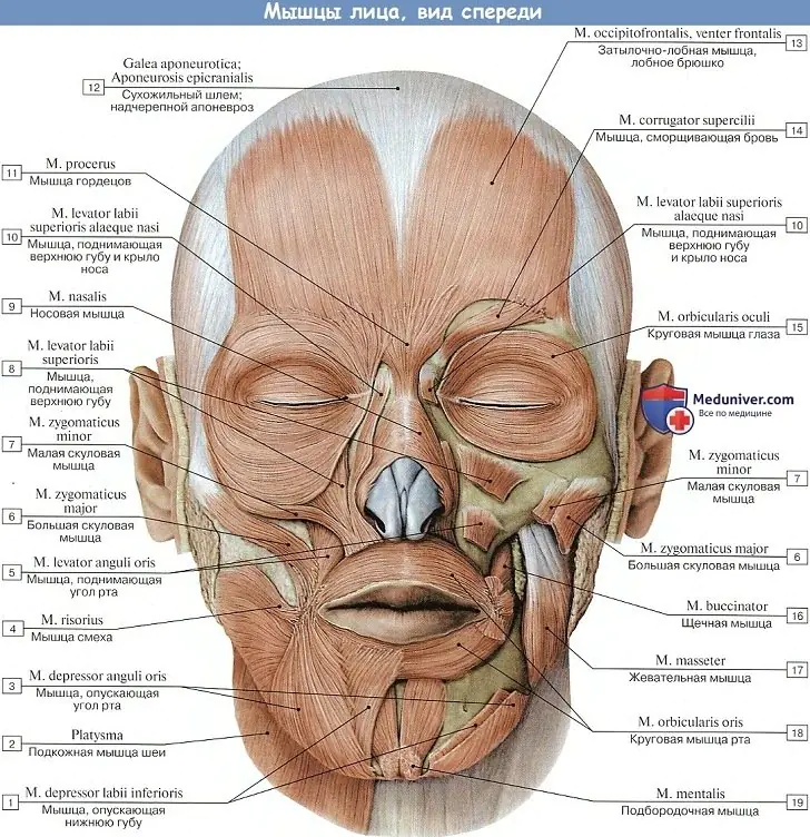
Facial muscles are the muscles of the face. Their specificity is that they are attached to bones at one end and to the skin or other muscles at the other. Each muscle is encased in fascia - a connective membrane (thin capsule) that all muscles have. What's happened fascia, every housewife can imagine - when cutting meat, we get rid of white films, which, due to their density, worsen its soft consistency. In relation to the facial muscles of the face, in comparison with the muscles of the body, these membranes are so transparent and thin that, from the point of view of classical anatomy, it is believed that the facial muscles do not have fascia. In any case, the surface of each muscle fiber on the face has a denser structure than its interior. These connective tissue membranes are woven into the structure of the entire fascial system of the body (through aponeuroses).
It is the contractions of the facial muscles that give our face a variety of expressions, as a result of which the facial skin shifts and our face takes on one expression or another.
1. Muscles of the cranial vault
Epicranial muscle
Transverse nuchalis muscle
Anterior auricularis
Superior auricular muscle
Posterior auricular muscle
2. Muscles of the eye circumference
2.1. Corrugator muscle
2.2. Muscle of the proud
2.3. Orbicularis oculi muscle
4. Muscular system of the nose
4.1. Nasal muscle: alar part, transverse part
4.2. Depressor septum muscle
4.3. Levator labii and ala nasi muscle
5. Cheek muscles:
Zygomatic major muscle
Zygomatic minor muscle
Muscles of the cranial vault
A large percentage of the muscles of the cranial vault are complex in structure supracranial muscle, which covers the main part of the skull and has a rather complex muscle structure. The epicranial muscle consists of tendon And muscular parts, while the muscular part, in turn, is represented by the entire muscle structure. The tendon part is formed from connective tissue, so it is very strong and virtually non-stretchable. There is a tendon part in order to maximally stretch the muscle part in the areas of its attachment to the bones.
Schematically, epicranial muscle can be represented as the following diagram:
The tendon part is very extensive and is otherwise called the tendon helmet or supracranial aponeurosis. The muscular part consists of three separate muscle bellies:
1) frontal abdomen located under the skin in the forehead area. This muscle consists of vertically running bundles that begin above the frontal tubercles, and, heading down, are woven into the skin of the forehead at the level of the brow ridges.
2) occipital abdomen formed by short muscle bundles. These muscle bundles originate in the region of the highest nuchal line, then rise upward and are woven into the posterior sections of the tendon helmet. In some sources, the frontal and occipital abdomen are combined into fronto-occipital muscle.
Figure 1. Frontal, occipital abdomen. Tendon helmet.
3) lateral abdomen is located on the lateral surface of the skull and is poorly developed, being a remnant of the ear muscles. It is divided into three small muscles suitable for the front of the ear:
- Anterior auricularis moves the auricle forward and upward.
- Superior auricular muscle moves the auricle upward, tightens the tendon helmet. A bundle of fibers of the superior auricular muscle, which intertwined in a tendon helmet, called temporoparietal muscle . The anterior and superior muscles are covered by the temporal fascia, so their depiction in anatomy textbooks is often difficult to find.
- Posterior auricular muscle pulls the ear back.
Figure 2. Lateral belly: anterior, superior, posterior auricular muscles
Muscles of the eye circumference
Corrugator muscle, starts from the frontal bone above the lacrimal bone, then goes up and attaches to the skin of the eyebrows. The action of the muscle is to bring the eyebrows to the midline, forming vertical folds in the area of the bridge of the nose.
Figure 3. Corrugator muscle.
Muscle of the proud (pyramidalis muscle) - originates from the nasal bone on the back of the nose and attaches at the other end to the skin. During contraction of the procerus muscle, transverse folds are formed at the root of the nose.
Figure 4. Proud Muscle
The orbicularis oculi muscle is divided into three parts:
- Orbital, which starts from the frontal process of the maxilla, and follows along the upper and lower edges of the orbit, forming a ring consisting of muscle;
- Century-old – it is a continuation of the circular muscle and is located under the skin of the eyelid; has two parts - upper and lower. They begin at the medial ligament of the eyelids - the upper and lower edges and go to the lateral corner of the eye, where they attach to the lateral (side) ligament of the eyelids.
- tearful – starting from the posterior crest of the lacrimal bone, it is divided into 2 parts. They cover the lacrimal sac in front and behind and are lost among the muscle bundles of the peripheral part. The peripheral part of this part narrows the palpebral fissure and also smoothes the transverse folds of the skin of the forehead; the inner part closes the palpebral fissure; the lacrimal part expands the lacrimal sac.
Figure 5. Orbicularis oculi muscle
Orbicularis oris muscle
The orbicularis oris muscle has the appearance of a flat muscle plate, in which two layers are distinguished - superficial and deep. The muscle bundles are very tightly fused with the skin. The muscle fibers of the deep layer run radially towards the center of the mouth.
Figure 6. Orbicularis oris muscle
The superficial layer consists of two arcuate bundles surrounding the border of the lips and repeatedly intertwined with other muscles approaching the oral fissure. That is, in the corners of our mouth, in addition to the fibers of the circular muscles of the lip themselves, the muscle fibers of the triangular and buccal muscles are also woven. This is very important for understanding the biomechanics of aging of the lower part of the face in the section “Spasms of facial muscles”.
The main function of the orbicularis oris muscle is to narrow the oral cavity and extend the lips.
Muscular system of the nose
The muscular system of the nose is formed by the following muscles - the nasal muscle, the muscle that lowers the septum of the nose, the muscle that elevates the upper lip and the ala of the nose.
Nasalis muscle It is represented by a transverse and wing part, which perform different functions.
A) Outer or transverse part, goes around the wing of the nose, widens somewhat and at the midline passes into a tendon, which connects here with the tendon of the muscle of the same name on the opposite side. The transverse part narrows the openings of the nostrils. Let's look at the picture:
b) The inner, or wing part, attaches to the posterior end of the cartilage of the nasal wing. The wing part lowers the wing of the nose.>
Figure 7. Transverse and alar parts of the nasal muscle.
Depressor septum muscle, most often included in the alar part of the nose. This muscle lowers the nasal septum and lowers the middle of the upper lip. Its bundles are attached to the cartilaginous part of the nasal septum.
Figure 8. Depressor septum muscle.
Levator labii and ala nasi muscle plays a significant role in the formation of nasal folds in a team with the nasal muscle and the muscle that lowers the nasal septum. It starts from the upper jaw and is attached to the skin of the wing of the nose and upper lip.
Figure 10. Muscle that lifts the upper lip and ala nasi.
In the cheekbone area there are the zygomatic minor and major muscles, the main function of which is to move the corners of the mouth up and to the sides, forming a smile. Like all facial muscles, both zygomatic muscles have a hard point of upper attachment - the zygomatic bone. At the other end they are attached to the skin of the corner of the mouth and the orbicularis oris muscle.
Zygomatic minor muscle starts from the buccal surface of the zygomatic bone and is attached to the thickness of the nasolabial fold. By contracting, it raises the corner of the mouth and changes the shape of the nasolabial fold itself, although this change is not as strong as with the contraction of the zygomatic major muscle.
Figure 11. Zygomatic minor muscle
Zygomatic major muscle is the main muscle of laughter. It is attached simultaneously to both the zygomatic bone and the zygomatic arch. The zygomaticus major muscle pulls the corner of the mouth outward and upward, greatly deepening the nasolabial fold. Moreover, this muscle is involved in every movement in which a person needs to lift the upper lip and pull it to the side.
Figure 12. Zygomaticus major muscle
The buccal muscle originates from the upper and lower jaws and is woven with another, narrower end into the muscles surrounding the oral cavity. The surface of the buccal muscle on the side of the oral cavity is covered with a thick layer of fatty and connective tissue.
Figure 13. Buccal muscle
Depressor anguli oris muscle (triangular muscle)
Figure 14. Triangular muscle
Depressor labii muscle
The depressor labii inferioris (or quadrangularis inferior lip) is located under the lip, in the middle of the chin. She twists her lower lip, and it gives our face an expression of disgust. With bilateral contraction, this muscle is even capable of turning the entire lower lip inside out.
The depressor labii muscle is partially covered by the triangularis oris muscle. This means that these two muscles are in an interdependent position. When one muscle is deformed, the location of another changes, which is important for understanding the biomechanics of aging.
The depressor labii muscle starts from the lower part of the jaw and attaches directly to the skin of the lower lip.
Figure 15. Muscle depressing the lower lip.
Levator labii muscle
The levator labii superioris muscle is often confused with another muscle, the levator labii superioris and ala nasi. However, these are different muscles that perform different functions. The bundles of the first muscle in the lower part enter into the thickness of the muscle that lifts the upper lip and the wing of the nose.
The levator labii muscle, like all facial muscles, has a fixed end attached to the bone - the outer surface of the zygomatic arch, and a movable end that is woven into the thickness of the skin of the nasolabial fold. True to its name, the muscle raises the area of the upper lip under the nostrils.
Figure 16. Levator labii superioris muscle.
The mentalis muscle forms a bulge in the chin area and is a bundle of muscle fibers collected in the shape of a cone. This muscle originates on the lower jaw, and the other end is woven into the skin of the chin. By contracting, the mentalis muscle tightens the skin of the chin and protrudes the lower lip, which gives the face a certain arrogance. That is why strengthening this muscle can correct the shape of the lower lip.
Figure 17. Mental muscle.
Like everything else in our body and appearance, the forehead ages over time. Age-related changes appear on it, more often they are expressed in changes in the quality of the skin and in the appearance of wrinkles on it. It is generally accepted that the forehead muscles weaken over time, which is why folds form in this area. However, the real reason for the formation of wrinkles on the forehead, on the contrary, lies in the hypertonicity of the frontal muscles, which, when clenched, form those very hated folds. To relieve this spasm, training that includes exercises for the forehead against wrinkles is well suited.
Causes of forehead wrinkles
There are different approaches to performing forehead gymnastics. For example, face-building for the forehead suggests strengthening and pumping up weakened muscles; Cantienika, Revitonics and Osmionics, on the contrary, recommend relaxing them. Having studied the works of gurus in various areas of facial practice, we came to the conclusion that it is better not to pump up already spasmodic muscles, but, on the contrary, to help them relax.
Due to the fact that during active facial movements the forehead muscles actively work, muscle spasms occur in these places, for example, between the eyebrows and in the center of the forehead. Deep folds are formed there and creases appear on the skin.
In general, in addition to the frontal muscles, other muscles in the face, head and neck also spasm, leading to an imbalance in the entire muscular system. Hypertonicity of some muscles causes weakening of the tone of others. Due to such changes, a person’s appearance changes, reflected in all areas of the face and body as a whole: swelling appears, eyes droop, eyebrows and corners of lips droop, wrinkles appear around the mouth, facial features are distorted.
Therefore, to prevent and slow down the aging process, as well as to restore the tone of the forehead muscles, it is good to carry out relaxation, relaxation of the forehead muscles with the help of exercises, taping and massage with vacuum cans, and not, as is often said, pumping them up.
Contraindications to performing gymnastics for the forehead
If you have any open wounds or problems with facial muscles, then the practice is contraindicated for you. Otherwise, doing forehead relaxation exercises will benefit everyone.
Forehead massage should be done with caution for those who have blocked the functioning of its muscles with Botox injections. In this case, it is recommended to first consult a cosmetologist.
A set of exercises for the forehead
Gymnastics for the forehead against wrinkles includes exercises for all areas of the forehead and affects not only the forehead itself, but also other muscles of the face and head as a whole.
So, let's get started and find out how to relax the forehead muscles with exercises and massage.
Relax your forehead
Spasms of the muscles of the frontal zone leads to wrinkles between the eyebrows on the bridge of the nose, lowering the level of the eyebrows, narrowing of the orbicularis oculi muscle and drooping of the eyelids. This exercise against wrinkles on the forehead helps to relax cramped muscles.
- Place one palm above your neck at the back of your head. The second is on the forehead.
- Mentally transfer the energy, following it with your inner gaze, from the forehead to the back of the head and in the opposite direction for 4 seconds in each direction. Think of it as a vibration. Execution time is from 30 seconds to a minute.
Photo from the book by N.B. Osminina "The world of the face and its secrets"
Raising the frontalis muscle
- Place your elbows on the table. Place the bottom of your palms on the bone above your eye. Lower your face so that your fingers are on your hair and your little fingers are in the center of your forehead.
- Mentally transmit energy in the form of vibration with your inner gaze, following it from the wrist to the fingertips and back for 4 seconds, three approaches in each direction.
- Place your palms on your forehead, feel that they are moisturized and glued to the forehead muscle. Slowly move your palms up, trying to straighten wrinkles and lift the frontal muscle.
Stretching the temporal muscles
- Place the bases of your palms at the temples from the eyebrows and further to the back of the head. With your fingers spread out like a fan, clasp your head along the entire surface of the temporalis muscle. The tips of the fingers are directed towards the coronal (highest) area of the head
- Stretch your palms slightly towards each other towards the crown of your head. Transfer the energy between your palms with your mind's eye following this vibrational water for 4 seconds in one direction, 4 seconds in the other. Perform 2-3 times on each side.
- Gradually the skin between the palms and scalp will become moisturized. Slide over the muscle underneath, smoothing and pulling the temporalis muscle away from the temples and toward the coronal region.
It feels similar to when you pull your hair into a ponytail at the highest area of your head.
Raising eyebrows - horseshoe technique
- Grab both sides of your eyebrow with your index fingers and thumbs.
- Squeeze your eyebrows in the center into a horseshoe with your fingers and pull the captured part slightly forward from the bone for thirty seconds.
- Then stretch your eyebrow in an arc for eight seconds, straightening it from the center to the side. Perform once for each eye.
Removing vertical wrinkles between the eyebrows
At the inner edge of the eyebrow there are muscles responsible for wrinkling the eyebrows and the formation of vertical wrinkles. Let's look at two techniques for smoothing them out.
- Place one index finger above the other horizontally in the area between the eyebrows and move them in different directions, as if erasing a wrinkle. Massage this way for 30 seconds.
- Pinch the inner surface of each eyebrow with the index finger and thumb of the corresponding hand, stretch it slightly and hold the pinch for 30 seconds.
Photo from the book by N. Osminina “Fitness for the face. Revitonica system."
- Place the first phalanges of the index and middle fingers towards the beginning of the eyebrows at the bridge of the nose.
- Make pulsating movements with your fingers to the sides. The forehead muscles react quickly and begin to stretch to the sides.
- At the same time, you need to imagine how the tip of your nose stretches forward and downward in the same rhythm. The nostrils do not widen.
- The ears can also participate in this movement; move them in the same rhythm to the back of the head.
- At the end of the exercise, place the fingers of your palm on your forehead so that your fingers touch in the center along the hairline and begin to pulse towards the back of your head, imagining that your nose and chin are stretching forward, creating counteraction with the movements of the root of your tongue and ears. Perform 30 times at any time.
Erase horizontal wrinkles between the eyebrows
- Squeeze your fingers between the eyebrows on the right and left sides. Hold for 30 seconds.
- Smooth the work area from the bridge of the nose to the hair. Do this 3 times.
Removing horizontal wrinkles on the forehead
These wrinkles will begin to disappear after relaxing the forehead and performing the previous techniques, since all our muscles are interconnected, and by bringing some to normal tone, we normalize the condition of others. In addition to the above techniques, forehead massage in this exercise will help:
- straightening and disappearance of wrinkles in the frontal area;
- smoothing the skin of the forehead;
- alignment of eyebrows;
- if you do it before bed, it will improve the quality of your sleep.
It is best to perform the exercise with your forehead wrinkled and your eyebrows raised.
- Place the index finger of one hand with the entire side surface above the eyebrow.
Use your thumb to place pressure on your temple, placing it to the side, ensuring the skin is stretched horizontally. Your palm should form a kind of visor with which you cover your eyes from the sun. - Throw your other hand over your head and draw spirals, starting from the eyebrow to the hairline. It is necessary that with each spiral movement the skin is pulled up and the eyebrow rises higher.
- Do the same on the second eyebrow.
- Apply to the middle of the forehead. Place your palms on your face so that your fingers touch your forehead and your little fingers touch each other and press against the midline of your forehead. Using massage spiral movements, move from bottom to top, rolling out wrinkle rollers.
The direction of movement is from bottom to top and from center to periphery. We emphasize the movement of the little fingers up along the midline of the forehead, pushing them with a mimic raising of the eyebrows.
Photo from the book “Fitness for the face. Revitonica system"
Facial tension from the center to the back of the head
With age, all facial tissues are pulled towards the center, so you need to learn to move them back, try to pull your temples to the back of your head and imagine how the skin from the bridge of the nose stretches like rays across the entire face. Try to keep the tension so that it becomes a habit. This lifting exercise can be called ear wiggle.
- Place your index fingers at the point where there is a depression behind the earlobes and pulse, as if transferring energy, towards the coronal region (the highest point of the head) 60-100 times.
- Place your finger in the groove in front of the top edge of your ear, with your middle and ring fingers at the hairline at your temple. Pulse.
Pulling your hair, relaxing your muscles and improving blood circulation
- Start lightly patting movements with your fingertips from the bottom of the cheeks up the cheeks;
- In the temple area, place the bases of your palms on your temples and move your palms up to the top of your head;
- On either side of the highest part of your head, grab and pull your hair, guiding your scalp alternately forward and backward, right and left;
- Next, grab the hair at the hairline and pull it for 7 seconds, release the hair and grab it somewhere else. Go through the entire head in this manner.
In conclusion of the set of exercises, we would like to recommend you an excellent set of exercises for a smooth forehead in the video:
Tips on how to stop frowning
Focusing on the forehead area when doing exercises will help you start tracking your facial expressions.
Since it is difficult for people with active facial expressions not to wrinkle their forehead, it is recommended to stick a patch to this area while at home. With it, any unconscious muscle work in this area will remind you of unwanted wrinkles and force you to consciously straighten your forehead.
In order not to consciously wrinkle your forehead, you can use the device as in the picture:
You can also use muscle relaxant creams, which, like injections, block muscle movement, but in a gentler and lighter form. One of these creams with a 100% natural composition from a Russian scientific laboratory is the muscle relaxant cream from Mirra.
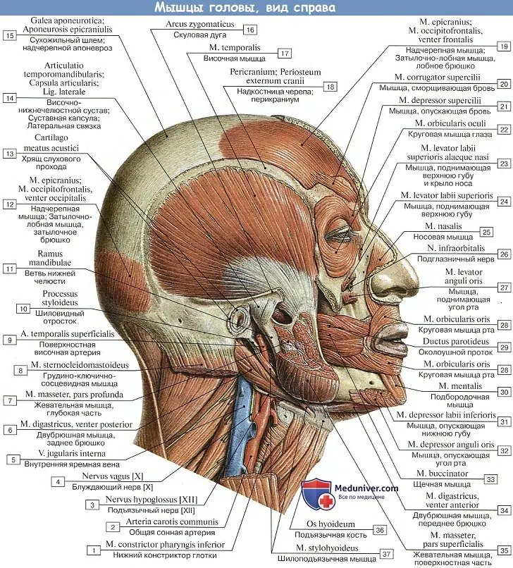
The visceral muscles of the head, which were previously related to the viscera located in the head and neck region, gradually turned in part into the cutaneous muscles of the neck, and from it, through differentiation into separate thin bundles, into the facial muscles of the face. This explains the close relationship between the facial muscles and the skin, which they set in motion. This also explains other features of the structure and function of these muscles.
So, facial muscles unlike skeletal ones, they do not have a double attachment to the bones, but are necessarily woven into the skin or mucous membrane with two or one end. As a result, they do not have fascia and, when contracting, move the skin. When their skin relaxes, due to its elasticity, it returns to its previous state, so the role of antagonists here is much less than that of skeletal muscles.
Facial muscles represent thin and small muscle bundles that are grouped around natural openings: the mouth, nose, palpebral fissure and ear, taking part in one way or another in closing or, conversely, expanding these openings.
Contactors (sphincters) are usually located around the holes in a ring shape, and expanders (dilators) are located radially. By changing the shape of the holes and moving the skin to form different folds, the facial muscles give the face a certain expression corresponding to a particular experience. These kinds of facial changes are called facial expressions, which is where the name of the muscles comes from. In addition to the main function of expressing sensations, facial muscles take part in speech, chewing, etc.
The shortening of the jaw apparatus and the participation of the lips in articulate speech led to a special development of facial muscles around the mouth, and, conversely, the ear muscles, well developed in animals, in humans were reduced and preserved only in the form of rudimentary muscles.
Facial muscles or facial muscles. Muscles of the eye circumference
2. M. procerus, proud muscle, starts from the bony dorsum of the nose and aponeurosis m. nasalis and ends in the skin of the glabellae area, connecting with the frontal muscle. By lowering the skin of the named area downwards, it causes the formation of transverse folds above the bridge of the nose.
3. M. orbicularis oculi, circular muscle of the eye, surrounds the palpebral fissure, located with its peripheral part, pars orbitalis, on the bony edge of the orbit, and its internal part, pars palpebralis, on the eyelids. There is also a third small part, pars lacrimals, which arises from the wall of the lacrimal sac and, expanding it, affects the absorption of tears through the lacrimal canaliculi.
Pars palpebralis closes the eyelids. orbital part, pars orbitalis, with a strong contraction produces squinting of the eye.
In m. orbicularis oculi isolate another small part lying under pars orbitalis and called m. corrugator supercilii, eyebrow wrinkler. This part of the orbicularis oculi muscle brings the eyebrows together and causes the formation of vertical wrinkles in the space between the eyebrows above the bridge of the nose. Often, in addition to vertical folds, short transverse wrinkles form above the bridge of the nose in the middle third of the forehead, caused by the simultaneous action venter frontalis. This position of the eyebrows occurs during suffering, pain and is characteristic of difficult emotional experiences.
Facial muscles or facial muscles. Muscles of the mouth circumference
4. M. levator labii superioris, muscle that lifts the upper lip, starts from the infraorbital edge of the upper jaw and ends mainly in the skin of the nasolabial fold. A bundle splits off from it, going to the wing of the nose and therefore receiving its own name - m. levator labii superioris alaeque nasi. When contracting, it raises the upper lip, deepening the sulcus nasolabialis; pulls the wing of the nose upward, widening the nostrils.
5. M. zygomaticus minor, zygomatic minor muscle, It starts from the zygomatic bone and is woven into the nasolabial fold, which it deepens during contraction.
6. M. zygomaticus major, zygomaticus major muscle, goes from the facies lateralis of the zygomatic bone to the corner of the mouth and partly to the upper lip. Pulls the corner of the mouth upward and laterally, and the nasolabial fold deepens greatly. With this action of the muscle, the face becomes laughing, so m. The zygomaticus is primarily the muscle of laughter.
7. M. risorius, muscle of laughter, a small transverse tuft going to the corner of the mouth is often absent. Stretches the mouth when laughing; In some people, due to the attachment of the muscle to the skin of the cheek, when it contracts, a small dimple is formed on the side of the corner of the mouth.
8. M. depressor anguli oris, muscle depressor anguli oris, begins on the lower edge of the lower jaw lateral to the tuberculum mentale and attaches to the skin of the corner of the mouth and upper lip. Pulls the corner of the mouth downwards and makes the nasolabial fold straight. Lowering the corners of the mouth gives the face an expression of sadness.
9. M. levator anguli oris, the levator anguli oris muscle, lies under the m. levator labii superioris and m. zygomaticus major - originates from fossa canina (which is why it was previously called m. caninus) below the foramen infraorbitale and attaches to the corner of the mouth. Pulls the corner of the mouth upward.
10. M. depressor labii inferioris, muscle that lowers the lower lip. It begins at the edge of the lower jaw and attaches to the skin of the entire lower lip. Pulls the lower lip down and somewhat laterally, as, by the way, is observed during facial expressions of disgust.
11. M. mentalis, the mentalis muscle arises from the juga alveolaria of the lower incisors and canines, and is attached to the skin of the chin. Raises the skin of the chin upward, and small dimples form on it, and moves the lower lip upward, pressing it towards the upper.
12. M. buccinator, buccal muscle, forms the lateral wall of the oral cavity. At the level of the second upper molar, the duct of the parotid gland, ductus parotideus, passes through the muscle. Outer surface m. buccinator is covered with fascia buccopharyngea, on top of which lies a fatty lump of the cheek. Its beginning is the alveolar process of the upper jaw, the buccal ridge and the alveolar part of the lower jaw, the pterygomandibular suture. Attachment - to the skin and mucous membrane of the corner of the mouth, where it passes into the orbicularis oris muscle. Pulls the corners of the mouth to the sides, presses the cheeks to the teeth, compresses the cheeks, and protects the oral mucosa from biting when chewing.
13. M. orbicularis oris, orbicularis oris muscle, lying in the thickness of the lips around the oral fissure. With contraction of the peripheral part of m. orbicularis oris the lips tighten and move forward, as when kissing; when the part lying under the red border of the lips contracts, the lips, tightly approaching each other, are wrapped inward, as a result of which the red border is hidden.
M. orbicularis oris, located around the mouth, performs the function of a sphincter (sphincter), i.e., a muscle that closes the opening of the mouth. In this regard, it is an antagonist to the radiar muscles of the mouth, i.e., the muscles that radiate from it and open the mouth (mm. levatores lab. sup. et anguli oris, depressores lab. infer, et anguli oris, etc.).
Facial muscles or facial muscles. Muscles of the nasal circumference
14. M. nasalis, the nasal muscle itself, poorly developed, partially covered by the levator labii muscle, compresses the cartilaginous part of the nose. Her pars alaris lowers her wing. nose, and the so-called depressor septi (nasi) lowers the cartilaginous part of the nasal septum.
