Pemphigus is a very serious dermatological disease that can lead to serious consequences and complications. The development of pemphigus in newborns is especially dangerous; in this case, even death cannot be ruled out.
People of all ages are susceptible to this disease, however, in most cases it is observed in adults 40-60 years old. Children suffer from this disease extremely rarely. Since the disease is an autoimmune pathology, it is difficult to treat and is chronic.
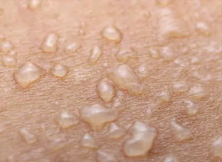
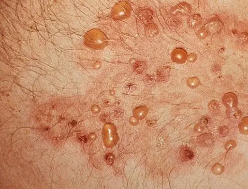
The disease is expressed by the appearance of blisters filled with exudate on the skin and mucous membrane. New growths merge with each other and quickly spread throughout the body.
Causes of the disease
Specific causes of the occurrence of vesicles have not been identified, but provoking factors are identified that influence the development of the disease, namely:
- hereditary predisposition;
- viral infections;
- harmful working conditions;
- allergic reaction to certain types of products, metals, chemicals;
- disruption of metabolic processes in the body;
- endocrine diseases;
- long-term use of certain types of medications.
An autoimmune cause of the disease is also being considered. In children, the cause of the disease can be viruses and bacteria, the main one being Staphylococcus aureus.
Types of pemphigus
First of all, the pathological process of the disease is classified into:
- true pemphigus (acantholic);
- benign pemphigus (neacantholic).
True pemphigus, depending on the manifestation of the pathological process, is classified into the following subtypes:
- Pemphigus vulgaris (ordinary). The most common type. It is characterized by the appearance of blisters on the skin without signs of inflammation.
If there is no proper treatment, there is a rapid spread of bubbles throughout the body. When the disease is advanced, death often occurs. - Erythematous, aka seborrheic pemphigus. Difficult to treat, characterized by the appearance of red spots with dense crusts.
- Leaf-shaped. Pemphigus is characterized by the appearance of flat blisters and many crusts overlapping each other, which create the effect of leaves (photo on the right).
- Brazilian. A similar pathology is common in Brazil. As a rule, all family members suffer. Both children and elderly people are susceptible.
Symptoms of the disease
Regardless of the type, the disease has similar symptoms. The disease develops very rapidly, so at the first manifestations you should immediately consult a doctor.
Pemphigus is characterized by a wavy course, but a rapid spread of foci and a progressive course. There are remissions of varying degrees and durations.
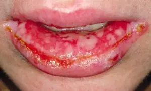
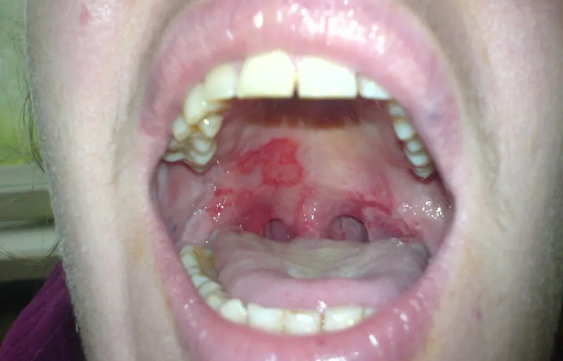
Pemphigus in the mouth does not appear in the form of ulcers immediately, several days after infection. The first symptoms may be malaise, fever and redness of the throat. In such cases, the disease is classified as a simple cold, and treatment is prescribed incorrectly.
Only the manifestation of ulcers and blisters in the oral cavity indicates the development of a pathological process. In this case, the patient experiences bad breath.
Specifics of pemphigus of the oral mucosa
According to medical dental statistics, the oral mucosa is most often affected by pemphigus vulgaris. It is this type of disease that first affects the mucous membrane of the mouth and larynx, and then spreads to the face and body.
The spread of blisters on the skin may begin several months after the first blisters appear in the mouth, or they may appear immediately, after one or two days.
With a strong immune system, timely and competent treatment, the spread of blisters over the skin may not begin.
Erosion that appears on the mucous membrane does not bleed, but due to constant friction and contact with food, it quickly opens.
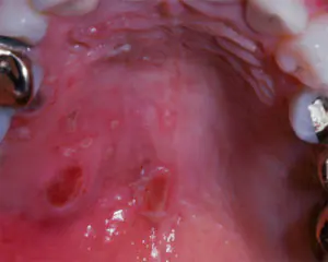
In this case, when examining the oral cavity, it is rarely possible to detect the presence of blisters. In place of the blisters, oval or round ulcers remain, which take a long time to heal. As a rule, erosions of the oral mucosa heal without scars.
However, without proper treatment, erosions do not heal, but on the contrary, they increase and merge into large affected areas. Most often, ulcers are localized on the inside of the cheeks, the lower surface of the tongue and the palate. A white coating appears on the ulcers, which can be easily removed by the dentist using a medical spatula.
The patient's pain is quite strong, especially intense when talking and eating. Ulcers become infected quite quickly. If the oral cavity is not sanitized, the likelihood of infection of erosions and the addition of additional fungal and viral infections increases. A dental examination is required.
With oral pemphigus, the patient often suffers from stomatitis. In addition to the mouth, the disease can spread to other organs covered with mucous membrane: the larynx, digestive tract and others.
Diagnosis and severity of the disease
Since at the initial stage of the development of the disease the symptoms are similar to many other dermatological diseases, to accurately determine the diagnosis it is necessary to conduct a comprehensive examination, namely:
- examination of the patient - the nature of the lesion and location;
- studying the patient’s medical history and the presence of concomitant diseases;
- performing Nikolsky's test, which will distinguish pemphigus from similar pathological processes;
- carrying out histological and cytological studies;
- the use of immunological methods that confirm or refute the autoimmune nature of the disease.
As the blisters develop and spread, the disease is characterized by varying degrees of severity: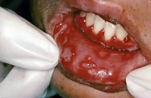
- Lightweight. The pathological process appears on the mucous membranes or skin gradually with a minimum number of foci.
- Average. With a moderate course of the disease, an increase in the spread of blisters on the skin and mucous membrane is observed.
- Heavy. Most of the skin and oral cavity are affected. The ulcers merge into large lesions. Complications and pathologies begin.
What are the methods of treatment and prevention?
Without proper competent treatment, the outcome of the disease is unfavorable. Almost two years later, if the disease is advanced, death can occur.
The treatment process is quite complex and lengthy. Due to the rapid development of the disease, the functioning of many organs is disrupted, which can lead to the appearance of additional diseases.
Drug treatment
While taking any drug, you should see a doctor and monitor your blood pressure and sugar levels by regularly taking laboratory blood and urine tests.
Drug therapy includes taking the following drugs:
- antibiotics - used for purulent infectious processes, contribute to the death of the pathogen;
- corticosteroid drugs - help reduce the manifestations and speed of spread of blisters over the skin;
- cytostatics - used to reduce the possibility of complications.

The dose of the drug and duration of administration are selected by the doctor based on the severity of the disease. After a course of treatment, the patient may be prescribed corticosteroid drugs to prevent relapse. Many medications have side effects.
Local treatment
Local therapy plays only a supporting role in drug treatment. During the treatment of pemphigus of the oral mucosa, it is advisable to follow the following recommendations as local therapy:
- carry out timely sanitation of the oral cavity;

- before and after eating, rinse the affected oral mucosa with disinfectants and deodorants, antibacterial solutions based on chamomile and St. John's wort;
- bubbles can be treated with aloe juice, tincture of plantain, calendula and celandine;
- apply lotions from chamomile decoction;
- Apply nettle leaf juice to damaged skin;
- exclude spicy and salty foods from the diet.
Local treatment and folk remedies will only help temporarily reduce the symptoms of the disease and reduce pain, but will not cure the disease.
Prevention methods
Patients who have been diagnosed with this disease should be registered with a dermatologist. The disease requires constant medical supervision and compliance with preventive measures:
- observe a gentle work schedule and moderate physical activity;
- to refuse from bad habits;
- avoid exposure to sunlight on the skin;
- avoid stressful situations;
- monitor the cleanliness of bed linen and underwear.
In the case of an advanced form of pemphigus and the absence of timely and competent treatment, the prognosis of the disease is unfavorable. Various infections may occur, as a result of which the patient may die from complications.
Possible complications and consequences
In the case of a complicated course of the disease, a large area of the skin or mucous membrane is affected, ulcerative erosions are formed, which become covered with crusts during the healing process.

The most dangerous complication is infection of damaged skin or the addition of secondary infections.
For children, the complications and consequences of pemphigus pose a great threat to their further development. The disease has a detrimental effect on the child’s immune system and his health.
The most severe consequences arise due to the lack of timely diagnosis and necessary treatment, namely:
- the patient’s general condition deteriorates significantly and the protective immune system decreases;
- bubbles of varying densities spread quickly, causing the patient severe pain and inconvenience;
- dense crusts of a large area are formed, resembling lichen in appearance;
- Serious secondary diseases develop, for example, heart failure, cerebral edema and many others.
Any form of pemphigus is a dangerous and serious pathology that should not be neglected. The disease develops very quickly, so any delay is fraught with serious consequences and complications that can lead to death. At the first symptoms of the disease, you need to urgently undergo examination and monitor the course of the disease.
Pemphigus is a group of bullous dermatoses in which the pathogenetic role belongs to circulating autoantibodies directed against antigens of the desmosomal apparatus system of stratified squamous epithelium (skin, mucous membranes of the oral cavity, esophagus and other organs).
The disease develops under the influence of various factors (taking medications containing thiol groups; insolation; infectious agents; stress; consumption of certain foods; physical factors, etc.), but often it is not possible to determine the provoking factor.
During the disease process, antigen-presenting cells recognize their own molecules that make up desmosomes, cancel T- and B-cell tolerance to their own autoantigens, and synthesize autoantibodies.
What it is?
Pemphigus is a chronic autoimmune disease characterized by the appearance of a special type of blisters on the surface of previously healthy skin and mucous membranes. Among the types of pemphigus can be distinguished: vulgar, vegetative, erythematous and foliate. Pemphigus can be diagnosed if acantholytic cells are detected, which are detected in a smear taken or as part of blisters in the epidermis itself (during histological examination).
Causes of the disease
The causes of the disease are unclear. Under the influence of unstudied factors, the body begins to produce antibodies to the proteins of special plates that connect cells - desmosomes. The reaction between antibodies and desmoglein proteins leads to the destruction of intercellular connections in the surface layer of the skin. This phenomenon is called "acantholysis". As a result of acantholysis, detachment of the epidermis occurs and the formation of numerous blisters.
There are several theories about the origin of the disease:
- Viral. In support of this, some scientists cite the fact that the contents of the bubbles can infect chicken embryos, laboratory mice or rabbits. In addition, there is a similar effect on the tissue of the discharge of blisters in pemphigus and Dühring's dermatitis, which is of viral origin. However, this theory has not yet been confirmed.
- Neurogenic. It was put forward in the 19th century by P.V. Nikolsky, who studied this disease in detail. He believed that the cause of neurogenic pemphigus is a change in nerve cells leading to disruption of the innervation of the skin. To support this theory, the scientist cited cases of the disease occurring after emotional shocks. In patients who died from pemphigus, changes in the spinal cord are sometimes noted. Today, scientists believe that these changes are involved in the development of the disease, but are not its cause.
- Exchange. In patients, the function of the adrenal glands, secreting glucocorticoids, is altered, up to its depletion; water, protein and salt metabolism are disrupted. To prove this hypothesis, cases of the appearance of the disease during pregnancy and its spontaneous disappearance after childbirth are cited. However, it is more likely that these disorders are secondary and appear under the influence of an unknown factor. In particular, isolated cases of inherited transmission of the disease have been described.
Proposed mechanisms of immune disorders that can cause pemphigus:
- Damage to the entire immune system, including the thymus gland, which can be genetically programmed.
- Secondary inhibition of the body’s immune response under the influence of external factors (foreign substances, toxins, solar radiation).
- Damage to the epidermis itself, in which antibodies are formed against the intercellular substance of the skin. They bind to epidermal cells, which, when destroyed, release an enzyme that dissolves proteins. Acantholysis develops under its influence.
People experiencing this pathology may wonder how the disease is transmitted. They cannot be infected from humans.
True (autoimmune) pemphigus accounts for up to 1.5% of all skin diseases (dermatoses). There are other diseases that are also called pemphigus, but unlike the true one, their cause has been established, and the prognosis is more favorable.
Symptoms and photos of pemphigus
Three types of disease can be distinguished: pemphigus vulgaris, which occurs due to autoimmune disorders; viral pemphigus, which is caused by the coxsackie virus; Pemphigus of newborns is an infectious disease caused by Staphylococcus aureus. In addition, depending on the clinical picture, erythematous, foliaceous and vegetative pemphigus are distinguished - all these forms of pemphigus disease occur with different symptoms.
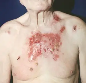
- Pemphigus vulgaris (pemphigus vulgaris) is the most common. The main symptoms of pemphigus are the appearance of watery blisters on the mucous membrane of the mouth, followed by a rash on the skin throughout the body, including the inguinal folds and armpits. Sometimes patients may not notice the appearance of small bubbles and not pay attention to them. Later, the blisters can reach the size of a walnut and, when ruptured, release clear or bloody contents. The covering on the blisters is very thin and flabby. When tires dry out, brown crusts form. And when the apparently healthy skin located between the blisters is rubbed, the upper layers of the epidermis are rejected. Pemphigus vulgaris is accompanied by general malaise, weakness, fever, sore throat during eating and when talking. The disease can last for years and have a severe chronic course with damage to the kidneys, heart, and liver. Pemphigus vulgaris is sometimes complicated by the malignant course of the disease and, despite treatment, death is possible.
- Pemphigus foliaceus is a relatively rare disease. Can occur on any part of the body. Most often, rashes appear on the scalp and face in the form of flaccid, flat blisters that have a thin covering and protrude above the surface. When tires tear, erosions are exposed that heal very slowly. The tires shrink and form thin lamellar scales that grow on top of each other. The clinical feature of pemphigus foliaceus is newly appearing blisters that merge with adjacent affected areas of the skin, forming an extensive wound surface. With this disease, the mucous membranes are usually not affected. The severity of the patient's general condition depends on the area of skin damage; there may be an increase in temperature, disturbance of salt and water metabolism. The disease can last for years and develop into a chronic form if treatment is not started in time.
- Pemphigus vegetans. Vegetating pemphigus at the very beginning of its development is very similar to common pemphigus. It also appears initially on the oral mucosa. Blisters then form around natural orifices, behind the ears, under the breasts (in women), and in the armpits. At the site of the opening of the blisters, erosions covered with a purulent coating are formed, with the release of a large amount of exudate. The lesions often merge and form large wound surfaces. Patients complain of pain during active movement and burning, exhaustion of the whole body. The course of the disease with pemphigus vegetans is benign; patients have been feeling satisfactorily for years.
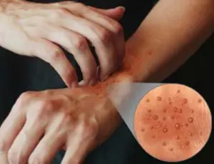
- Seborrheic pemphigus is a severe skin disease. Seborrheic pemphigus blisters, unlike ordinary pemphigus, are small in size. They dry out quickly and form yellow or brown crusts that look like scales. First of all, they appear on the face and scalp, then down to the back and chest. They appear extremely rarely on the oral mucosa. With pemphigus, seborrheic crusts form very quickly. After removal, wet erosion can be seen underneath them. Very often, the bubbles are not immediately noticeable and it is very difficult to identify the primary nature of the crusts. The disease lasts a long time, the course is benign.
Pemphigus disease can only be diagnosed by a dermatologist, based on the results of examination, immunological, cytological, and histological examinations.
Pemphigus in newborns
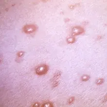
Pemphigus in newborns is an acute infectious skin disease, which clinically manifests itself in the form of pustules that quickly spread throughout the entire skin.
Pemphigus in newborns is often bacterial in nature. Its causative agent is Staphylococcus aureus. Speaking about the pathogenesis of pemphigus among newborns, the skin reaction of children occupies a significant place. The skin reaction will intensify in the event of birth trauma or prematurity, as well as the incorrect lifestyle of the pregnant woman herself. Blisters will form on the child's skin due to exposure to bacterial factors.
The epidemiology of pemphigus in newborns indicates poor hygiene in the maternity hospital, the presence of chronic infections among maternity hospital staff, and the possible occurrence of autoinfected pemphigus (if a newborn develops purulent types of navel diseases).
Pemphigus in newborns forms in the first days of life, but the development of the disease is possible even after one to two weeks. On healthy skin, small blisters with thin walls and serous contents appear. After a few hours, the process will generalize, the bubbles will increase in size and open. In place of the blisters, there will be painful erosions with remaining particles of the epidermis located at the edges. Such erosions will be covered with a serous-purulent crust. If pemphigus occurs in newborns, there will be intoxication, fever and lack of appetite.
If pemphigus is not treated in the early stages, the newborn will develop inflammatory processes in the internal organs (phlegmon, otitis, pneumonia). In weak newborns or premature infants, a septic form of pemphigus cannot be excluded. With the latter, the mortality rate is very high.
Pemphigus in newborns can be diagnosed based on visual examination. Pemphigus in newborns should be distinguished from the syphilistic form of pemphigus, which is a symptom of congenital syphilis. With the latter, the bubbles are located on the palms.
Antibiotic therapy can reduce the percentage of deaths from pemphigus among newborns. With timely treatment of pemphigus among newborns, the favorable outcome of the disease is significantly higher than with other types. Doctors may also prescribe the use of aniline dyes and various types of non-aggressive antiseptics.
Diagnostics
Clinical manifestations, especially in the initial stages of the disease, are uninformative, and therefore interviewing the patient allows one to avoid an erroneous diagnosis.
Laboratory tests allow one to suspect pemphigus, as acantholytic cells are found in fingerprint smears during cytological examination. Histological examination reveals an intraepidermal location of blisters.
Complications
If left untreated, pemphigus provokes inflammation of internal organs, pneumonia, phlegmon, and otitis. In newborns, severe septic form of the disease can be fatal.
In adults, there is a high probability of secondary infection. Pemphigus vulgaris can cause damage to the kidneys, liver, and cardiovascular system, while pemphigus foliaceus can cause sepsis and death.
Pemphigus treatment
The principle of treating pemphigus in children and adults is to suppress the pathological activity of the immune system.
For this purpose, glucocorticosteroids (Prednisolone, Methylprednisolone) are used. Often, the doctor begins treatment of the patient immediately with large doses in order to influence the pathological process as quickly as possible. The patient must take this dose for four to six weeks, then the dose is gradually reduced. With a small dose of the drug, relapses of the disease occur.
In addition, glucocorticoids can be used in combination with immunosuppressive drugs (Azathioprine, Methotrexate). The use of immunosuppressive drugs allows you to use a lower dose of glucorticoids and reduce their side effects.
In the most severe form of the disease, pulse therapy is used. This is a short-term administration of high doses of glucocorticoids and Cyclophosphamide according to a specific scheme. As an additional method, plasmapheresis is performed, which allows you to quickly reduce the level of antibodies in the blood. If an infection occurs, the patient is prescribed antibiotics.
As an external treatment, blisters on the skin are treated with solutions of aniline dyes (fucorcin). To treat the oral cavity, rinsing with solutions with an anti-inflammatory effect is practiced.
The prognosis for acantholytic pemphigus is conditionally unfavorable. On the one hand, in the absence of effective treatment, there is a high probability of complications and death. On the other hand, patients with pemphigus are forced to take glucocorticosteroids for a long time, and sometimes for life, which is fraught with the development of side effects. But hasty refusal of drugs leads to immediate relapse of the disease. Glucocorticosteroids do not eliminate the cause of the disease, but inhibit the pathological process and prevent its progression.
Nutrition
Products that can cause allergies, rough foods, simple carbohydrates, salt, and canned food are excluded from the diet. The menu includes foods high in protein and vitamins. When blisters form in the oral cavity, pureed soups and soft porridges are recommended to prevent mechanical damage to the mucous membrane.
Prevention
Depending on the cause of pemphigus disease, various preventive measures are taken.
Pemphigus vulgaris is difficult to prevent, because... this is an autoimmune disease - just like to prevent many other diseases, it is necessary to constantly maintain immunity. To prevent relapses, monitor the condition of the skin, monitor the level of prothrombin, blood sugar, urine, blood pressure at least 2-3 times a week, take vitamins and calcium supplements.
To prevent viral pemphigus and pemphigus of newborns, you should follow basic rules of hygiene and antiseptics.

There are several varieties of Enterovirus, and the diseases that these viruses cause differ in their symptoms. Enteroviral fever in children is perhaps the most common type of disease, but other types of disease are also quite dangerous for the child’s health.
">
