When a person develops a myocardial infarction, blood flow in one or more coronary vessels is disrupted. This leads to an imbalance between the need of myocardiocytes for oxygen and its supply. Changes in metabolism due to lack of nutrients aggravate the condition of the affected tissue. As a result, cardiac muscle cells begin to necrotize and die. In place of the dead tissue, a scar forms. In this article I want to talk about the mechanism and possible consequences of such a “replacement”.
Development mechanism
At the time of development of an acute infarction, a sharp disruption of the blood supply to the myocardium occurs for the following reasons:
- Rupture of an atherosclerotic plaque under the influence of a sharp jump in pressure, increased heart rate and acceleration, and accelerated blood flow through the coronary vessels.
- Blockage of blood vessels due to blood thickening (acceleration of platelet aggregation, activation of the coagulation system, decreased rate of blood clot lysis).
- Spasm of the coronary artery (vasoconstriction).
I often observed patients in whom several factors were identified as the cause of the disease with myocardial damage. In young patients, vasospasm is often the basis of pathological disorders, which is not possible to determine after the start of treatment.
Expert advice
I strongly recommend starting treatment in a hospital immediately after an acute attack, since only in this case is it possible to limit the further spread of necrosis and minimize irreversible changes in the myocardium.
The study of histological samples confirms the destruction of the cardiac myocyte 20 minutes after the development of ischemia. After 2-3 hours of lack of oxygen, their glycogen reserves are depleted, which marks their irreversible death. Replacement of myocarditis with granulation tissue occurs within 1-2 months.
As my practice and the observations of colleagues show, the scar on the heart is finally consolidated after six months from the moment the first symptoms of acute infarction appear and is a section of coarse collagen fibers.
Classification
Heart scars can be classified according to their location and extent of distribution.
They can be located along the coronary vessels:
- Impairment of blood flow in the anterior interventricular artery leads to ischemia with the subsequent appearance of a scar in the area of the septum between the ventricles, involving the papillae and lateral wall, as well as on the anterior surface and apex of the left ventricle.
- The infero-posterior and lateral part is affected when the left circumflex coronary artery is blocked.
- Problems with the blood supply to the myocardium in the right artery results in irreversible changes in the right ventricle and can affect the posterior inferior part of the left ventricle and the septum. But such a violation is extremely rare.
According to the type of distribution, scars can be local (focal), which can be compared to a scar on the body, or diffuse (multiple). Experts call the second option dystrophic changes in the myocardium.
How does a scar manifest itself?
The acute period of a heart attack is characterized by a variety of clinical manifestations. The main symptom is pain, which can be relieved exclusively with narcotic analgesics and can be observed from an hour to 2-3 days. Then the pain syndrome disappears and the formation of an area of necrosis begins, which takes another 2-3 days. Then comes a period of replacing the affected area with loose connective tissue fibers.
If the correct treatment tactics are used, the following symptoms are noted:
- development of compensatory hypertrophy;
- rhythm disturbance (which often accompanies the acute period) is eliminated;
- tolerance to stress gradually increases.
If a scar that appears on the heart crosses the conduction paths along which the impulse travels, a conduction disorder is recorded, such as a complete or partial blockade.
In the case of successful recovery after a primary small-focal infarction, I did not notice any significant disturbances associated with the functioning of the heart in my patients.
If patients have formed a large scar or many small ones, the following deviations are observed:
- dyspnea;
- increased heart rate;
- the appearance of edema;
- enlargement of the left chambers of the heart;
- pressure fluctuations.
How dangerous is this?
The most dangerous is the development of a scar as a result of large-focal or transmural infarctions, as well as several repeated violations in different basins of the coronary vessels with diffuse multiple lesions.
In the case of a large area of damage or widespread cardiosclerosis, the remaining healthy cells cannot fully compensate for the work of damaged cardiomyocytes. The frequency and strength of contractions increases in order to provide organs and tissues with oxygen and necessary substances.
As a result, tachycardia develops; with its appearance, the load on the heart becomes even greater, which leads to dilatation of the left ventricle and atrium. As it progresses, blood stagnation appears in the right side with the development of heart failure.
I also observed another version of the complication: a scar on the heart after a heart attack with extensive and deep lesions of all layers of the organ caused the formation of an aneurysm due to the thinning of its wall.
The reasons for the appearance of such a defect are:
- transmural lesion;
- increased blood pressure;
- increased blood pressure inside the ventricle;
- excessive physical activity of the patient, refusal to comply with the regimen.
An aneurysm leads to the rapid development of heart failure, the formation of a parietal thrombus, and severe stagnation in the systemic circulation. Often complicated by severe rhythm disturbances leading to death (paroxysmal tachycardia and ventricular fibrillation).
Diagnostics
In order to establish a diagnosis, I conduct a survey and study the medical history (mainly, it includes ischemic heart disease with a history of heart attack). External examination usually reveals an increase in respiratory rate, weakening of heart sounds during auscultation, the presence of edema, and various rhythm disturbances. I will definitely take a blood pressure measurement.
Then I send you to the following research:
- general and biochemical blood test, coagulogram (will help determine concomitant diseases, cholesterol levels and clotting time);
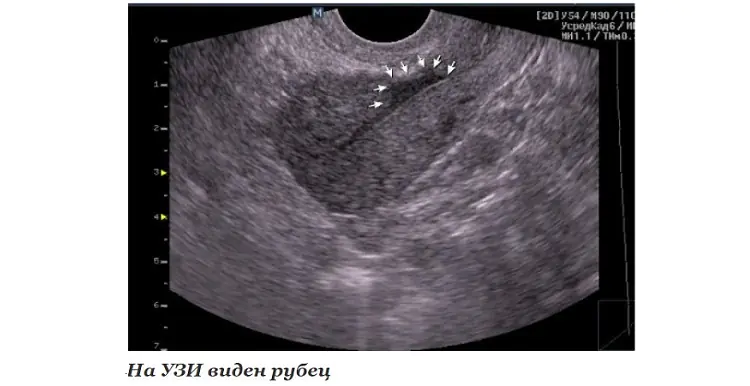
- EchoCG or ultrasound of the heart helps to establish the presence of localized or diffuse areas of connective tissue, allows you to clarify the location and extent of distribution;
- MRI helps to visualize and reliably assess the affected area;
- scintigraphy is required to determine dysfunctional areas of the myocardium.
With the help of an ECG after a transmural and large-focal infarction, it is possible to clarify where the scar is located on the diseased heart.
It is determined by the presence of a Q wave in different leads, as can be seen in the table.
Localization of post-infarction scar in the left ventricle
Under the influence of various unfavorable factors, the process of death of heart cells can begin. As a result, they are replaced by scar tissue, characterized by a high content of protein and collagen. In medicine, the pathology is usually called cardiosclerosis. It is important to understand that a scar on the heart is a condition that poses a danger not only to the health, but also to the life of the patient. In this regard, when the first alarming signs occur, you should contact a cardiologist. The specialist will issue a referral for comprehensive diagnostics, based on the results of which he will create the most effective treatment regimen. Therapy may include both conservative and surgical techniques.
Pathogenesis
It is important to understand that a scar on the heart is a protective reaction of the body that occurs when necrotic foci form. In most cases, death of heart muscle cells occurs after a heart attack.
As soon as the process of cell death begins, connective tissue begins to form in this area. In this way, the body tries to prevent an increase in the area of necrosis. However, the scar on the heart after a heart attack cannot perform the functions of the organ. That is why the formation of connective tissue is only a temporary solution to the problem, which often leads to the development of life-threatening pathologies.
It is important to understand that a scar on the heart is a condition that prevents the development of acute myocardial failure and death. But it also delays the development of all kinds of complications. This is due to the fact that heart failure takes on a chronic form, characterized by constant alternation between periods of remission and relapse.
Etiology
A scar always forms in the area of muscle fiber rupture or in areas of necrosis. The body starts the synthesis of fibrin protein, which quickly fills the damage.
Causes of scars on the heart:
- Thrombosis and embolism of blood vessels. According to statistics, half of the world's population aged 40 years and older suffer from pathological changes. For example, a combination of increased blood clotting and even the initial stage of atherosclerosis leads to thrombosis. The resulting clot of liquid connective tissue partially narrows the lumen of the vessel. As a result, heart cells do not receive the required amount of nutrients and oxygen and begin to die. This situation is life-threatening, so fibrotic changes occur very quickly.
- Myocarditis. One of the most common causes of heart scars. Under the influence of unfavorable factors (allergy, infection, etc.), the myocardial muscle tissue becomes inflamed. As a result, dilatation develops, causing the heart to wear out and become damaged. Microtraumas are subsequently replaced by connective tissue.
- Cardiac ischemia. This term refers to a pathological condition characterized by chronic oxygen starvation of the myocardium. As a result, the process of degenerative-dystrophic changes is launched.
- Heart attack. A scar on the heart appears after it most often. The danger is that sometimes a heart attack is asymptomatic, and changes are detected only on an ECG.
Doctors identify myocardial dystrophy as a separate cause of scar formation. This is a pathological condition in which atrophic changes are noticeable in the heart, that is, the tissue is weaker and thinner than it should be.
- Vitamin deficiency in the body.
- Lack of magnesium, calcium and potassium.
- Excess body weight.
- Frequent and high-intensity physical activity.
Doctors say that if at least one close relative has a heart scar after a heart attack, it is necessary to visit a cardiologist annually for prevention.
Types of scars
Against the background of various pathologies, fibrosis of one of three types can form:
- Focal. It has clear boundaries and a specific location. For example, the scar may be on the back wall of the heart muscle.
- Diffuse. It differs in that it affects all tissues.
- Diffuse-focal. This form is mixed. It is characterized by the presence of small pathological foci that are evenly distributed over the entire surface of the heart. Sometimes scars grow together.
Cardiologists say that scars on the heart are a pathology, the treatment of which is not only complex, but also lengthy. In most cases, doctors create a treatment plan aimed at maintaining the functioning of the organ.
Clinical manifestations
Symptoms and their severity directly depend on what disease caused the damage to muscle tissue. Cardiologists say that scars on the heart after a heart attack (a photo of the affected organ is shown schematically below) can form over several years. In this case, the process is often asymptomatic.
The absence of clinical manifestations is due to the fact that the organ manages to maintain contractility and compensate for the volume of normal tissue. When it is no longer able to function fully, the following symptoms appear:
- Painful sensations in the chest.
- Severe shortness of breath.
- Swelling of the face and limbs.
- Severe fatigue even after minor physical exertion.
- Increased degree of fatigue.
Over time, the fingertips on both the upper and lower extremities acquire a bluish tint. This is a specific sign of severe heart failure. At this stage, doctors take measures to prevent further damage to the heart. Often the only way to save the patient’s life is surgery.
Diagnostics
If the first alarming signs occur, you should contact a cardiologist as soon as possible. The specialist will collect anamnesis, conduct a physical examination and issue a referral for a comprehensive diagnosis, including the following studies:
- ECG.
- Dopplerography.
- EchoCG.
- X-ray.
- Coronary angiography.
Based on the diagnostic results, the doctor draws up the most effective treatment regimen. In severe cases, he evaluates the feasibility of surgical intervention.
Drug treatment
Conservative therapy involves taking medications whose active components help maintain heart function. In addition, patients need to follow the principles of a healthy lifestyle.
The choice of medications is made by the attending physician based on the diagnostic results. The cardiologist prescribes medications that improve heart function by accelerating metabolic processes and restoring the circulation of fluid connective tissue.
An effective method is stem cell treatment. Against the background of their use, natural processes of restoration of affected tissues are launched in the body. They are noticeable soon after the introduction of a cardiomyoblast (a specific cellular element). During treatment, the contractility of the organ is restored and blood circulation improves. In addition, atherosclerotic plaques dissolve, vessel walls are strengthened and necrosis is prevented.
If a heart attack develops as a result of ischemic disease, urgent medical treatment is indicated, which involves taking or intravenously administering the following drugs:
- Beta blockers.
- Diuretics.
- Metabolites.
- Nitrates.
- Acetylsalicylic acid.
If a scar on the heart was discovered during an ECG, you need to be prepared for the fact that it will increase in size for several more months. This information is also relevant for patients who have already undergone treatment. If your health suddenly deteriorates, you must call an ambulance. It is possible that emergency surgery will be required.
Self-medication is strictly prohibited. The wrong choice of drug can be fatal.
Installation of a pacemaker
This is a type of surgical treatment during which the surgeon implants a device in the patient whose task is to maintain normal heart conduction and rhythm. Installation of a pacemaker has no contraindications. In other words, the operation can be performed even on children.
In rare cases, the device is rejected by the body. Typically, this occurs in 2-8% of elderly patients.
Donor organ transplantation
This is a radical operation, which is performed only if it is impossible to save the patient’s life using other methods. Donor organ transplantation is performed only on persons under 65 years of age.
Contraindications are serious pathologies of internal organs, which in practice is very rare, since, for example, both atherosclerosis and ischemia are on the list of restrictions.
Bypass surgery
The essence of the operation is to expand the lumen of the affected blood vessels. As a rule, this type of surgical intervention is prescribed for severe atherosclerosis. This is a disease in which plaques consisting of “bad” cholesterol settle on the walls of blood vessels. They narrow the lumen, as a result of which the heart does not receive the required amount of oxygen and nutritional components. The natural consequence is tissue necrosis.
If the lumen is completely blocked by plaques, the surgeon creates a new vessel to bypass the affected one. This can significantly improve tissue nutrition and, accordingly, heart function.
Aneurysm removal
This is a specific bulge that most often forms in the area of the left ventricle or the posterior wall. After the aneurysm is removed, the blood stops stagnating, and the heart muscle again receives the necessary amount of nutrients and oxygen.
Why are scars dangerous?
Many patients are interested in how long they live with a scar on the heart. It is important to understand that the prognosis depends not only on the underlying disease, but also on the timeliness of seeing a doctor. What is it, the causes of scars on the heart, how to treat the pathology - the cardiologist provides all information regarding the disease during the appointment.
The most unfavorable prognosis is considered if the scar has formed in the area of the left ventricle. This area is subject to the greatest load, which means its damage will invariably lead to the development of heart failure. In addition, other organs (including the brain) will begin to suffer from hypoxia, not receiving the required amount of oxygen.
A condition in which both the left ventricle and the mitral valve are affected is also a life-threatening condition. In this case, a life-threatening pathology develops - aortic stenosis.
If you consult a doctor in a timely manner and follow all recommendations, the patient has every chance of living a very long time.
Prevention
Cardiosclerosis is a disease of the cardiovascular system. In this regard, both primary and secondary prevention consists of observing the following rules:
- Balanced diet.
- Regular but moderate physical activity.
- Quitting smoking and drinking alcoholic beverages.
- Avoiding getting into stressful situations.
- Frequent walks.
- Spa treatment.
In addition, it is necessary to be examined annually by a cardiologist in order to prevent pathologies of the cardiovascular system.
Finally
Sometimes, based on research results, the doctor diagnoses a “scar on the heart.” What does this concept mean? A heart scar is a pathological condition that is a kind of protective reaction of the body to myocardial damage. The formation of dense connective tissue is triggered when the integrity of the muscle is damaged or when areas of necrosis appear on it. Despite this, the pathology needs treatment. It is important to understand that scar tissue cannot perform the functions of the heart, which means that sooner or later it will cause the development of other diseases. The doctor draws up a treatment regimen based on the results of instrumental diagnostics. The treatment plan may include both conservative and surgical methods.
What is macrofocal myocardial infarction?
Have you been struggling with HYPERTENSION for many years without success?
Head of the Institute: “You will be amazed at how easy it is to cure hypertension by taking it every day.
One of the most dangerous cardiac pathologies, often leading to death, is large-focal myocardial infarction. Patients with this disease require urgent medical attention and long-term restorative treatment.
Detailed description
Myocardium is the heart muscle. It makes up the bulk of the volume of the human heart. Through the myocardium, rhythmic automatic contractions of the heart and its periodic relaxations of a natural nature are formed. In fact, it is a healthy myocardium that is the main condition for the normal functioning of the heart and the entire body.
Our readers successfully use ReCardio to treat hypertension. Seeing how popular this product is, we decided to bring it to your attention.
Read more here...
Myocardial infarction is one of the clinical manifestations of coronary heart disease. It is a consequence of impaired blood supply to the heart. For example, in case of obstruction of the coronary artery, its spasm or complete blockage.
During myocardial infarction, necrosis (death) of the muscle tissue of the heart is observed. Based on the volume of tissue covered by necrosis, small-focal and large-focal myocardial infarction is diagnosed. The latter is also called Q-infarction. Myocardial infarction differs in the nature of the anatomical lesions, in the localization of the necrotic focus, and in the nature of the course of the disease.
Possible risk factors
The risk of large-focal myocardial infarction increases in the presence of factors such as:
- arterial hypertension;
- rheumatic heart disease;
- strepto- or staphylococcal infection;
- atherosclerosis;
- cardiovascular diseases;
- a sharp change in blood cholesterol levels;
- active smoking;
- alcohol and drug abuse;
- inactive lifestyle, hypotension;
- elderly and advanced age;
- poor environmental conditions;
- diabetes;
- overweight, obesity;
- low level of immunity.
Recently, the risk of large myocardial infarctions in young and middle-aged people has increased sharply. This is usually due to an unhealthy lifestyle and poor environment.
Basically, large myocardial infarctions occur after 50 years in women and after 40 years in men. At the same time, men, as a rule, suffer from heart attacks much more often than women. The main reason for this is atherosclerosis. Its prevalence among males is much higher than among females.
Causes
In addition to the risk factors described above, the cause of large-focal myocardial infarction is:
- cardiac ischemia;
- vascular thrombosis;
- increased blood viscosity;
- angina pectoris;
- severe arterial spasms;
- diseases of the central nervous system;
- frequent stress and emotional overstrain.
Main periods
In the process of formation and progression of large-focal myocardial infarction, separate stages of the disease are distinguished. The initial stage is acute, lasting no more than 120 minutes. At this time, tissue necrosis is not yet observed.
The next stage of the disease is the acute period. It can last from 2 to 10 days, in some cases it extends to 2 weeks. The beginning of this stage is considered to be the moment of formation of a necrotic focus. Next comes softening of the muscle tissue of the heart - myomalacia.
During the acute stage, the patient usually experiences:
- hyperthermia;
- signs of AHF;
- hypotension;
- absence of severe pain.
After a large-focal myocardial infarction, scarring of tissues affected by necrotic changes begins. At this point, the disease enters stage 3 - the subacute period. The duration of this phase is usually 4 weeks. The pain gradually disappears, the temperature returns to normal. The patient feels a noticeable improvement in his general condition.
Then the last stage of a major myocardial infarction begins—the post-infarction period. Its duration is usually from 3 to 5 months. At this stage, tissue compaction and gradual formation of a scar occur. The myocardium has time to adapt to functioning in new conditions. Previously observed symptoms gradually disappear. My health is returning to normal.
Symptoms
The development of myocardial infarction occurs very quickly. The main symptoms of a large focal infarction are:
- severe chest pain radiating to the left ear area;
- pathological muscle weakness;
- pain in the area of the shoulder blades and collarbone;
- profuse sweating;
- high blood pressure;
- difficulty breathing, shortness of breath;
- pathological pallor of the skin;
- panic fear.
The patient needs urgent medical care in a clinical setting. Before the ambulance arrives, first aid must be provided. The patient should be seated in a chair with a high inclined back or put to bed. Provide access to fresh air by opening a window. Loosen tight clothing or carefully remove it.
The main thing at this moment is the patient’s calm. Therefore, someone close to him should be next to him. You can give the patient nitroglycerin, acetylsalicylic acid, and sedatives.
In case of sudden cardiac arrest, loss of consciousness and rhythmic breathing disorder, without waiting for the pulse to disappear, you should immediately perform chest compressions and mouth-to-mouth breathing. These procedures must be continued until the ambulance specialists arrive.
Diagnostics
If a large-focal myocardial infarction is suspected, the patient immediately undergoes an electrocardiogram. To clarify some features of the clinical picture of the disease, an ultrasound, echogram and laboratory blood test are performed. A blood test can determine indicators such as:
- leukocyte level;
- rate of erythrocyte falling - ESR;
- enzymatic activity;
- level of content and activity of individual enzymes.
An ECG for a large myocardial infarction allows one to determine its exact location, the duration of the pathological process and the volume of affected tissue. In addition, through an ECG, doctors receive some special data that allows them to determine the onset and nature of a specific stage of the disease.
At the acute stage of development of a large infarction, the ECG shows the formation of such abnormalities as:
- pathological Q wave or QS complex;
- location of the RS-T segment relative to the isoline;
- merging of the RS-T segment with the T wave, first with a positive one, then with a negative one;
- isoelectric character of the RS - T segment:
- deepening of the coronary T wave and sharpening of its end.
In the subacute period, the ECG shows not only signs of necrosis and ischemia of the heart, but also the following data:
- gradual decrease in the amplitude of the coronary T wave;
- combination of the RS-T segment with the isoline.
An ECG performed in the post-infarction period shows:
- preservation of the pathological Q wave and QS complex;
- the character of the T wave is positive, smoothed, slightly negative.
Treatment
Medical treatment should begin immediately after a heart attack is discovered. The deadline is 12 hours after the onset of the disease.
It is best if assistance is provided within the first 4 hours, this will avoid serious post-infarction pathologies.
The main directions of treatment are pain relief and restoration of coronary artery patency. If there is a blood clot, this is done in 2 ways:
- the clot can be dissolved with medications;
- The patency of the artery is restored surgically by removing the thrombus.
In addition to thrombolytic therapy, the patient undergoes surgical dilation of blood vessels using balloon angioplasty or coronary artery bypass grafting. The patient remains in the clinic for some time. He is then discharged home. Until the post-infarction stage is completed, the patient is under constant supervision of the attending physician.
Special diet
Dietary nutrition is an important condition for treatment after a heart attack. The main goals of the diet in this case:
- reducing the total calorie content of consumed foods;
- avoid flatulence in the intestines;
- prevent stimulation of the cardiovascular system and central nervous system;
- prevent blood sugar levels from rising.
Therefore, the diet should not include:
- fresh bread and pastries;
- milk;
- legumes;
- carbonated drinks;
- cocoa, coffee, chocolate;
- herbs, spices.
The patient’s meals are organized in fractions; food is given in small portions, warm. In the first time after a heart attack, it is better to give pureed food. Hot dishes are prepared by boiling in the usual way or by steaming. Salt, sugar and the total amount of liquid in the diet should be limited.
Possible complications
The consequences of large-focal myocardial infarction are usually divided into early and late complications. Early complications include:
- OSN;
- cardiogenic shock;
- heart rhythm disturbance;
- deterioration of cardiac conductivity;
- myocardial rupture.
Later complications may appear such as:
- protrusion of the heart wall;
- dysfunction of the heart muscle;
- thromboembolism;
- CHF;
- left ventricular aneurysm;
- cardiosclerosis.
The main cause of concern after a large focal myocardial infarction is heart pain. When they appear, you should definitely see a specialist. This will help prevent the possibility of another myocardial infarction. The doctor will determine the nature of the pain and the cause of its occurrence. If necessary, he will prescribe the necessary treatment and give recommendations for correcting the regimen.
Shortness of breath that occurs after strenuous exercise or intense exercise may be due to AHF. After myocardial infarction, it occurs due to a disturbance in the rhythm of heart contractions. The reason for this is the scar formed.
In this case it is also observed:
- tachycardia;
- general weakness;
- cough at night;
- mild heart pain;
- increased urination;
- sudden mood swings.
All of the above-described ailments in the post-infarction period are associated with a gradual increase in physical activity as the patient returns to his usual lifestyle. Therefore, it is necessary to carefully monitor your health. If any additional symptoms and signs appear, consultation with a specialist is necessary. Self-medication in this situation can be dangerous.
To avoid dangerous consequences, the patient must follow a regimen, eat right, strictly follow all doctor’s prescriptions, undergo regular medical examinations and do an additional ECG if necessary.
Prevention
Prevention of myocardial infarction is helped, first of all, by a correct lifestyle. Therefore, it is extremely important to give up bad habits, organize a balanced diet without overeating and excessive consumption of irritating foods, and spend more time in the fresh air. Lack of excess weight significantly reduces the risk of developing many diseases, for example, atherosclerosis, which often causes cardiac ischemia.
Properly organized physical activity is of particular importance. They help not only strengthen the heart, but also significantly improve the condition of the body. During physical activity, the main thing is not to overexert yourself. Loads should be increased gradually.
If you have no experience, it is better to start classes under the guidance of a physical therapy inspector. A specialist will help you choose an individual set of exercises and determine the permissible level of load.
If you have any chronic diseases, you can start physical exercise only after passing the necessary medical examination. Based on the data received, the doctor will make the necessary prescriptions, according to which the exercise therapy instructor will organize classes.
