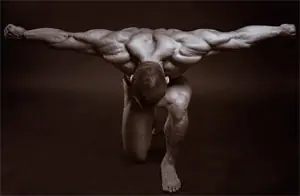
To successfully engage in physical exercises with weights and on machines, you must have a clear understanding of human musculoskeletal system.
The support of all human tissues and organs is skeleton, consisting of many bones. Movable joints in the bone skeleton - there are up to 230 of them - are called joints. The ends of the articulating bones are tightly covered by a connective membrane called the articular capsule.
Play a major role in strengthening joints ligaments - strong and elastic strands of connective tissue. They, merging with the connecting bag, strengthen it. Of great importance in strengthening joints are tendons, attached to bones. For a variety of movements, some joints have special plates or discs made of connective tissue fibrous substance. The viscous fluid (synovium) secreted into the joint cavity by the inner layers of tissue of the joint capsule reduces friction between the contacting surfaces of the bones. The main key movements in the joints are:
- a) bending
- b) extension,
- c) casting,
- d) abduction,
- e) rotation (rotation),
- e) circular movements.
Strength training increases joint strength, they become more mobile. However, with prohibitive (excessive) load and a significant excess of the degree of freedom, injuries are likely - dislocations, sometimes even with rupture of tissues and blood vessels.
A person performs all movements thanks to contractile activity more than six hundred skeletal muscles. There are two types of muscles - smooth, which contracts against the will (stomach, walls of blood vessels), and striated, which moves the body in space due to human-controlled muscle contraction. The striated muscle consists of thin filaments of the protein actin and thick filaments of myosin, which, when combined, form sarcomeres - elementary motor units where chemical energy is converted into mechanical energy, causing human movement.
There is an assumption that the contractile process of the muscle occurs as a result of the mutual penetration of threads actin And myosin. In this regard, the energy level of the sarcomere depends on the position of these threads in it. Combining into groups, sarcomeres form more than a thousand thin threads - fibrils, which make up muscle fiber. The fibers form muscle bundles, and when they unite, they form the muscle itself. The contractile fibers of the muscle end at the connective tissue, which passes into the tendon and transfers tension during contraction. Connective tissue has high strength.
Content- Types of muscles
- Mechanics of human movements
- Fast and slow muscle fibers
- Anatomy of movements
- Newton's third law
- Muscles of the shoulder girdle.
- Chest muscles.
- Back muscles.
- Abdominal muscles.
- Leg muscles.
Types of muscles
Depending on the muscle appearance received the following names:
- long,
- short,
- wide,
- ring-shaped.
Almost all the broad muscles are located on the body, the long muscles are located mainly on the limbs, and the short muscles are located between individual vertebrae. Visually, the long muscles resemble a spindle. The middle part of such a muscle is called "belly", the beginning is called "head", and the second end (which is longer) - "tail".
Some muscles have several heads or are stretched in the middle by tendon formations, dividing them into several parts. Muscle tendons are attached to all sorts of roughness, tuberosity and various protrusions of bones, firmly woven into the periosteum and even partially penetrating deep into the bone substance, and in some cases to the joint capsule, fascia or skin.
Mechanics of human movements
When a muscle contracts, it moves bones that act as leverage, in the joints. Shortening relatively slightly, it develops quite a lot of effort. Therefore, in the human musculoskeletal system, there are usually bone levers with a loss of force when the muscle works, but with a gain in the way of applying this force. The magnitude of the moment of force depends on the angle at which the force acts on the lever. The greatest effect is achieved when the force acts at right angles to the lever.
Various tubercles and protrusions on the bones of the skeleton, as well as sesamoid bones (for example, the kneecap) contribute to a more rational effect of the muscle on the bone levers. Muscles that, when contracting, cause movement of body parts in only one joint are called single-joint, and attached by their ends simultaneously to the bones and individual parts of the skeleton and leading to changes in angles in many joints at once - multi-joint.
When performing joint movement due to contraction of certain groups synergistic muscles - it is always possible (except for the presence of counteraction from external forces) to return the moving link to its original position due to the presence antagonist muscles.
The strength of a muscle depends on its anatomical structure. There are muscles that have a feathery structure, spindle-shaped with parallel fibers. It has been established that the muscles of the feathery structure are short and adapted to the development of tension of great strength (for example, the gastrocnemius), and the muscles with parallel and fusiform fibers are longer and provide fast, dexterous and sweeping movements (sartorius, biceps brachii).
Fast and slow muscle fibers
The strength of muscles is greater, the larger their cross-sectional area, and the magnitude of contraction is greater, the longer the muscle fibers. Some muscles can shorten to a third or half of their original length. Muscles have fast and slow muscle fibers. The former, presented mainly in the pennate muscles, for example in the gastrocnemius, contract faster than the slow ones, all other things being equal. Contraction also depends on external load, on the activity of the central nervous system and on the strength of the muscle itself.
The relationship between the strength of a muscle and its diameter is determined by the number of its constituent fibers. For example, a single striated fiber can develop a tension of 0.1 - 0.2 g.
Anatomy of movements
Contractility is characterized absolute force, developed by the entire muscle per 1 cm2 cross section (physiological diameter). This allows you to compare the strength of different muscles, regardless of their size. For example, the absolute strength of a) the gastrocnemius muscle in total with the soleus is 6.24, b) the biceps brachii - 11.4, c) the triceps brachii - 16.8, d) the brachialis - 12.1 kg/cm2. The physiological diameter of some muscles significantly exceeds the anatomical diameter.
The muscle contracts due to impulse, coming from the central nervous system (for a single impulse - a single contraction). The higher the load, the longer the latent period from the moment the impulse arrives until the moment of contraction. The magnitude of this contraction depends on the applied external load: the greater it is, the less the muscle shortens.
Having reached a maximum contraction after a single stimulation, the muscle relaxes again and lengthens to its original level. But this does not happen instantly, but over a period of time. Therefore, if, without allowing the muscle to completely relax, you repeat the irritation, it will contract again, but even faster and more powerfully than the first time. With frequent impulses of irritation, single contractions merge into one, called tetanus.
IN not working the muscle always has some tension, and it is slightly reduced due to incoming weak impulses. This circumstance largely determines the muscle relief, which is especially pronounced in athletically built athletes.
Each state of the muscle corresponds to its specific length. If there are no obstacles from external factors, then with a change in its physiological state, the muscle tends to take on a length corresponding to this state. In the case when, due to external conditions, the length and physiological state of the muscle do not correspond to each other (if the length of the muscle is greater than its length in an unloaded state), it is deformed relative to its own length, i.e., stretched. Considering the elastic properties of the muscle, we can talk about the presence of potential energy of elastic deformation, due to which, when external conditions change, work can be done to move the surrounding bone levers and other bodies associated with them.
Newton's third law
Muscle traction is born as a result of direct interaction of our motor apparatus with all kinds of external objects. The type of muscle work is determined by the nature of this interaction - relationship between internal and external forces. If the main moment of force of a muscle group exceeds the moment of force opposing the thrust, they carry out overcoming work, and otherwise - inferior. At the same time, when the moments of muscle traction forces are equal to resistance, we are dealing with a holding type of muscular work. In the position of the main stance, the leg muscles work in a static mode, during a squat - in a yielding mode, and when straightening the legs - in an overcoming mode.
Thus, physical work static or dynamic nature is always preceded by a change in the potential energy of elastic muscle deformation.
Each muscle in the body performs a strictly specific function. motor function. Let's look at the most basic of them:
Muscles of the shoulder girdle.
- The sternocleidomastoid muscle is attached to the manubrium of the sternum, the inner end of the clavicle and to the temporal bone of the skull (the so-called mastoid process). With the simultaneous contraction of the right and left muscles, the person’s head tilts forward; with unilateral contraction, the head rotates and tilts, respectively, towards the involved muscle.
- The deltoid muscle is a powerful superficial muscle that is attached to the deltoid tuberosity, located in the upper part of the humerus. Depending on the other attachments and functions, it is divided into clavicular, humeral and scapular, and all three parts are capable of independent contraction. The front part of the muscle takes the arm forward and turns inward; the middle part abducts the arm to the side, abducts it forward and upward; but the back one moves the arm up, back and rotates outward.
- The teres minor muscle attaches to the inferior and superior edges of the scapula and to the greater tuberosity of the humerus. Provides external rotation of the shoulder and adduction of the arm.
- The teres major extends from the inferior angle of the scapula to the crest of the lesser tuberosity of the humerus. Participates in downward and backward pull of the shoulder and in its rotation.
- The biceps brachii muscle (biceps) has two heads and one tail. It originates in the fossa of the shoulder joint and the so-called coracoid process and is attached to the radius. The biceps flexes the shoulder, as well as the forearm at the elbow joint, and is involved in outward rotation of the forearm.
- The triceps brachii muscle (triceps) has 3 heads: the long one has its origin from the scapula, the inner and outer heads - from the humerus. As a result, all these 3 heads converge to a single tendon attached to the olecranon process of the ulna. The muscle extends the forearm.
- The muscles of the forearms are divided into muscles of the anterior and posterior groups. The muscles of the anterior group bend the hand and fingers into a fist, rotate the forearm inward, and bend at the elbow joint. The muscles of the posterior group extend the hand and fingers, and also rotate the forearm outward and straighten it.
- The pectoralis major muscle runs superficially and is triangular in shape. Starting from the outer portion of the clavicle, sternum, more specifically from the cartilages of the 2nd-7th ribs, it is attached to the humerus - more precisely to the crest of its greater tubercle. Participates in the movements of bringing the arm to the torso, and also rotates it inward.
- The pectoralis minor muscle is fan-shaped and located deeper than the major muscle. When contracting, it pulls the scapula forward and downward.
Back muscles.
- The trapezoidal group is located in the upper third of the back. Its upper part raises the shoulder blade, the lower part lowers it, and the middle part brings it closer to the spine. As a result of muscle contraction, the scapula is brought to the midline. Its upper part largely determines the external contour of the neck, since it originates directly in the neck area and extends to the 12th thoracic vertebra.
- The latissimus dorsi muscle covers the lower-lateral part of the human back and, rising upward, is attached to the crest of the humerus - again, its small tubercle. This muscle pulls the arm back with the shoulder and also simultaneously rotates it inward. It also brings the lower angle of the scapula of the back to the chest.
- The deep back muscles are located on both sides of the spine along almost its entire length and form the long extensor spinae.
Abdominal muscles.
- The external oblique muscle of the torso runs in a wide layer from the outside and from top to bottom. It begins with teeth from the 8th lower ribs. In front and below it flows into a wide, flat tendon called the aponeurosis. The oblique muscles of the torso provide oblique movements of the spine in all possible directions and turns it to the right and left.
- The rectus abdominis muscle lies outside the midline and runs longitudinally from top to bottom. It is divided into 4 parts by tendon formations and, therefore, has four bellies. Participates in bending the torso forward.
Leg muscles.
- The gluteus maximus and gluteus minimus muscles. The large one rotates the hip outward, while simultaneously extending it. Small - abducts the hip.
- The quadriceps muscle of the lower limb (quadriceps) - extends our lower leg at the knee joint and also flexes the thigh.
- The biceps femoris muscle is located on its posterior surface at the outer edge. It flexes the tibia at the knee joint, extends the hip joint, and rotates the tibia outward.
- Flexion of the lower leg is also carried out with the help of the semitendinosus, semimembranosus and gracilis muscles of the posterior surface of the thigh.
It is important to understand that without theory - no practice. Therefore, only by thoroughly studying how our musculoskeletal system works can one achieve outstanding achievements in fitness And bodybuilding. Only by clearly understanding how our body works can we begin to construction. So don’t be lazy to look into the theory once again. The more you know, the fewer mistakes you will make and the less time you will spend, and this is worth a lot...
Post Views: 123


