What is synostosis, causes and symptoms, types. Diagnosis, treatment, types of surgical operations. Complications and prevention.
The content of the article:- What is synostosis
- Reasons for development
- Main symptoms
- Synostosis of the forearm
- Craniostenosis
- Klippel-Feil syndrome
- Other types
- Diagnostic methods
- How to treat synostosis
Synostosis is a disease in which nearby bones fuse together. The pathology is often congenital in nature, less often acquired. Treatment of the disease occurs only surgically or with the help of physiotherapy in infancy.
What is synostosis?
Bone synostosis is a process in which adjacent bones fuse together. This often happens in infancy or as the child grows, as well as after injuries or infectious diseases.
The disease may be asymptomatic or pose a threat to the life or normal development of the child. Much depends on where the pathological focus is located and how it affects the basic vital functions of the body.
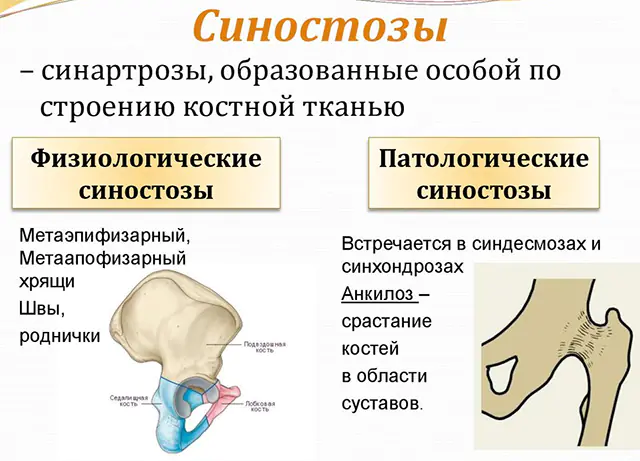
Types of synostosis are determined by the location and etiology of the pathological process. There is physiological synostosis, which is a normal condition that develops with age. As the child grows, fusion of the pelvic bones, sacral vertebrae, and bones at the base of the skull occurs. If the process goes beyond the norm and worsens the quality of life, it is defined as pathological.
Depending on the etiology, the following types of disease are distinguished:
- Congenital synostosis. Bone fusion occurs during pregnancy due to disturbances in the formation of connective tissue. The most common growth pathologies of the skull and arm bones become. Less common are syndactyly (fusion of the finger phalanges), synostosis of the ribs, vertebrae, or fusion of numerous bones.
- Acquired. The disease develops with age under the influence of infections and pathological conditions.
- Post-traumatic. Pathology that develops after injuries, damage in places of improper fusion of broken bones. The disease develops due to the absence or inadequate treatment of fractures. Synostosis of the vertebrae or lower leg bones is more often diagnosed.
- Artificial. The fusion is created during surgery to eliminate other pathology, for example, with multiple bone lesions. Artificial synostosis helps prevent the formation of false joints. More often the operation is performed on the tibia.
The following types of diseases are distinguished by location:
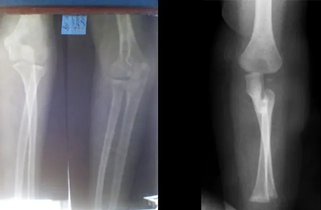
- Radioulnar (affects the bones of the forearm). A congenital disease in which the radius and ulna bones are fused. Symptoms of the disease may be completely absent or minor deviations may be observed. Muscular wasting gradually develops, patients are able to make complex movements with their hands. The minimum length of fusion is 1 cm, the maximum is 12 cm.
- Synostosis of the vertebrae in the neck or Klippel-Feil syndrome. A rare anomaly in which, as a result of fusion, the patient ends up with not 7 cervical vertebrae, but 4 or 5. In 1912, the disease was described by doctors from France Andre Feil and Maurice Klippel. With synostosis in the neck area, limited mobility of the upper vertebrae and low location of the hairline on the back of the head are observed. The disease is often combined with scoliosis and heart defects.
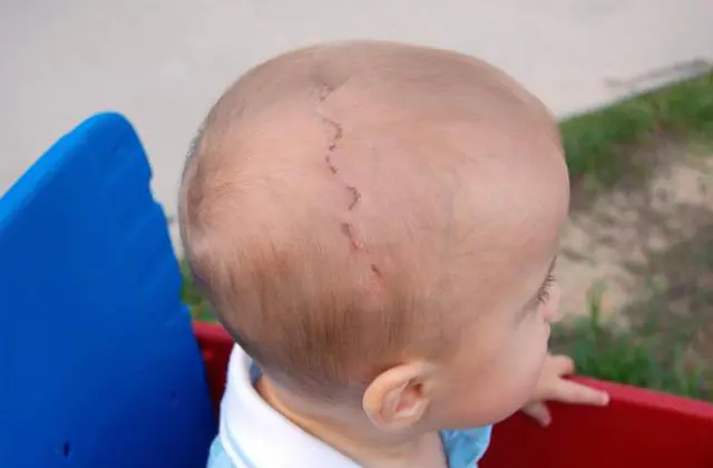
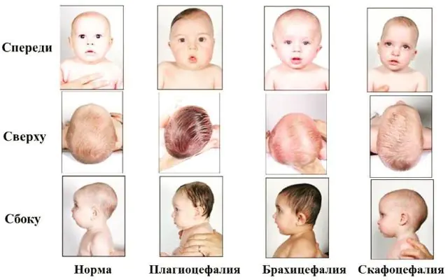
- Craniostenosis or synostosis of the skull. A common pathology in newborns, in which adjacent skull bones fuse along a bone suture. Depending on the location of the ossification line, the head is narrowed laterally or has a protrusion in the forehead area. Craniostenosis is diagnosed in 1 in 2000 newborns, with boys predominantly affected.
- Antley-Bixler syndrome. A rare disease characterized by the fusion of multiple bones.
- Rib synostosis. During the pathological process, the ribs in the back or front of the body fuse, sometimes several bones fuse completely.
- Lambdoid synostosis. A rare type of disease in which the occipital bones thicken and the front of the skull enlarges.
- Synostosis of the pelvic bone. The disease is characterized by fusion of the acetabulum and the head of the femur.
The photo shows trigonocephaly (metopic synostosis)
There are other types of synostosis depending on where the bones meet.
A serious consequence of synostosis is muscle atrophy. More often, deterioration of blood circulation in the hand area occurs, as a result of which it loses its functionality. When the vertebrae fuse, scoliosis and torticollis develop, posture is disturbed, and the risk of developing bronchopulmonary diseases increases.
Since the process of development of synostosis cannot be fully explained, it is impossible to develop clear preventive measures. Doctors recommend that women monitor their health during pregnancy, try to limit injuries and heavy physical activity, and be attentive to the treatment of bone injuries.
Reasons for the development of synostosis

Today, scientists cannot say unambiguously why bone synostosis develops. The main reason is called heredity. If one of your relatives has previously had similar abnormalities, the probability of their occurrence in the child ranges from 50 to 100%.
In most cases, synostosis in children occurs against the background of abnormal intrauterine development. But the disease can develop in childhood or adolescence for the following reasons:
- extensive injuries in which the integrity of bone and cartilage is disrupted;
- previous bone tuberculosis;
- severe osteochondrosis;
- typhoid fever, brucellosis;
- diseases associated with disruption of the endocrine system;
- dystrophy, degenerative changes in the spinal column.
Synostosis is not an independent disease, but develops against the background of injuries or infections. Sometimes there is no pathological basis and the etiology of the disease remains unknown.
Main symptoms of synostosis
Signs of synostosis vary depending on which part of the bone is affected. General symptoms include limited mobility of bones and joints, muscle wasting, and pain in the affected area.
Synostosis of the forearm
Symptoms of the disease are more pronounced with extensive adhesions. The patient's muscles in the hand and forearm gradually atrophy, and the size of the olecranon increases.
A person with synostosis has restrictions on joint mobility, in which he cannot:
- rotate the forearm;
- pick up objects;
- dress;
- use a spoon to eat;
- writing skills suffer.
To accurately make a diagnosis, a number of diagnostic measures are carried out.
Craniostenosis
Fusion of the skull bones is often combined with heart defects. Due to a decrease in the volume of the skull, a person’s intracranial pressure increases and frequent headaches occur.
Due to deformation, other symptoms are recorded:
- dizziness;
- vomit;
- convulsions;
- varicose veins in the head area;
- insomnia;
- chronic fatigue;
- strabismus;
- poor memory;
- mental disorders.
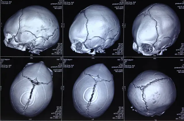
Diagnosis of the disease occurs without difficulties. The x-ray image clearly shows the bone fusion sites.
Klippel-Feil syndrome (short neck syndrome)
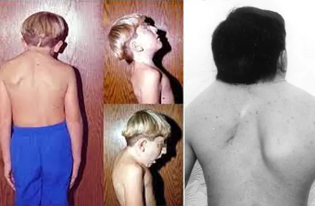
Synostosis of the spine in the neck area described by French authors is characterized by 3 symptoms:
- shortened neck;
- limited movements in the specified area;
- The hair on the back of the head is too low.
The disease leads to a decrease in the number of vertebrae in the cervical area or their fusion with the thoracic vertebrae.
Pathology is accompanied by other pathological conditions:
- scoliosis;
- high or wing-shaped arrangement of the scapula bones;
- fusion of fingers or toes;
- weakness of the arms due to impaired development of the chest muscles;
- absence of the ulna;
- flat feet.
Among the concomitant malformations of internal organs, kidney disease, defects of the gastric septum, lungs, and malposition of the aorta are diagnosed.
Other types of bone fusion
When the rib bones are deformed, the patient has problems with breathing and the cardiovascular system. Movement of the chest is limited and often causes pain. Along with fusion of the ribs, changes in the spine can be diagnosed.
Synostosis of the pelvic bones is manifested by lameness, pain when walking, and limited mobility of the lower extremities. If multiple bones fuse, a person has serious problems performing simple movements and meeting basic household needs.
Methods for diagnosing synostosis
Synostosis is treated by an orthopedic traumatologist, and in children by a pediatric surgeon. These specialists should be contacted if symptoms of the disease appear or if bone fusion is suspected in children. The doctor will prescribe the necessary tests to determine the location of the pathology.
After examining the patient, the doctor recommends a standard list of studies:
- x-ray of bones in the suspected affected area;
- MRI or CT (if x-ray does not reveal the location of the fusion);
- Ultrasound of the spine.
If the bones of the skull are affected, it is necessary to examine the fundus of the eye by an ophthalmologist and do craniometry (measure the volume of the skull). A neurologist evaluates the state of the brain and mental development.
If fused bones are concentrated in the cervical spine or forearm, an echocardiogram or cardiac ultrasound is recommended to identify cardiac pathologies.
How to treat synostosis?
If bone damage is diagnosed immediately after birth or in infancy, the situation can be corrected with the help of massages, therapeutic exercises and physiotherapy. The baby's bones are still soft and easy to handle. In older people, analgesics and drug administration using electrophoresis are used for pain relief.
There is no drug treatment for synostosis. To eliminate the defect, surgical intervention is used. The types of surgery depend on the affected area. More often, surgeons perform correction of the skull bones in children under 2-3 years of age to expand the volume of the skull and eliminate a cosmetic defect.
Surgeries to reconstruct the skull are performed under general anesthesia. For craniostenosis, the type of surgical intervention is determined by the nature of bone fusion:
- Linear craniotomy. The bones are cut along the line of the ossified suture. The same trajectory is chosen for cutting soft tissues. The edges of the bone are covered with a material that prevents further fusion, after which the soft tissue is sutured.
- Circular craniotomy. The technique differs from the previous one in that the incision is made around the circumference, then a strip of bone about 1 cm wide is removed.
- Fragmentation. The operation is carried out in 2 stages with an interval of 2-3 weeks. A flap of soft tissue is cut out from each side of the skull. Holes are made in the cranial bones and individual fragments are cut out to reduce intracranial pressure.
- Bilateral flap craniotomy. The soft tissue incision is made in an arc along the base of the scalp to the midline of the head. Using cutters, holes are made in the skull and a section of bone up to 2 cm wide is removed. The resulting oval flap is divided into 2 parts and bone bridges are formed to them.
- Fronto-orbital extension. During surgery, the frontal bones are moved forward and secured with titanium plates.
When the bones of the forearm are affected, the Ilizarov apparatus is used to stretch the interosseous membrane and move the forearm to a functionally comfortable position.
When fusion of cervical vertebrae occurs, massage, physiotherapy, and physical therapy are prescribed. They improve posture and prevent further fusion of bones.
In other cases, the need for surgery is determined individually, since the intervention entails possible complications. The help of a surgeon is sought if the defect significantly impairs the quality of life, and not just appearance.
Synostosis is almost impossible to treat. But with a properly performed surgical operation and rehabilitation period, it is possible to partially restore functionality to the bones and joints involved in the pathological process.
